The hair follicle is one of the most complex mini-organs in human biology, capable of cyclical regeneration, rapid cell differentiation, and the production of a highly specialized fiber. Central to this process is the inner root sheath (IRS), a transient but indispensable epithelial structure that molds, guides, and stabilizes the growing hair shaft. Although the IRS is destroyed before the hair fiber emerges from the skin surface, its influence on hair shape, strength, and integrity is profound. Defects in IRS formation or differentiation can lead to fragile, misshapen, or poorly anchored hair, underscoring its critical biological role.
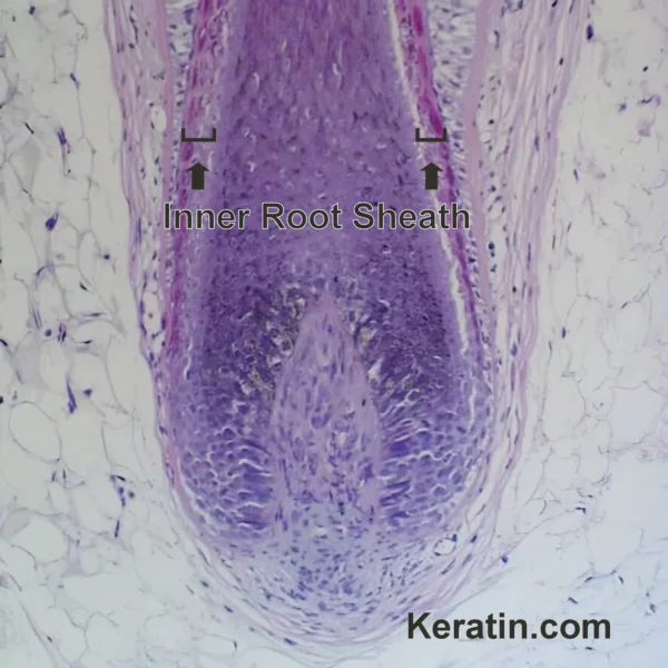
Anatomical Position and Developmental Context: The inner root sheath lies between the hair shaft and the outer root sheath (ORS) in the lower portion of the follicle. It originates from rapidly proliferating matrix keratinocytes adjacent to the dermal papilla. As these progenitor cells move upward, they undergo a tightly regulated program of differentiation that produces the concentric layers of the IRS.
The IRS extends from the hair bulb upward to approximately the level of the isthmus, where it undergoes abrupt keratinization and disintegration. Above this point, the hair shaft is no longer encased by the IRS and instead interfaces directly with the follicular canal. This spatial restriction highlights the IRS as a developmental scaffold rather than a permanent structure.
Structural Organization of the Inner Root Sheath: The IRS is composed of three morphologically and molecularly distinct layers arranged from outermost to innermost:
Henle’s Layer: Henle’s layer is the outermost IRS component, consisting of a single layer of flattened cells adjacent to the outer root sheath. It is the first IRS layer to undergo terminal differentiation and hardening. Early keratinization of Henle’s layer contributes to mechanical rigidity, stabilizing the follicle architecture during active hair growth.Huxley’s Layer: Beneath Henle’s layer lies Huxley’s layer, typically composed of one to three cell layers. These cells contain characteristic trichohyalin granules, which play a central role in IRS keratinization. Huxley’s layer is particularly important for maintaining adhesion between IRS layers and for transmitting mechanical forces during fiber elongation.Inner Root Sheath Cuticle: The innermost layer of the IRS interlocks directly with the cuticle of the hair shaft. This layer consists of a single layer of flattened cells whose cuticular scales are oriented oppositely to those of the hair shaft cuticle. This complementary “gear-like” arrangement is essential for anchoring the growing hair fiber and ensuring correct alignment as it ascends through the follicle. Molecular Composition and Keratinization: The inner root sheath is molecularly distinct from both the hair shaft and the outer root sheath. Its cells express a specific repertoire of type I and type II keratins, including keratins such as K25, K27, and K28, which are largely restricted to IRS lineages. These keratins form intermediate filament networks that provide structural integrity but differ from the harder, sulfur-rich keratins of the hair shaft cortex.
A defining molecular feature of the IRS is trichohyalin, a large, histidine-rich protein that accumulates in cytoplasmic granules, particularly within Huxley’s layer. During terminal differentiation, trichohyalin is enzymatically modified and cross-linked to keratin filaments by transglutaminases. This process creates a rigid, insoluble protein matrix that reinforces the IRS and supports the growing hair fiber.
Unlike epidermal keratinization, IRS cornification does not result in a classic stratum corneum. Instead, IRS cells undergo a specialized form of programmed hardening that preserves their shape long enough to fulfill their molding function before disintegrating.
Functional Roles of the Inner Root Sheath: The inner root sheath has a variety of important functions for hair follicle structure and hair fiber production
Hair Shaft Molding and Shape Determination: One of the most critical roles of the IRS is to define the shape and symmetry of the hair shaft. The geometry of the IRS, particularly the contour of the inner root sheath cuticle, constrains the developing hair fiber. Variations in IRS thickness, curvature, or differentiation are thought to contribute to differences in hair shape, including straight, wavy, and curly phenotypes.Mechanical Support and Guidance: During anagen, the hair shaft elongates rapidly and would be mechanically unstable without structural guidance. The IRS acts as a rigid sleeve that maintains alignment, prevents buckling, and transmits mechanical forces evenly along the follicle axis. This is especially important in long terminal hairs, where mechanical stress is substantial.Anchoring and Frictional Coupling: The interlocking cuticles of the IRS and hair shaft create frictional resistance that helps anchor the fiber within the follicle. This coupling ensures that the hair shaft remains properly positioned relative to the dermal papilla and matrix during growth. Disruption of this interface can result in loosely anchored hairs that are easily shed.Barrier and Compartmentalization Functions: Although not a classic permeability barrier, the IRS contributes to compartmentalization within the follicle. By separating the hair shaft from the metabolically active outer root sheath, it helps maintain distinct biochemical microenvironments necessary for orderly differentiation. Inner Root Sheath Dynamics During the Hair Cycle: The IRS is present only during the anagen (growth) phase of the hair cycle. As the follicle transitions into catagen, matrix cell proliferation ceases, and IRS production stops. The existing IRS undergoes rapid degeneration, coinciding with follicular regression. During telogen, the IRS is absent, reflecting the quiescent state of the follicle.
This cyclical regeneration underscores the IRS as a highly dynamic structure, rebuilt anew with each growth cycle. Errors in IRS reformation during early anagen can have lasting effects on hair quality throughout the entire cycle.
Relevance to Cosmetic and Regenerative Science: Understanding the biology of the inner root sheath has implications beyond clinical dermatology. In cosmetic science, IRS integrity influences fiber smoothness, alignment, and resistance to mechanical damage. Although cosmetic products cannot directly modify the IRS in vivo, insights into its function help explain why certain hair types are more prone to breakage or distortion.
In regenerative medicine and hair follicle bioengineering, successful recreation of a functional IRS is a major challenge. Any attempt to generate hair follicles ex vivo or through cell-based therapies must reproduce the precise spatial and molecular organization of the IRS to yield structurally normal hair fibers.
Clinical and Pathological Relevance: Abnormalities of the inner root sheath are implicated in several hair disorders, particularly those characterized by hair fragility or abnormal anchoring. Genetic defects affecting IRS-specific keratins or trichohyalin processing can result in structural hair shaft anomalies observable by light or electron microscopy.
Inner Root Sheath Alterations in Hair Shaft Disorders: From a clinical perspective, abnormalities of the inner root sheath are most clearly appreciated in congenital and acquired hair shaft disorders. Conditions such as loose anagen hair syndrome, trichorrhexis nodosa–like fragility patterns, and certain forms of hypotrichosis demonstrate impaired anchoring of the hair shaft that can often be traced to defective IRS–hair cuticle interlocking. In these disorders, premature IRS disintegration or abnormal cuticular differentiation results in hair fibers that lack adequate frictional coupling within the follicle, leading to effortless extraction and increased shedding. Recognition of IRS involvement helps distinguish true hair cycle abnormalities from primary shaft structural defects.Diagnostic Significance in Dermatopathology: In scalp biopsy interpretation, the integrity and differentiation state of the inner root sheath provide valuable contextual information, particularly when evaluating non-scarring versus early scarring alopecias. Preservation of IRS architecture during anagen favors a diagnosis of non-scarring alopecia, whereas premature loss, distortion, or inflammatory disruption of the IRS – especially below the isthmus – may signal early follicular damage with potential for permanent loss. Subtle IRS abnormalities can be overlooked if attention is focused exclusively on the outer root sheath or perifollicular inflammation, emphasizing the importance of systematic evaluation of all follicular compartments.Inner Root Sheath Vulnerability in Inflammatory Alopecias: The IRS appears particularly susceptible to inflammatory and immune-mediated injury due to its transient nature and high metabolic activity during anagen. In conditions such as alopecia areata, early inflammatory infiltrates may disrupt matrix differentiation programs that give rise to the IRS, resulting in structurally weakened follicles even before overt hair shedding becomes apparent. Similarly, in certain cicatricial alopecias, early IRS damage may precede irreversible outer root sheath stem cell loss, suggesting that IRS involvement could represent a critical early window in disease progression.Therapeutic and Prognostic Implications: Although current therapies do not directly target the inner root sheath, its condition has important implications for treatment response and prognosis. Effective anagen restoration – whether through anti-inflammatory, immunomodulatory, or regenerative interventions – necessarily involves re-establishment of normal IRS differentiation. In this context, clinical improvement in hair caliber, anchoring strength, and shaft uniformity may serve as indirect indicators of successful IRS regeneration. Conversely, persistent fragility or abnormal regrowth despite apparent disease control may suggest incomplete restoration of IRS function. Conclusion: The inner root sheath is a transient yet essential component of the hair follicle, acting as a biological mold, mechanical guide, and anchoring interface for the developing hair shaft. Its highly specialized structure, unique molecular composition, and tightly regulated life cycle reflect the remarkable sophistication of follicular biology. Although invisible once the hair emerges from the skin, the IRS leaves an enduring imprint on hair shape, strength, and integrity. Continued research into its development and pathology will remain central to advancing both medical and cosmetic approaches to hair disorders.
Bibliography
11711645 {11711645:JACCEB5G},{11711645:JI8HBGGW},{11711645:ECHG2URN},{11711645:EEEKG5X4},{11711645:DK7F4KF7},{11711645:YH24RHGQ},{11711645:RLQTY6RN},{11711645:Z5K38QX7},{11711645:I884PWLI},{11711645:DPPRT78B},{11711645:UFBR6R5M},{11711645:RD45M82E},{11711645:L69KDZIF},{11711645:8DT47LFR},{11711645:Y5NMY5WH},{11711645:PTV4CSWD},{11711645:R4S7RIFR},{11711645:83PBHH5H},{11711645:9PMLLKVZ},{11711645:9SLVDBEX},{11711645:BFIYXNK7},{11711645:N6HGRNMU},{11711645:I2GGA643},{11711645:R4YZ6DDH},{11711645:W42LDIDM},{11711645:ZJRTPA8E},{11711645:B8L28VAG},{11711645:YLDSSIEB},{11711645:EHJCAB8P},{11711645:KENLRRRW} 1 vancouver 50 date asc 2083 https://www.keratin.com/wp-content/plugins/zotpress/ %7B%22status%22%3A%22success%22%2C%22updateneeded%22%3Afalse%2C%22instance%22%3Afalse%2C%22meta%22%3A%7B%22request_last%22%3A0%2C%22request_next%22%3A0%2C%22used_cache%22%3Atrue%7D%2C%22data%22%3A%5B%7B%22key%22%3A%22DK7F4KF7%22%2C%22library%22%3A%7B%22id%22%3A11711645%7D%2C%22meta%22%3A%7B%22creatorSummary%22%3A%22Birbeck%20and%20Mercer%22%2C%22parsedDate%22%3A%221957-03-25%22%2C%22numChildren%22%3A0%7D%2C%22bib%22%3A%22%26lt%3Bdiv%20class%3D%26quot%3Bcsl-bib-body%26quot%3B%20style%3D%26quot%3Bline-height%3A%201.35%3B%20%26quot%3B%26gt%3B%5Cn%20%20%26lt%3Bdiv%20class%3D%26quot%3Bcsl-entry%26quot%3B%20style%3D%26quot%3Bclear%3A%20left%3B%20%26quot%3B%26gt%3B%5Cn%20%20%20%20%26lt%3Bdiv%20class%3D%26quot%3Bcsl-left-margin%26quot%3B%20style%3D%26quot%3Bfloat%3A%20left%3B%20padding-right%3A%200.5em%3B%20text-align%3A%20right%3B%20width%3A%201em%3B%26quot%3B%26gt%3B1.%26lt%3B%5C%2Fdiv%26gt%3B%26lt%3Bdiv%20class%3D%26quot%3Bcsl-right-inline%26quot%3B%20style%3D%26quot%3Bmargin%3A%200%20.4em%200%201.5em%3B%26quot%3B%26gt%3BBirbeck%20MS%2C%20Mercer%20EH.%20The%20electron%20microscopy%20of%20the%20human%20hair%20follicle.%20III.%20The%20inner%20root%20sheath%20and%20trichohyaline.%20J%20Biophys%20Biochem%20Cytol.%201957%20Mar%2025%3B3%282%29%3A223%26%23x2013%3B30.%26lt%3B%5C%2Fdiv%26gt%3B%5Cn%20%20%20%26lt%3B%5C%2Fdiv%26gt%3B%5Cn%26lt%3B%5C%2Fdiv%26gt%3B%22%2C%22data%22%3A%7B%22itemType%22%3A%22journalArticle%22%2C%22title%22%3A%22The%20electron%20microscopy%20of%20the%20human%20hair%20follicle.%20III.%20The%20inner%20root%20sheath%20and%20trichohyaline%22%2C%22creators%22%3A%5B%7B%22creatorType%22%3A%22author%22%2C%22firstName%22%3A%22M.%20S.%22%2C%22lastName%22%3A%22Birbeck%22%7D%2C%7B%22creatorType%22%3A%22author%22%2C%22firstName%22%3A%22E.%20H.%22%2C%22lastName%22%3A%22Mercer%22%7D%5D%2C%22abstractNote%22%3A%221.%20The%20three%20cylinders%20of%20cells%2C%20each%20one%20cell%20thick%2C%20which%20together%20constitute%20the%20inner%20root%20sheath%2C%20arise%20from%20the%20peripheral%20portions%20of%20the%20undifferentiated%20matrix.%20These%20cells%2C%20like%20the%20hair%20cuticle%2C%20are%20stabilised%20by%20the%20spread%20of%20adhesive%20contacts%20between%20their%20plasma%20membranes%20which%20occurs%20in%20the%20mid-bulb%20and%20upper%20bulb%20of%20the%20hair%20follicle.%202.%20The%20characteristic%20intracellular%20product%20of%20all%20three%20cell%20layers%20is%20trichohyaline.%20This%20substance%20is%20formed%20in%20the%20first%20place%20as%20amorphous%20droplets%20which%20subsequently%20transform%20into%20a%20birefringent%20form.%203.%20This%20transformation%2C%20involving%20the%20formation%20of%20a%20birefringent%20product%20from%20an%20amorphous%20precursor%2C%20is%20in%20contrast%20to%20the%20formation%20in%20the%20cortex%20of%20keratin%20which%20originates%20in%20a%20fibrous%20form.%204.%20Trichohyaline%20appears%20first%20and%20transforms%20first%20in%20the%20cells%20of%20Henle%20which%20are%20nearest%20the%20outer%20sheath%20and%20the%20dermal%20supply%20vessels.%20This%20transformation%20occurs%20at%20the%20level%20of%20the%20neck%20of%20the%20follicle.%20Synthesis%20and%20transformation%20in%20the%20cells%20of%20Huxley%20and%20the%20sheath%20cuticle%20lag%20behind%20the%20similar%20events%20in%20the%20cells%20of%20Henle.%20The%20transformation%20does%20not%20begin%20until%20the%20lower%20prekeratinous%20zone%20in%20the%20Huxley%20and%20cuticle%20cells.%205.%20The%20amorpous-fibrous%20transformation%20occurs%20rapidly%20cell%20by%20cell%20and%20involves%20the%20conversion%20of%20all%20the%20trichohyaline%20droplets.%20In%20longitudinal%20sections%20the%20birefringent%20modification%20can%20be%20seen%20extending%20from%20the%20droplets%20in%20both%20directions%20parallel%20to%20the%20axis%20of%20the%20hair.%20In%20cross-sections%20the%20images%20of%20the%20transformed%20material%20are%20difficult%20to%20interpret.%20They%20may%20be%20seen%20as%20sections%20of%20corrugated%20sheets%20%28%20approximately%20100%20A%20thick%29%20or%20condensed%20fibrils%20approximately%20100%20A%20in%20width.%206.%20At%20the%20same%20time%20that%20the%20trichohyaline%20transforms%2C%20the%20spacing%20between%20the%20cell%20membranes%20increases%20and%20a%20dark%20deposit%20appears%20centrally%20between%20them.%20This%20membrane%20complex%2C%20and%20the%20similar%20complex%20of%20the%20hair%20cuticle%20cells%20described%20in%20Part%202%2C%20may%20be%20specialised%20formations%20whose%20purpose%20is%20to%20hold%20the%20hardened%20cells%20together.%22%2C%22date%22%3A%221957-03-25%22%2C%22section%22%3A%22%22%2C%22partNumber%22%3A%22%22%2C%22partTitle%22%3A%22%22%2C%22DOI%22%3A%2210.1083%5C%2Fjcb.3.2.223%22%2C%22citationKey%22%3A%22%22%2C%22url%22%3A%22%22%2C%22PMID%22%3A%22%22%2C%22PMCID%22%3A%22%22%2C%22ISSN%22%3A%220095-9901%22%2C%22language%22%3A%22eng%22%2C%22collections%22%3A%5B%22JS9K5C2J%22%5D%2C%22dateModified%22%3A%222026-02-18T15%3A45%3A04Z%22%7D%7D%2C%7B%22key%22%3A%22YH24RHGQ%22%2C%22library%22%3A%7B%22id%22%3A11711645%7D%2C%22meta%22%3A%7B%22creatorSummary%22%3A%22Jarrett%22%2C%22parsedDate%22%3A%221958%22%2C%22numChildren%22%3A0%7D%2C%22bib%22%3A%22%26lt%3Bdiv%20class%3D%26quot%3Bcsl-bib-body%26quot%3B%20style%3D%26quot%3Bline-height%3A%201.35%3B%20%26quot%3B%26gt%3B%5Cn%20%20%26lt%3Bdiv%20class%3D%26quot%3Bcsl-entry%26quot%3B%20style%3D%26quot%3Bclear%3A%20left%3B%20%26quot%3B%26gt%3B%5Cn%20%20%20%20%26lt%3Bdiv%20class%3D%26quot%3Bcsl-left-margin%26quot%3B%20style%3D%26quot%3Bfloat%3A%20left%3B%20padding-right%3A%200.5em%3B%20text-align%3A%20right%3B%20width%3A%201em%3B%26quot%3B%26gt%3B1.%26lt%3B%5C%2Fdiv%26gt%3B%26lt%3Bdiv%20class%3D%26quot%3Bcsl-right-inline%26quot%3B%20style%3D%26quot%3Bmargin%3A%200%20.4em%200%201.5em%3B%26quot%3B%26gt%3BJarrett%20A.%20The%20chemistry%20of%20inner%20root%20sheath%20and%20hair%20keratins.%20Br%20J%20Dermatol.%201958%3B70%288%26%23x2013%3B9%29%3A271%26%23x2013%3B83.%26lt%3B%5C%2Fdiv%26gt%3B%5Cn%20%20%20%26lt%3B%5C%2Fdiv%26gt%3B%5Cn%26lt%3B%5C%2Fdiv%26gt%3B%22%2C%22data%22%3A%7B%22itemType%22%3A%22journalArticle%22%2C%22title%22%3A%22The%20chemistry%20of%20inner%20root%20sheath%20and%20hair%20keratins%22%2C%22creators%22%3A%5B%7B%22creatorType%22%3A%22author%22%2C%22firstName%22%3A%22A.%22%2C%22lastName%22%3A%22Jarrett%22%7D%5D%2C%22abstractNote%22%3A%22%22%2C%22date%22%3A%221958%22%2C%22section%22%3A%22%22%2C%22partNumber%22%3A%22%22%2C%22partTitle%22%3A%22%22%2C%22DOI%22%3A%2210.1111%5C%2Fj.1365-2133.1958.tb13339.x%22%2C%22citationKey%22%3A%22%22%2C%22url%22%3A%22%22%2C%22PMID%22%3A%22%22%2C%22PMCID%22%3A%22%22%2C%22ISSN%22%3A%220007-0963%22%2C%22language%22%3A%22eng%22%2C%22collections%22%3A%5B%22JS9K5C2J%22%5D%2C%22dateModified%22%3A%222026-02-18T15%3A44%3A39Z%22%7D%7D%2C%7B%22key%22%3A%22RLQTY6RN%22%2C%22library%22%3A%7B%22id%22%3A11711645%7D%2C%22meta%22%3A%7B%22creatorSummary%22%3A%22Rogers%22%2C%22parsedDate%22%3A%221958-04%22%2C%22numChildren%22%3A0%7D%2C%22bib%22%3A%22%26lt%3Bdiv%20class%3D%26quot%3Bcsl-bib-body%26quot%3B%20style%3D%26quot%3Bline-height%3A%201.35%3B%20%26quot%3B%26gt%3B%5Cn%20%20%26lt%3Bdiv%20class%3D%26quot%3Bcsl-entry%26quot%3B%20style%3D%26quot%3Bclear%3A%20left%3B%20%26quot%3B%26gt%3B%5Cn%20%20%20%20%26lt%3Bdiv%20class%3D%26quot%3Bcsl-left-margin%26quot%3B%20style%3D%26quot%3Bfloat%3A%20left%3B%20padding-right%3A%200.5em%3B%20text-align%3A%20right%3B%20width%3A%201em%3B%26quot%3B%26gt%3B1.%26lt%3B%5C%2Fdiv%26gt%3B%26lt%3Bdiv%20class%3D%26quot%3Bcsl-right-inline%26quot%3B%20style%3D%26quot%3Bmargin%3A%200%20.4em%200%201.5em%3B%26quot%3B%26gt%3BRogers%20GE.%20Some%20aspects%20of%20the%20structure%20of%20the%20inner%20root%20sheath%20of%20hair%20follicles%20revealed%20by%20light%20and%20electron%20microscopy.%20Exp%20Cell%20Res.%201958%20Apr%3B14%282%29%3A378%26%23x2013%3B87.%26lt%3B%5C%2Fdiv%26gt%3B%5Cn%20%20%20%26lt%3B%5C%2Fdiv%26gt%3B%5Cn%26lt%3B%5C%2Fdiv%26gt%3B%22%2C%22data%22%3A%7B%22itemType%22%3A%22journalArticle%22%2C%22title%22%3A%22Some%20aspects%20of%20the%20structure%20of%20the%20inner%20root%20sheath%20of%20hair%20follicles%20revealed%20by%20light%20and%20electron%20microscopy%22%2C%22creators%22%3A%5B%7B%22creatorType%22%3A%22author%22%2C%22firstName%22%3A%22G.%20E.%22%2C%22lastName%22%3A%22Rogers%22%7D%5D%2C%22abstractNote%22%3A%22%22%2C%22date%22%3A%221958-04%22%2C%22section%22%3A%22%22%2C%22partNumber%22%3A%22%22%2C%22partTitle%22%3A%22%22%2C%22DOI%22%3A%2210.1016%5C%2F0014-4827%2858%2990195-2%22%2C%22citationKey%22%3A%22%22%2C%22url%22%3A%22%22%2C%22PMID%22%3A%22%22%2C%22PMCID%22%3A%22%22%2C%22ISSN%22%3A%220014-4827%22%2C%22language%22%3A%22eng%22%2C%22collections%22%3A%5B%22JS9K5C2J%22%5D%2C%22dateModified%22%3A%222026-02-18T15%3A44%3A26Z%22%7D%7D%2C%7B%22key%22%3A%22Z5K38QX7%22%2C%22library%22%3A%7B%22id%22%3A11711645%7D%2C%22meta%22%3A%7B%22creatorSummary%22%3A%22Rogers%22%2C%22parsedDate%22%3A%221958-07%22%2C%22numChildren%22%3A0%7D%2C%22bib%22%3A%22%26lt%3Bdiv%20class%3D%26quot%3Bcsl-bib-body%26quot%3B%20style%3D%26quot%3Bline-height%3A%201.35%3B%20%26quot%3B%26gt%3B%5Cn%20%20%26lt%3Bdiv%20class%3D%26quot%3Bcsl-entry%26quot%3B%20style%3D%26quot%3Bclear%3A%20left%3B%20%26quot%3B%26gt%3B%5Cn%20%20%20%20%26lt%3Bdiv%20class%3D%26quot%3Bcsl-left-margin%26quot%3B%20style%3D%26quot%3Bfloat%3A%20left%3B%20padding-right%3A%200.5em%3B%20text-align%3A%20right%3B%20width%3A%201em%3B%26quot%3B%26gt%3B1.%26lt%3B%5C%2Fdiv%26gt%3B%26lt%3Bdiv%20class%3D%26quot%3Bcsl-right-inline%26quot%3B%20style%3D%26quot%3Bmargin%3A%200%20.4em%200%201.5em%3B%26quot%3B%26gt%3BRogers%20GE.%20Some%20observations%20on%20the%20proteins%20of%20the%20inner%20root%20sheath%20cells%20of%20hair%20follicles.%20Biochim%20Biophys%20Acta.%201958%20July%3B29%281%29%3A33%26%23x2013%3B43.%26lt%3B%5C%2Fdiv%26gt%3B%5Cn%20%20%20%26lt%3B%5C%2Fdiv%26gt%3B%5Cn%26lt%3B%5C%2Fdiv%26gt%3B%22%2C%22data%22%3A%7B%22itemType%22%3A%22journalArticle%22%2C%22title%22%3A%22Some%20observations%20on%20the%20proteins%20of%20the%20inner%20root%20sheath%20cells%20of%20hair%20follicles%22%2C%22creators%22%3A%5B%7B%22creatorType%22%3A%22author%22%2C%22firstName%22%3A%22G.%20E.%22%2C%22lastName%22%3A%22Rogers%22%7D%5D%2C%22abstractNote%22%3A%22%22%2C%22date%22%3A%221958-07%22%2C%22section%22%3A%22%22%2C%22partNumber%22%3A%22%22%2C%22partTitle%22%3A%22%22%2C%22DOI%22%3A%2210.1016%5C%2F0006-3002%2858%2990143-4%22%2C%22citationKey%22%3A%22%22%2C%22url%22%3A%22%22%2C%22PMID%22%3A%22%22%2C%22PMCID%22%3A%22%22%2C%22ISSN%22%3A%220006-3002%22%2C%22language%22%3A%22eng%22%2C%22collections%22%3A%5B%22JS9K5C2J%22%5D%2C%22dateModified%22%3A%222026-02-18T15%3A44%3A03Z%22%7D%7D%2C%7B%22key%22%3A%22I884PWLI%22%2C%22library%22%3A%7B%22id%22%3A11711645%7D%2C%22meta%22%3A%7B%22creatorSummary%22%3A%22Sugiyama%22%2C%22parsedDate%22%3A%221989-04%22%2C%22numChildren%22%3A0%7D%2C%22bib%22%3A%22%26lt%3Bdiv%20class%3D%26quot%3Bcsl-bib-body%26quot%3B%20style%3D%26quot%3Bline-height%3A%201.35%3B%20%26quot%3B%26gt%3B%5Cn%20%20%26lt%3Bdiv%20class%3D%26quot%3Bcsl-entry%26quot%3B%20style%3D%26quot%3Bclear%3A%20left%3B%20%26quot%3B%26gt%3B%5Cn%20%20%20%20%26lt%3Bdiv%20class%3D%26quot%3Bcsl-left-margin%26quot%3B%20style%3D%26quot%3Bfloat%3A%20left%3B%20padding-right%3A%200.5em%3B%20text-align%3A%20right%3B%20width%3A%201em%3B%26quot%3B%26gt%3B1.%26lt%3B%5C%2Fdiv%26gt%3B%26lt%3Bdiv%20class%3D%26quot%3Bcsl-right-inline%26quot%3B%20style%3D%26quot%3Bmargin%3A%200%20.4em%200%201.5em%3B%26quot%3B%26gt%3BSugiyama%20S.%20Cytochemistry%20of%20trichohyalin%20granules%3A%20a%20possible%20role%20for%20cornification%20of%20inner%20root%20sheath%20cell%20in%20the%20hair%20follicle.%20J%20Dermatol.%201989%20Apr%3B16%282%29%3A89%26%23x2013%3B97.%26lt%3B%5C%2Fdiv%26gt%3B%5Cn%20%20%20%26lt%3B%5C%2Fdiv%26gt%3B%5Cn%26lt%3B%5C%2Fdiv%26gt%3B%22%2C%22data%22%3A%7B%22itemType%22%3A%22journalArticle%22%2C%22title%22%3A%22Cytochemistry%20of%20trichohyalin%20granules%3A%20a%20possible%20role%20for%20cornification%20of%20inner%20root%20sheath%20cell%20in%20the%20hair%20follicle%22%2C%22creators%22%3A%5B%7B%22creatorType%22%3A%22author%22%2C%22firstName%22%3A%22S.%22%2C%22lastName%22%3A%22Sugiyama%22%7D%5D%2C%22abstractNote%22%3A%22We%20studied%20ultracytochemically%20how%20trichohyalin%20granules%20%28TGs%29%20were%20involved%20in%20the%20cornification%20of%20the%20inner%20root%20sheath%20%28IRS%29%20cells%20in%20the%20hair%20follicle.%20After%20storing%20unfixed%20mouse%20skin%20samples%20in%20glycerol%20and%20then%20low%20ionic%20strength%20salt%20solution%20%28G-LISS%29%2C%20the%20TGs%20reduced%20electron-dense%20amorphous%20material%20%28EDM%29%20to%20various%20degrees%2C%20with%20the%20appearance%20of%20filamentous%20material%20in%20routine%20staining.%20As%20the%20IRS%20cells%20were%20differentiating%2C%20these%20changes%20in%20the%20TGs%20became%20successively%20prominent%3B%20Some%20TGs%20consisted%20of%20the%20filamentous%20material%20and%20residual%20granular%20EDM%2C%20showing%20a%20%26quot%3Bfilamentogranular%20structure%26quot%3B.%20In%20the%20cells%20in%20transition%20to%20cornified%20cells%2C%20or%20transitional%20cells%2C%20the%20EDM%20of%20the%20TGs%20disappeared%20and%20consisted%20of%20only%20filamentous%20material%20which%20was%20intertwined%20with%20keratin%20filaments%20%28KFs%29.%20The%20internal%20substructure%20of%20the%20TGs%20induced%20by%20the%20G-LISS%20treatment%20stained%20well%20with%20ethanolic%20phosphotungstic%20acid%2C%20which%20reacts%20with%20basic%20proteins%2C%20and%20the%20KFs%20associated%20with%20the%20TGs%20also%20stained%20well.%20In%20skin%20samples%20not%20treated%20with%20G-LISS%2C%20the%20TGs%20of%20the%20transitional%20cells%20often%20exhibited%20fibrogranular%20structure%20after%20PTA%20staining%20to%20detect%20tissue%20basic%20proteins%2C%20suggesting%20a%20release%20of%20PTA%20stainable%20proteins%28s%29%20by%20the%20TGs%20in%20living%20transitional%20cells.%20The%20KFs%20of%20the%20cornified%20cells%20were%20more%20intensively%20stained%20and%20thicker%20%28about%2020%20nm%20wide%29%20than%20those%20of%20the%20differentiating%20IRS%20cells.%20These%20findings%20suggest%20that%20the%20TGs%20may%20contain%20at%20least%20two%20kinds%20of%20basic%20proteins%3B%20One%20is%20G-LISS-soluble%2C%20and%20the%20other%20is%20a%20very%20insoluble%20filamentous%20protein.%20These%20protein%20materials%20may%20be%20added%20to%20or%20incorporated%20into%20the%20KFs%20in%20the%20cornified%20cells%20with%20the%20disappearance%20of%20the%20TGs%2C%20since%20the%20KFs%20are%20markedly%20thick%20with%20PTA%20staining.%22%2C%22date%22%3A%221989-04%22%2C%22section%22%3A%22%22%2C%22partNumber%22%3A%22%22%2C%22partTitle%22%3A%22%22%2C%22DOI%22%3A%2210.1111%5C%2Fj.1346-8138.1989.tb01228.x%22%2C%22citationKey%22%3A%22%22%2C%22url%22%3A%22%22%2C%22PMID%22%3A%22%22%2C%22PMCID%22%3A%22%22%2C%22ISSN%22%3A%220385-2407%22%2C%22language%22%3A%22eng%22%2C%22collections%22%3A%5B%22JS9K5C2J%22%5D%2C%22dateModified%22%3A%222026-02-18T15%3A43%3A41Z%22%7D%7D%2C%7B%22key%22%3A%22JI8HBGGW%22%2C%22library%22%3A%7B%22id%22%3A11711645%7D%2C%22meta%22%3A%7B%22creatorSummary%22%3A%22Stark%20et%20al.%22%2C%22parsedDate%22%3A%221990-08%22%2C%22numChildren%22%3A0%7D%2C%22bib%22%3A%22%26lt%3Bdiv%20class%3D%26quot%3Bcsl-bib-body%26quot%3B%20style%3D%26quot%3Bline-height%3A%201.35%3B%20%26quot%3B%26gt%3B%5Cn%20%20%26lt%3Bdiv%20class%3D%26quot%3Bcsl-entry%26quot%3B%20style%3D%26quot%3Bclear%3A%20left%3B%20%26quot%3B%26gt%3B%5Cn%20%20%20%20%26lt%3Bdiv%20class%3D%26quot%3Bcsl-left-margin%26quot%3B%20style%3D%26quot%3Bfloat%3A%20left%3B%20padding-right%3A%200.5em%3B%20text-align%3A%20right%3B%20width%3A%201em%3B%26quot%3B%26gt%3B1.%26lt%3B%5C%2Fdiv%26gt%3B%26lt%3Bdiv%20class%3D%26quot%3Bcsl-right-inline%26quot%3B%20style%3D%26quot%3Bmargin%3A%200%20.4em%200%201.5em%3B%26quot%3B%26gt%3BStark%20HJ%2C%20Breitkreutz%20D%2C%20Limat%20A%2C%20Ryle%20CM%2C%20Roop%20D%2C%20Leigh%20I%2C%20et%20al.%20Keratins%201%20and%2010%20or%20homologues%20as%20regular%20constituents%20of%20inner%20root%20sheath%20and%20cuticle%20cells%20in%20the%20human%20hair%20follicle.%20Eur%20J%20Cell%20Biol.%201990%20Aug%3B52%282%29%3A359%26%23x2013%3B72.%26lt%3B%5C%2Fdiv%26gt%3B%5Cn%20%20%20%26lt%3B%5C%2Fdiv%26gt%3B%5Cn%26lt%3B%5C%2Fdiv%26gt%3B%22%2C%22data%22%3A%7B%22itemType%22%3A%22journalArticle%22%2C%22title%22%3A%22Keratins%201%20and%2010%20or%20homologues%20as%20regular%20constituents%20of%20inner%20root%20sheath%20and%20cuticle%20cells%20in%20the%20human%20hair%20follicle%22%2C%22creators%22%3A%5B%7B%22creatorType%22%3A%22author%22%2C%22firstName%22%3A%22H.%20J.%22%2C%22lastName%22%3A%22Stark%22%7D%2C%7B%22creatorType%22%3A%22author%22%2C%22firstName%22%3A%22D.%22%2C%22lastName%22%3A%22Breitkreutz%22%7D%2C%7B%22creatorType%22%3A%22author%22%2C%22firstName%22%3A%22A.%22%2C%22lastName%22%3A%22Limat%22%7D%2C%7B%22creatorType%22%3A%22author%22%2C%22firstName%22%3A%22C.%20M.%22%2C%22lastName%22%3A%22Ryle%22%7D%2C%7B%22creatorType%22%3A%22author%22%2C%22firstName%22%3A%22D.%22%2C%22lastName%22%3A%22Roop%22%7D%2C%7B%22creatorType%22%3A%22author%22%2C%22firstName%22%3A%22I.%22%2C%22lastName%22%3A%22Leigh%22%7D%2C%7B%22creatorType%22%3A%22author%22%2C%22firstName%22%3A%22N.%22%2C%22lastName%22%3A%22Fusenig%22%7D%5D%2C%22abstractNote%22%3A%22Morphological%20maturation%20of%20the%20inner%20root%20sheath%20%28IRS%29%20and%20cuticle%20of%20the%20human%20hair%20follicle%20reveals%20analogies%20to%20differentiation%20processes%20in%20other%20keratinizing%20epithelia.%20Detailed%20biochemical%20analysis%20of%20respective%20differentiation%20products%2C%20however%2C%20has%20been%20largely%20restricted%20by%20their%20low%20solubility.%20Herein%20we%20provide%20further%20evidence%20for%20the%20existence%20of%20K1%20and%20K10-derivatives%20in%20IRS%20and%20hair%20cuticle%20based%20on%20protein%20analysis%20of%20isolated%20fractions%20and%20immunofluorescence%20in%20situ%2C%20substantiating%20our%20earlier%20data%20%28Stark%2C%20H.%20J.%2C%20et%20al.%20Differentiation%2035%2C%20236-248%20%281987%29%29.%20Extracts%20from%20both%20compartments%20showed%20on%20two-dimensional%20%282D%29-polyacrylamide%20gels%20a%20group%20of%20presumptive%20K1%20and%20K10-turnover%20products%20in%20a%20wide%20pI%20%28basic%20to%20acidic%29%20and%20Mr%20range%20%2856%2C000-65%2C000%29%2C%20named%20IC-I%20to%20III%20and%20IC-IV%2C%20respectively.%20These%20components%20%28also%20found%20in%20nail%20plate%29%20reacted%20with%20specific%20antibodies%20%28to%20K1%20and%20K10%29%20on%20Western%20blots.%20Weak%20but%20distinctive%20radiolabeling%20of%20presumptive%20precursor%20spots%20close%20to%20authentic%20K1%20and%20K10%2C%20respectively%2C%20and%20their%20presence%20in%20lower%20follicle%20fractions%20%28distant%20from%20infundibulum%29%20largely%20precluded%20epidermal%20contamination.%20Two-dimensional%20tryptic%20peptide%20maps%20of%20excised%202D%20spots%20from%20the%20IC-I%20to%20III%20series%20revealed%20high%20homology%20to%20K1%2C%20and%20those%20from%20IC-IV%20components%20to%20K10.%20Immunodetection%20in%20frozen%20sections%20was%20improved%20by%20trypsin%20pretreatment%20and%20showed%20distinguished%20staining%20for%20K1%20and%20K10%20in%20IRS%20ranging%20from%20the%20lower%20bulbus%20region%20up%20to%20the%20%26quot%3Bkeratinizing%20zone%26quot%3B%20of%20the%20follicle.%20Above%2C%20the%20reaction%20was%20abruptly%20abolished%20which%20coincides%20with%20ultrastructural%20%26quot%3Bmelting%26quot%3B%20of%20distinct%20filaments%20in%20the%20intracellular%20matrix.%20Thus%2C%20our%20data%20suggest%20that%20differentiation%20in%20these%20follicular%20compartments%20%28IRS%20and%20cuticle%29%20might%20follow%20common%20principles%20of%20keratinization.%22%2C%22date%22%3A%221990-08%22%2C%22section%22%3A%22%22%2C%22partNumber%22%3A%22%22%2C%22partTitle%22%3A%22%22%2C%22DOI%22%3A%22%22%2C%22citationKey%22%3A%22%22%2C%22url%22%3A%22%22%2C%22PMID%22%3A%22%22%2C%22PMCID%22%3A%22%22%2C%22ISSN%22%3A%220171-9335%22%2C%22language%22%3A%22eng%22%2C%22collections%22%3A%5B%22JS9K5C2J%22%5D%2C%22dateModified%22%3A%222026-02-18T15%3A47%3A16Z%22%7D%7D%2C%7B%22key%22%3A%22Y5NMY5WH%22%2C%22library%22%3A%7B%22id%22%3A11711645%7D%2C%22meta%22%3A%7B%22creatorSummary%22%3A%22Clemmensen%20et%20al.%22%2C%22parsedDate%22%3A%221991-06%22%2C%22numChildren%22%3A0%7D%2C%22bib%22%3A%22%26lt%3Bdiv%20class%3D%26quot%3Bcsl-bib-body%26quot%3B%20style%3D%26quot%3Bline-height%3A%201.35%3B%20%26quot%3B%26gt%3B%5Cn%20%20%26lt%3Bdiv%20class%3D%26quot%3Bcsl-entry%26quot%3B%20style%3D%26quot%3Bclear%3A%20left%3B%20%26quot%3B%26gt%3B%5Cn%20%20%20%20%26lt%3Bdiv%20class%3D%26quot%3Bcsl-left-margin%26quot%3B%20style%3D%26quot%3Bfloat%3A%20left%3B%20padding-right%3A%200.5em%3B%20text-align%3A%20right%3B%20width%3A%201em%3B%26quot%3B%26gt%3B1.%26lt%3B%5C%2Fdiv%26gt%3B%26lt%3Bdiv%20class%3D%26quot%3Bcsl-right-inline%26quot%3B%20style%3D%26quot%3Bmargin%3A%200%20.4em%200%201.5em%3B%26quot%3B%26gt%3BClemmensen%20OJ%2C%20Hainau%20B%2C%20Hansted%20B.%20The%20ultrastructure%20of%20the%20transition%20zone%20between%20specialized%20cells%20%28%26%23x201C%3BFl%26%23xFC%3Bgelzellen%26%23x201D%3B%29%20of%20Huxley%26%23x2019%3Bs%20layer%20of%20the%20inner%20root%20sheath%20and%20cells%20of%20the%20outer%20root%20sheath%20of%20the%20human%20hair%20follicle.%20Am%20J%20Dermatopathol.%201991%20June%3B13%283%29%3A264%26%23x2013%3B70.%26lt%3B%5C%2Fdiv%26gt%3B%5Cn%20%20%20%26lt%3B%5C%2Fdiv%26gt%3B%5Cn%26lt%3B%5C%2Fdiv%26gt%3B%22%2C%22data%22%3A%7B%22itemType%22%3A%22journalArticle%22%2C%22title%22%3A%22The%20ultrastructure%20of%20the%20transition%20zone%20between%20specialized%20cells%20%28%5C%22Fl%5Cu00fcgelzellen%5C%22%29%20of%20Huxley%27s%20layer%20of%20the%20inner%20root%20sheath%20and%20cells%20of%20the%20outer%20root%20sheath%20of%20the%20human%20hair%20follicle%22%2C%22creators%22%3A%5B%7B%22creatorType%22%3A%22author%22%2C%22firstName%22%3A%22O.%20J.%22%2C%22lastName%22%3A%22Clemmensen%22%7D%2C%7B%22creatorType%22%3A%22author%22%2C%22firstName%22%3A%22B.%22%2C%22lastName%22%3A%22Hainau%22%7D%2C%7B%22creatorType%22%3A%22author%22%2C%22firstName%22%3A%22B.%22%2C%22lastName%22%3A%22Hansted%22%7D%5D%2C%22abstractNote%22%3A%22We%20studied%20biopsy%20specimens%20taken%20from%20the%20scalp%20of%20four%20normal%20volunteers%20by%20transmission%20electron%20microscopy%20%28TEM%29.%20At%20the%20suprabulbous%20level%20of%20the%20follicle%2C%20where%20the%20cells%20of%20Henle%26%23039%3Bs%20layer%20are%20fully%20keratinized%20but%20the%20cells%20of%20Huxley%26%23039%3Bs%20layer%20are%20not%2C%20the%20cytoplasmic%20processes%20of%20Huxley%26%23039%3Bs%20layer%20%28%26quot%3BFl%5Cu00fcgelzellen%26quot%3B%29%20reach%20the%20cells%20of%20the%20outer%20root%20sheath%20through%20the%20keratinized%20cylinder%20of%20Henle%26%23039%3Bs%20layer.%20This%20junction%20between%20the%20outer%20root%20sheath%20cells%20and%20the%20%26quot%3BFl%5Cu00fcgelzellen%26quot%3B%20has%20not%20been%20previously%20characterized%20ultrastructurally.%20It%20is%20peculiar%20in%20its%20total%20lack%20of%20desmosomes%2C%20and%20it%20may%20have%20the%20features%20of%20a%20gap%20junction.%20The%20function%20of%20this%20structure%20and%20its%20possible%20role%20in%20hair%20growth%20is%20unknown.%20It%20may%20be%20related%20to%20nutrition%2C%20differentiation%2C%20or%20both.%22%2C%22date%22%3A%221991-06%22%2C%22section%22%3A%22%22%2C%22partNumber%22%3A%22%22%2C%22partTitle%22%3A%22%22%2C%22DOI%22%3A%2210.1097%5C%2F00000372-199106000-00008%22%2C%22citationKey%22%3A%22%22%2C%22url%22%3A%22%22%2C%22PMID%22%3A%22%22%2C%22PMCID%22%3A%22%22%2C%22ISSN%22%3A%220193-1091%22%2C%22language%22%3A%22eng%22%2C%22collections%22%3A%5B%22JS9K5C2J%22%5D%2C%22dateModified%22%3A%222026-02-18T15%3A38%3A13Z%22%7D%7D%2C%7B%22key%22%3A%22W42LDIDM%22%2C%22library%22%3A%7B%22id%22%3A11711645%7D%2C%22meta%22%3A%7B%22creatorSummary%22%3A%22Tarcsa%20et%20al.%22%2C%22parsedDate%22%3A%221997-10-31%22%2C%22numChildren%22%3A0%7D%2C%22bib%22%3A%22%26lt%3Bdiv%20class%3D%26quot%3Bcsl-bib-body%26quot%3B%20style%3D%26quot%3Bline-height%3A%201.35%3B%20%26quot%3B%26gt%3B%5Cn%20%20%26lt%3Bdiv%20class%3D%26quot%3Bcsl-entry%26quot%3B%20style%3D%26quot%3Bclear%3A%20left%3B%20%26quot%3B%26gt%3B%5Cn%20%20%20%20%26lt%3Bdiv%20class%3D%26quot%3Bcsl-left-margin%26quot%3B%20style%3D%26quot%3Bfloat%3A%20left%3B%20padding-right%3A%200.5em%3B%20text-align%3A%20right%3B%20width%3A%201em%3B%26quot%3B%26gt%3B1.%26lt%3B%5C%2Fdiv%26gt%3B%26lt%3Bdiv%20class%3D%26quot%3Bcsl-right-inline%26quot%3B%20style%3D%26quot%3Bmargin%3A%200%20.4em%200%201.5em%3B%26quot%3B%26gt%3BTarcsa%20E%2C%20Marekov%20LN%2C%20Andreoli%20J%2C%20Idler%20WW%2C%20Candi%20E%2C%20Chung%20SI%2C%20et%20al.%20The%20fate%20of%20trichohyalin.%20Sequential%20post-translational%20modifications%20by%20peptidyl-arginine%20deiminase%20and%20transglutaminases.%20J%20Biol%20Chem.%201997%20Oct%2031%3B272%2844%29%3A27893%26%23x2013%3B901.%26lt%3B%5C%2Fdiv%26gt%3B%5Cn%20%20%20%26lt%3B%5C%2Fdiv%26gt%3B%5Cn%26lt%3B%5C%2Fdiv%26gt%3B%22%2C%22data%22%3A%7B%22itemType%22%3A%22journalArticle%22%2C%22title%22%3A%22The%20fate%20of%20trichohyalin.%20Sequential%20post-translational%20modifications%20by%20peptidyl-arginine%20deiminase%20and%20transglutaminases%22%2C%22creators%22%3A%5B%7B%22creatorType%22%3A%22author%22%2C%22firstName%22%3A%22E.%22%2C%22lastName%22%3A%22Tarcsa%22%7D%2C%7B%22creatorType%22%3A%22author%22%2C%22firstName%22%3A%22L.%20N.%22%2C%22lastName%22%3A%22Marekov%22%7D%2C%7B%22creatorType%22%3A%22author%22%2C%22firstName%22%3A%22J.%22%2C%22lastName%22%3A%22Andreoli%22%7D%2C%7B%22creatorType%22%3A%22author%22%2C%22firstName%22%3A%22W.%20W.%22%2C%22lastName%22%3A%22Idler%22%7D%2C%7B%22creatorType%22%3A%22author%22%2C%22firstName%22%3A%22E.%22%2C%22lastName%22%3A%22Candi%22%7D%2C%7B%22creatorType%22%3A%22author%22%2C%22firstName%22%3A%22S.%20I.%22%2C%22lastName%22%3A%22Chung%22%7D%2C%7B%22creatorType%22%3A%22author%22%2C%22firstName%22%3A%22P.%20M.%22%2C%22lastName%22%3A%22Steinert%22%7D%5D%2C%22abstractNote%22%3A%22Trichohyalin%20%28THH%29%20is%20a%20major%20structural%20protein%20of%20the%20inner%20root%20sheath%20cells%20and%20medulla%20layer%20of%20the%20hair%20follicle%20and%2C%20to%20a%20lesser%20extent%2C%20of%20other%20specialized%20epithelia.%20THH%20is%20a%20high%20molecular%20weight%20insoluble%20alpha-helix-rich%20protein%20that%20forms%20rigid%20structures%20as%20a%20result%20of%20postsynthetic%20modifications%20by%20two%20Ca2%2B-dependent%20enzymes%2C%20transglutaminases%20%28TGases%29%20%28protein%20cross-linking%29%20and%20peptidyl-arginine%20deiminase%20%28conversion%20of%20arginines%20to%20citrullines%20with%20loss%20of%20organized%20structure%29.%20The%20modified%20THH%20is%20thought%20to%20serve%20as%20a%20keratin%20intermediate%20filament%20matrix%20protein%20and%5C%2For%20as%20a%20constituent%20of%20the%20cell%20envelope.%20In%20this%20paper%2C%20we%20have%20explored%20in%20vitro%20the%20order%20of%20processing%20of%20THH%20to%20fulfill%20these%20functions%2C%20using%20an%20expressed%20truncated%2C%20more%20soluble%20form%20THH-8.%20THH-8%20is%20a%20complete%20substrate%20for%20three%20known%20TGases%20expressed%20in%20epithelia%2C%20but%20the%20kinetic%20efficiency%20with%20TGase%203%20is%20by%20far%20the%20greatest.%20Following%20maximal%20conversion%20of%20its%20arginines%20to%20citrullines%2C%20THH-8%20is%20cross-linked%20even%20more%20efficiently%20by%20TGase%203%2C%20using%20most%20glutamines%20partially%20and%20all%20lysines.%20In%20addition%2C%20we%20show%20that%20insoluble%20aggregates%20of%20THH-8%20or%20native%20pig%20tongue%20THH%20can%20be%20solubilized%20following%20peptidyl-arginine%20deiminase%20modification.%20Together%2C%20these%20data%20suggest%20an%20in%20vivo%20model%20in%20which%20THH%20located%20in%20insoluble%20cytoplasmic%20droplets%20is%20first%20modified%20by%20peptidyl-arginine%20deiminase%20which%20denatures%20it%20and%20makes%20it%20more%20soluble.%20This%20renders%20it%20available%20for%20efficient%20cross-linking%20by%20TGase%203%20to%20form%20highly%20cross-linked%20rigid%20structures%20in%20the%20cells.%20This%20temporal%20order%20of%20reaction%20is%20supported%20by%20the%20observation%20that%20THH%20is%20expressed%20in%20hair%20follicle%20cells%20before%20the%20TGase%203%20enzyme.%22%2C%22date%22%3A%221997-10-31%22%2C%22section%22%3A%22%22%2C%22partNumber%22%3A%22%22%2C%22partTitle%22%3A%22%22%2C%22DOI%22%3A%2210.1074%5C%2Fjbc.272.44.27893%22%2C%22citationKey%22%3A%22%22%2C%22url%22%3A%22%22%2C%22PMID%22%3A%22%22%2C%22PMCID%22%3A%22%22%2C%22ISSN%22%3A%220021-9258%22%2C%22language%22%3A%22eng%22%2C%22collections%22%3A%5B%22JS9K5C2J%22%5D%2C%22dateModified%22%3A%222026-02-18T15%3A27%3A42Z%22%7D%7D%2C%7B%22key%22%3A%22KENLRRRW%22%2C%22library%22%3A%7B%22id%22%3A11711645%7D%2C%22meta%22%3A%7B%22creatorSummary%22%3A%22Paus%20and%20Cotsarelis%22%2C%22parsedDate%22%3A%221999-08-12%22%2C%22numChildren%22%3A0%7D%2C%22bib%22%3A%22%26lt%3Bdiv%20class%3D%26quot%3Bcsl-bib-body%26quot%3B%20style%3D%26quot%3Bline-height%3A%201.35%3B%20%26quot%3B%26gt%3B%5Cn%20%20%26lt%3Bdiv%20class%3D%26quot%3Bcsl-entry%26quot%3B%20style%3D%26quot%3Bclear%3A%20left%3B%20%26quot%3B%26gt%3B%5Cn%20%20%20%20%26lt%3Bdiv%20class%3D%26quot%3Bcsl-left-margin%26quot%3B%20style%3D%26quot%3Bfloat%3A%20left%3B%20padding-right%3A%200.5em%3B%20text-align%3A%20right%3B%20width%3A%201em%3B%26quot%3B%26gt%3B1.%26lt%3B%5C%2Fdiv%26gt%3B%26lt%3Bdiv%20class%3D%26quot%3Bcsl-right-inline%26quot%3B%20style%3D%26quot%3Bmargin%3A%200%20.4em%200%201.5em%3B%26quot%3B%26gt%3BPaus%20R%2C%20Cotsarelis%20G.%20The%20biology%20of%20hair%20follicles.%20N%20Engl%20J%20Med.%201999%20Aug%2012%3B341%287%29%3A491%26%23x2013%3B7.%26lt%3B%5C%2Fdiv%26gt%3B%5Cn%20%20%20%26lt%3B%5C%2Fdiv%26gt%3B%5Cn%26lt%3B%5C%2Fdiv%26gt%3B%22%2C%22data%22%3A%7B%22itemType%22%3A%22journalArticle%22%2C%22title%22%3A%22The%20biology%20of%20hair%20follicles%22%2C%22creators%22%3A%5B%7B%22creatorType%22%3A%22author%22%2C%22firstName%22%3A%22R.%22%2C%22lastName%22%3A%22Paus%22%7D%2C%7B%22creatorType%22%3A%22author%22%2C%22firstName%22%3A%22G.%22%2C%22lastName%22%3A%22Cotsarelis%22%7D%5D%2C%22abstractNote%22%3A%22%22%2C%22date%22%3A%221999-08-12%22%2C%22section%22%3A%22%22%2C%22partNumber%22%3A%22%22%2C%22partTitle%22%3A%22%22%2C%22DOI%22%3A%2210.1056%5C%2FNEJM199908123410706%22%2C%22citationKey%22%3A%22%22%2C%22url%22%3A%22%22%2C%22PMID%22%3A%22%22%2C%22PMCID%22%3A%22%22%2C%22ISSN%22%3A%220028-4793%22%2C%22language%22%3A%22eng%22%2C%22collections%22%3A%5B%22JS9K5C2J%22%5D%2C%22dateModified%22%3A%222026-02-18T15%3A24%3A08Z%22%7D%7D%2C%7B%22key%22%3A%22DPPRT78B%22%2C%22library%22%3A%7B%22id%22%3A11711645%7D%2C%22meta%22%3A%7B%22creatorSummary%22%3A%22Nakatake%20et%20al.%22%2C%22parsedDate%22%3A%222001-02-16%22%2C%22numChildren%22%3A0%7D%2C%22bib%22%3A%22%26lt%3Bdiv%20class%3D%26quot%3Bcsl-bib-body%26quot%3B%20style%3D%26quot%3Bline-height%3A%201.35%3B%20%26quot%3B%26gt%3B%5Cn%20%20%26lt%3Bdiv%20class%3D%26quot%3Bcsl-entry%26quot%3B%20style%3D%26quot%3Bclear%3A%20left%3B%20%26quot%3B%26gt%3B%5Cn%20%20%20%20%26lt%3Bdiv%20class%3D%26quot%3Bcsl-left-margin%26quot%3B%20style%3D%26quot%3Bfloat%3A%20left%3B%20padding-right%3A%200.5em%3B%20text-align%3A%20right%3B%20width%3A%201em%3B%26quot%3B%26gt%3B1.%26lt%3B%5C%2Fdiv%26gt%3B%26lt%3Bdiv%20class%3D%26quot%3Bcsl-right-inline%26quot%3B%20style%3D%26quot%3Bmargin%3A%200%20.4em%200%201.5em%3B%26quot%3B%26gt%3BNakatake%20Y%2C%20Hoshikawa%20M%2C%20Asaki%20T%2C%20Kassai%20Y%2C%20Itoh%20N.%20Identification%20of%20a%20novel%20fibroblast%20growth%20factor%2C%20FGF-22%2C%20preferentially%20expressed%20in%20the%20inner%20root%20sheath%20of%20the%20hair%20follicle.%20Biochim%20Biophys%20Acta.%202001%20Feb%2016%3B1517%283%29%3A460%26%23x2013%3B3.%26lt%3B%5C%2Fdiv%26gt%3B%5Cn%20%20%20%26lt%3B%5C%2Fdiv%26gt%3B%5Cn%26lt%3B%5C%2Fdiv%26gt%3B%22%2C%22data%22%3A%7B%22itemType%22%3A%22journalArticle%22%2C%22title%22%3A%22Identification%20of%20a%20novel%20fibroblast%20growth%20factor%2C%20FGF-22%2C%20preferentially%20expressed%20in%20the%20inner%20root%20sheath%20of%20the%20hair%20follicle%22%2C%22creators%22%3A%5B%7B%22creatorType%22%3A%22author%22%2C%22firstName%22%3A%22Y.%22%2C%22lastName%22%3A%22Nakatake%22%7D%2C%7B%22creatorType%22%3A%22author%22%2C%22firstName%22%3A%22M.%22%2C%22lastName%22%3A%22Hoshikawa%22%7D%2C%7B%22creatorType%22%3A%22author%22%2C%22firstName%22%3A%22T.%22%2C%22lastName%22%3A%22Asaki%22%7D%2C%7B%22creatorType%22%3A%22author%22%2C%22firstName%22%3A%22Y.%22%2C%22lastName%22%3A%22Kassai%22%7D%2C%7B%22creatorType%22%3A%22author%22%2C%22firstName%22%3A%22N.%22%2C%22lastName%22%3A%22Itoh%22%7D%5D%2C%22abstractNote%22%3A%22We%20isolated%20cDNA%20encoding%20a%20novel%20fibroblast%20growth%20factor%20%28FGF-22%29%20%28170%20amino%20acids%29%20from%20human%20placenta.%20Of%20the%20FGF%20family%20members%2C%20FGF-22%2C%20which%20appears%20to%20be%20a%20secreted%20protein%2C%20is%20most%20similar%20to%20FGF-10%20and%20FGF-7%20%28approximately%2046%25%20and%20approximately%2040%25%20amino%20acid%20identities%2C%20respectively%29.%20The%20human%20FGF-22%20gene%20was%20localized%20on%20chromosome%2019p13.3.%20We%20also%20isolated%20mouse%20cDNA%20encoding%20FGF-22%20%28162%20amino%20acids%29%20from%20the%20skin.%20Mouse%20FGF-22%20shows%20high%20homology%20%2887%25%20amino%20acid%20identity%29%20to%20human%20FGF-22.%20Mouse%20FGF-22%20mRNA%20was%20found%20to%20be%20preferentially%20expressed%20in%20the%20skin%20among%20the%20mouse%20adult%20tissues%20examined%20by%20Northern%20blotting%20analysis.%20By%20in%20situ%20hybridization%2C%20FGF-22%20mRNA%20in%20the%20skin%20was%20found%20to%20be%20preferentially%20expressed%20in%20the%20inner%20root%20sheath%20of%20the%20hair%20follicle.%20Therefore%2C%20FGF-22%20is%20expected%20to%20be%20a%20unique%20FGF%20that%20plays%20a%20role%20in%20hair%20development.%22%2C%22date%22%3A%222001-02-16%22%2C%22section%22%3A%22%22%2C%22partNumber%22%3A%22%22%2C%22partTitle%22%3A%22%22%2C%22DOI%22%3A%2210.1016%5C%2Fs0167-4781%2800%2900302-x%22%2C%22citationKey%22%3A%22%22%2C%22url%22%3A%22%22%2C%22PMID%22%3A%22%22%2C%22PMCID%22%3A%22%22%2C%22ISSN%22%3A%220006-3002%22%2C%22language%22%3A%22eng%22%2C%22collections%22%3A%5B%22JS9K5C2J%22%5D%2C%22dateModified%22%3A%222026-02-18T15%3A43%3A27Z%22%7D%7D%2C%7B%22key%22%3A%22BFIYXNK7%22%2C%22library%22%3A%7B%22id%22%3A11711645%7D%2C%22meta%22%3A%7B%22creatorSummary%22%3A%22Ellis%20et%20al.%22%2C%22parsedDate%22%3A%222001-09-01%22%2C%22numChildren%22%3A0%7D%2C%22bib%22%3A%22%26lt%3Bdiv%20class%3D%26quot%3Bcsl-bib-body%26quot%3B%20style%3D%26quot%3Bline-height%3A%201.35%3B%20%26quot%3B%26gt%3B%5Cn%20%20%26lt%3Bdiv%20class%3D%26quot%3Bcsl-entry%26quot%3B%20style%3D%26quot%3Bclear%3A%20left%3B%20%26quot%3B%26gt%3B%5Cn%20%20%20%20%26lt%3Bdiv%20class%3D%26quot%3Bcsl-left-margin%26quot%3B%20style%3D%26quot%3Bfloat%3A%20left%3B%20padding-right%3A%200.5em%3B%20text-align%3A%20right%3B%20width%3A%201em%3B%26quot%3B%26gt%3B1.%26lt%3B%5C%2Fdiv%26gt%3B%26lt%3Bdiv%20class%3D%26quot%3Bcsl-right-inline%26quot%3B%20style%3D%26quot%3Bmargin%3A%200%20.4em%200%201.5em%3B%26quot%3B%26gt%3BEllis%20T%2C%20Gambardella%20L%2C%20Horcher%20M%2C%20Tschanz%20S%2C%20Capol%20J%2C%20Bertram%20P%2C%20et%20al.%20The%20transcriptional%20repressor%20CDP%20%28Cutl1%29%20is%20essential%20for%20epithelial%20cell%20differentiation%20of%20the%20lung%20and%20the%20hair%20follicle.%20Genes%20Dev.%202001%20Sept%201%3B15%2817%29%3A2307%26%23x2013%3B19.%26lt%3B%5C%2Fdiv%26gt%3B%5Cn%20%20%20%26lt%3B%5C%2Fdiv%26gt%3B%5Cn%26lt%3B%5C%2Fdiv%26gt%3B%22%2C%22data%22%3A%7B%22itemType%22%3A%22journalArticle%22%2C%22title%22%3A%22The%20transcriptional%20repressor%20CDP%20%28Cutl1%29%20is%20essential%20for%20epithelial%20cell%20differentiation%20of%20the%20lung%20and%20the%20hair%20follicle%22%2C%22creators%22%3A%5B%7B%22creatorType%22%3A%22author%22%2C%22firstName%22%3A%22T.%22%2C%22lastName%22%3A%22Ellis%22%7D%2C%7B%22creatorType%22%3A%22author%22%2C%22firstName%22%3A%22L.%22%2C%22lastName%22%3A%22Gambardella%22%7D%2C%7B%22creatorType%22%3A%22author%22%2C%22firstName%22%3A%22M.%22%2C%22lastName%22%3A%22Horcher%22%7D%2C%7B%22creatorType%22%3A%22author%22%2C%22firstName%22%3A%22S.%22%2C%22lastName%22%3A%22Tschanz%22%7D%2C%7B%22creatorType%22%3A%22author%22%2C%22firstName%22%3A%22J.%22%2C%22lastName%22%3A%22Capol%22%7D%2C%7B%22creatorType%22%3A%22author%22%2C%22firstName%22%3A%22P.%22%2C%22lastName%22%3A%22Bertram%22%7D%2C%7B%22creatorType%22%3A%22author%22%2C%22firstName%22%3A%22W.%22%2C%22lastName%22%3A%22Jochum%22%7D%2C%7B%22creatorType%22%3A%22author%22%2C%22firstName%22%3A%22Y.%22%2C%22lastName%22%3A%22Barrandon%22%7D%2C%7B%22creatorType%22%3A%22author%22%2C%22firstName%22%3A%22M.%22%2C%22lastName%22%3A%22Busslinger%22%7D%5D%2C%22abstractNote%22%3A%22The%20mammalian%20Cutl1%20gene%20codes%20for%20the%20CCAAT%20displacement%20protein%20%28CDP%29%2C%20which%20has%20been%20implicated%20as%20a%20transcriptional%20repressor%20in%20diverse%20processes%20such%20as%20terminal%20differentiation%2C%20cell%20cycle%20progression%2C%20and%20the%20control%20of%20nuclear%20matrix%20attachment%20regions.%20To%20investigate%20the%20in%20vivo%20function%20of%20Cutl1%2C%20we%20have%20replaced%20the%20C-terminal%20Cut%20repeat%203%20and%20homeodomain%20exons%20with%20an%20in-frame%20lacZ%20gene%20by%20targeted%20mutagenesis%20in%20the%20mouse.%20The%20CDP-lacZ%20fusion%20protein%20is%20retained%20in%20the%20cytoplasm%20and%20fails%20to%20repress%20gene%20transcription%2C%20indicating%20that%20the%20Cutl1%28lacZ%29%20allele%20corresponds%20to%20a%20null%20mutation.%20Cutl1%20mutant%20mice%20on%20inbred%20genetic%20backgrounds%20are%20born%20at%20Mendelian%20frequency%2C%20but%20die%20shortly%20after%20birth%20because%20of%20retarded%20differentiation%20of%20the%20lung%20epithelia%2C%20which%20indicates%20an%20essential%20role%20of%20CDP%20in%20lung%20maturation.%20A%20less%20pronounced%20delay%20in%20lung%20development%20allows%20Cutl1%20mutant%20mice%20on%20an%20outbred%20background%20to%20survive%20beyond%20birth.%20These%20mice%20are%20growth-retarded%20and%20develop%20an%20abnormal%20pelage%20because%20of%20disrupted%20hair%20follicle%20morphogenesis.%20The%20inner%20root%20sheath%20%28IRS%29%20is%20reduced%2C%20and%20the%20transcription%20of%20Sonic%20hedgehog%20and%20IRS-specific%20genes%20is%20deregulated%20in%20Cutl1%20mutant%20hair%20follicles%2C%20consistent%20with%20the%20specific%20expression%20of%20Cutl1%20in%20the%20progenitors%20and%20cell%20lineages%20of%20the%20IRS.%20These%20data%20implicate%20CDP%20in%20cell-lineage%20specification%20during%20hair%20follicle%20morphogenesis%2C%20which%20resembles%20the%20role%20of%20the%20related%20Cut%20protein%20in%20specifying%20cell%20fates%20during%20Drosophila%20development.%22%2C%22date%22%3A%222001-09-01%22%2C%22section%22%3A%22%22%2C%22partNumber%22%3A%22%22%2C%22partTitle%22%3A%22%22%2C%22DOI%22%3A%2210.1101%5C%2Fgad.200101%22%2C%22citationKey%22%3A%22%22%2C%22url%22%3A%22%22%2C%22PMID%22%3A%22%22%2C%22PMCID%22%3A%22%22%2C%22ISSN%22%3A%220890-9369%22%2C%22language%22%3A%22eng%22%2C%22collections%22%3A%5B%22JS9K5C2J%22%5D%2C%22dateModified%22%3A%222026-02-18T15%3A29%3A38Z%22%7D%7D%2C%7B%22key%22%3A%22N6HGRNMU%22%2C%22library%22%3A%7B%22id%22%3A11711645%7D%2C%22meta%22%3A%7B%22creatorSummary%22%3A%22Kaufman%20et%20al.%22%2C%22parsedDate%22%3A%222003-09-01%22%2C%22numChildren%22%3A0%7D%2C%22bib%22%3A%22%26lt%3Bdiv%20class%3D%26quot%3Bcsl-bib-body%26quot%3B%20style%3D%26quot%3Bline-height%3A%201.35%3B%20%26quot%3B%26gt%3B%5Cn%20%20%26lt%3Bdiv%20class%3D%26quot%3Bcsl-entry%26quot%3B%20style%3D%26quot%3Bclear%3A%20left%3B%20%26quot%3B%26gt%3B%5Cn%20%20%20%20%26lt%3Bdiv%20class%3D%26quot%3Bcsl-left-margin%26quot%3B%20style%3D%26quot%3Bfloat%3A%20left%3B%20padding-right%3A%200.5em%3B%20text-align%3A%20right%3B%20width%3A%201em%3B%26quot%3B%26gt%3B1.%26lt%3B%5C%2Fdiv%26gt%3B%26lt%3Bdiv%20class%3D%26quot%3Bcsl-right-inline%26quot%3B%20style%3D%26quot%3Bmargin%3A%200%20.4em%200%201.5em%3B%26quot%3B%26gt%3BKaufman%20CK%2C%20Zhou%20P%2C%20Pasolli%20HA%2C%20Rendl%20M%2C%20Bolotin%20D%2C%20Lim%20KC%2C%20et%20al.%20GATA-3%3A%20an%20unexpected%20regulator%20of%20cell%20lineage%20determination%20in%20skin.%20Genes%20Dev.%202003%20Sept%201%3B17%2817%29%3A2108%26%23x2013%3B22.%26lt%3B%5C%2Fdiv%26gt%3B%5Cn%20%20%20%26lt%3B%5C%2Fdiv%26gt%3B%5Cn%26lt%3B%5C%2Fdiv%26gt%3B%22%2C%22data%22%3A%7B%22itemType%22%3A%22journalArticle%22%2C%22title%22%3A%22GATA-3%3A%20an%20unexpected%20regulator%20of%20cell%20lineage%20determination%20in%20skin%22%2C%22creators%22%3A%5B%7B%22creatorType%22%3A%22author%22%2C%22firstName%22%3A%22Charles%20K.%22%2C%22lastName%22%3A%22Kaufman%22%7D%2C%7B%22creatorType%22%3A%22author%22%2C%22firstName%22%3A%22Ping%22%2C%22lastName%22%3A%22Zhou%22%7D%2C%7B%22creatorType%22%3A%22author%22%2C%22firstName%22%3A%22H.%20Amalia%22%2C%22lastName%22%3A%22Pasolli%22%7D%2C%7B%22creatorType%22%3A%22author%22%2C%22firstName%22%3A%22Michael%22%2C%22lastName%22%3A%22Rendl%22%7D%2C%7B%22creatorType%22%3A%22author%22%2C%22firstName%22%3A%22Diana%22%2C%22lastName%22%3A%22Bolotin%22%7D%2C%7B%22creatorType%22%3A%22author%22%2C%22firstName%22%3A%22Kim-Chew%22%2C%22lastName%22%3A%22Lim%22%7D%2C%7B%22creatorType%22%3A%22author%22%2C%22firstName%22%3A%22Xing%22%2C%22lastName%22%3A%22Dai%22%7D%2C%7B%22creatorType%22%3A%22author%22%2C%22firstName%22%3A%22Maria-Luisa%22%2C%22lastName%22%3A%22Alegre%22%7D%2C%7B%22creatorType%22%3A%22author%22%2C%22firstName%22%3A%22Elaine%22%2C%22lastName%22%3A%22Fuchs%22%7D%5D%2C%22abstractNote%22%3A%22Multipotent%20skin%20stem%20cells%20give%20rise%20to%20epidermis%20and%20its%20appendages%2C%20including%20the%20hair%20follicle.%20The%20Lef-1%5C%2FTcf%20family%20of%20Wnt-regulated%20transcription%20factors%20plays%20a%20major%20role%20in%20specification%20of%20the%20hair%20shaft%2C%20but%20little%20is%20known%20about%20how%20the%20equally%20important%20hair%20channel%2C%20the%20inner%20root%20sheath%20%28IRS%29%2C%20develops%20in%20concert%20to%20shape%20and%20guide%20the%20hair.%20In%20a%20microarray%20screen%20to%20search%20for%20transcriptional%20regulators%20of%20hair%20follicle%20morphogenesis%2C%20we%20identified%20GATA-3%2C%20a%20key%20regulator%20of%20T-cell%20lineage%20determination.%20Surprisingly%2C%20this%20transcription%20factor%20is%20essential%20for%20stem%20cell%20lineage%20determination%20in%20skin%2C%20where%20it%20is%20expressed%20at%20the%20onset%20of%20epidermal%20stratification%20and%20IRS%20specification%20in%20follicles.%20GATA-3-null%5C%2FlacZ%20knock-in%20embryos%20can%20survive%20up%20to%20embryonic%20day%2018.5%20%28E18.5%29%2C%20when%20they%20fail%20to%20form%20the%20IRS.%20Skin%20grafting%20unveiled%20additional%20defects%20in%20GATA-3-null%20hairs%20and%20follicles.%20IRS%20progenitors%20failed%20to%20differentiate%2C%20whereas%20cortical%20progenitors%20differentiated%2C%20but%20produced%20an%20aberrant%20hair%20structure.%20Curiously%2C%20some%20GATA-3-null%20progenitor%20cells%20expressed%20mixed%20IRS%20and%20hair%20shaft%20markers.%20Taken%20together%2C%20these%20findings%20place%20GATA-3%20with%20Lef-1%5C%2FWnts%20at%20the%20crossroads%20of%20the%20IRS%20versus%20hair%20shaft%20cell%20fate%20decision%20in%20hair%20follicle%20morphogenesis.%20This%20newfound%20function%20for%20GATA-3%20in%20skin%20development%20strengthens%20the%20parallels%20between%20the%20differentiation%20programs%20governing%20hair%20follicle%20and%20lymphocyte%20differentiation.%22%2C%22date%22%3A%222003-09-01%22%2C%22section%22%3A%22%22%2C%22partNumber%22%3A%22%22%2C%22partTitle%22%3A%22%22%2C%22DOI%22%3A%2210.1101%5C%2Fgad.1115203%22%2C%22citationKey%22%3A%22%22%2C%22url%22%3A%22%22%2C%22PMID%22%3A%22%22%2C%22PMCID%22%3A%22%22%2C%22ISSN%22%3A%220890-9369%22%2C%22language%22%3A%22eng%22%2C%22collections%22%3A%5B%22JS9K5C2J%22%5D%2C%22dateModified%22%3A%222026-02-18T15%3A29%3A11Z%22%7D%7D%2C%7B%22key%22%3A%22ZJRTPA8E%22%2C%22library%22%3A%7B%22id%22%3A11711645%7D%2C%22meta%22%3A%7B%22creatorSummary%22%3A%22Steinert%20et%20al.%22%2C%22parsedDate%22%3A%222003-10-17%22%2C%22numChildren%22%3A0%7D%2C%22bib%22%3A%22%26lt%3Bdiv%20class%3D%26quot%3Bcsl-bib-body%26quot%3B%20style%3D%26quot%3Bline-height%3A%201.35%3B%20%26quot%3B%26gt%3B%5Cn%20%20%26lt%3Bdiv%20class%3D%26quot%3Bcsl-entry%26quot%3B%20style%3D%26quot%3Bclear%3A%20left%3B%20%26quot%3B%26gt%3B%5Cn%20%20%20%20%26lt%3Bdiv%20class%3D%26quot%3Bcsl-left-margin%26quot%3B%20style%3D%26quot%3Bfloat%3A%20left%3B%20padding-right%3A%200.5em%3B%20text-align%3A%20right%3B%20width%3A%201em%3B%26quot%3B%26gt%3B1.%26lt%3B%5C%2Fdiv%26gt%3B%26lt%3Bdiv%20class%3D%26quot%3Bcsl-right-inline%26quot%3B%20style%3D%26quot%3Bmargin%3A%200%20.4em%200%201.5em%3B%26quot%3B%26gt%3BSteinert%20PM%2C%20Parry%20DAD%2C%20Marekov%20LN.%20Trichohyalin%20mechanically%20strengthens%20the%20hair%20follicle%3A%20multiple%20cross-bridging%20roles%20in%20the%20inner%20root%20shealth.%20J%20Biol%20Chem.%202003%20Oct%2017%3B278%2842%29%3A41409%26%23x2013%3B19.%26lt%3B%5C%2Fdiv%26gt%3B%5Cn%20%20%20%26lt%3B%5C%2Fdiv%26gt%3B%5Cn%26lt%3B%5C%2Fdiv%26gt%3B%22%2C%22data%22%3A%7B%22itemType%22%3A%22journalArticle%22%2C%22title%22%3A%22Trichohyalin%20mechanically%20strengthens%20the%20hair%20follicle%3A%20multiple%20cross-bridging%20roles%20in%20the%20inner%20root%20shealth%22%2C%22creators%22%3A%5B%7B%22creatorType%22%3A%22author%22%2C%22firstName%22%3A%22Peter%20M.%22%2C%22lastName%22%3A%22Steinert%22%7D%2C%7B%22creatorType%22%3A%22author%22%2C%22firstName%22%3A%22David%20A.%20D.%22%2C%22lastName%22%3A%22Parry%22%7D%2C%7B%22creatorType%22%3A%22author%22%2C%22firstName%22%3A%22Lyuben%20N.%22%2C%22lastName%22%3A%22Marekov%22%7D%5D%2C%22abstractNote%22%3A%22Trichohyalin%20is%20expressed%20in%20specialized%20epithelia%20that%20are%20unusually%20mechanically%20strong%2C%20such%20as%20the%20inner%20root%20sheath%20cells%20of%20the%20hair%20follicle.%20We%20have%20previously%20shown%20that%20trichohyalin%20is%20sequentially%20subjected%20to%20post-synthetic%20modifications%20by%20peptidylarginine%20deaminases%2C%20which%20convert%20many%20of%20its%20arginines%20to%20citrullines%2C%20and%20by%20transglutaminases%2C%20which%20introduce%20intra-%20and%20interprotein%20chain%20cross-links.%20Here%20we%20have%20characterized%20in%20detail%20the%20proteins%20to%20which%20it%20becomes%20cross-linked%20in%20vivo%20in%20the%20inner%20root%20sheath%20of%20the%20mouse%20hair%20follicle.%20We%20suggest%20that%20it%20has%20three%20principal%20roles.%20First%2C%20it%20serves%20as%20an%20interfilamentous%20matrix%20protein%20by%20becoming%20cross-linked%20both%20to%20itself%20and%20to%20the%20head%20and%20tail%20end%20domains%20of%20the%20inner%20root%20sheath%20keratin%20intermediate%20filament%20chains.%20A%20new%20antibody%20reveals%20that%20arginines%20of%20the%20tail%20domains%20of%20the%20keratins%20are%20modified%20to%20citrullines%20before%20cross-linking%2C%20which%20clarifies%20previous%20studies.%20Second%2C%20trichohyalin%20serves%20as%20a%20cross-bridging%20reinforcement%20protein%20of%20the%20cornified%20cell%20envelope%20of%20the%20inner%20root%20sheath%20cells%20by%20becoming%20cross-linked%20to%20several%20known%20or%20novel%20barrier%20proteins%2C%20including%20involucrin%2C%20small%20proline-rich%20proteins%2C%20repetin%2C%20and%20epiplakin.%20Third%2C%20it%20coordinates%20linkage%20between%20the%20keratin%20filaments%20and%20cell%20envelope%20to%20form%20a%20seamless%20continuum.%20Together%2C%20our%20new%20data%20document%20that%20trichohyalin%20is%20a%20multi-functional%20cross-bridging%20protein%20that%20functions%20in%20the%20inner%20root%20sheath%20and%20perhaps%20in%20other%20specialized%20epithelial%20tissues%20by%20conferring%20to%20and%20coordinating%20mechanical%20strength%20between%20their%20peripheral%20cell%20envelope%20barrier%20structures%20and%20their%20cytoplasmic%20keratin%20filament%20networks.%22%2C%22date%22%3A%222003-10-17%22%2C%22section%22%3A%22%22%2C%22partNumber%22%3A%22%22%2C%22partTitle%22%3A%22%22%2C%22DOI%22%3A%2210.1074%5C%2Fjbc.M302037200%22%2C%22citationKey%22%3A%22%22%2C%22url%22%3A%22%22%2C%22PMID%22%3A%22%22%2C%22PMCID%22%3A%22%22%2C%22ISSN%22%3A%220021-9258%22%2C%22language%22%3A%22eng%22%2C%22collections%22%3A%5B%22JS9K5C2J%22%5D%2C%22dateModified%22%3A%222026-02-18T15%3A27%3A00Z%22%7D%7D%2C%7B%22key%22%3A%22JACCEB5G%22%2C%22library%22%3A%7B%22id%22%3A11711645%7D%2C%22meta%22%3A%7B%22creatorSummary%22%3A%22Ahmed%20et%20al.%22%2C%22parsedDate%22%3A%222005-06%22%2C%22numChildren%22%3A0%7D%2C%22bib%22%3A%22%26lt%3Bdiv%20class%3D%26quot%3Bcsl-bib-body%26quot%3B%20style%3D%26quot%3Bline-height%3A%201.35%3B%20%26quot%3B%26gt%3B%5Cn%20%20%26lt%3Bdiv%20class%3D%26quot%3Bcsl-entry%26quot%3B%20style%3D%26quot%3Bclear%3A%20left%3B%20%26quot%3B%26gt%3B%5Cn%20%20%20%20%26lt%3Bdiv%20class%3D%26quot%3Bcsl-left-margin%26quot%3B%20style%3D%26quot%3Bfloat%3A%20left%3B%20padding-right%3A%200.5em%3B%20text-align%3A%20right%3B%20width%3A%201em%3B%26quot%3B%26gt%3B1.%26lt%3B%5C%2Fdiv%26gt%3B%26lt%3Bdiv%20class%3D%26quot%3Bcsl-right-inline%26quot%3B%20style%3D%26quot%3Bmargin%3A%200%20.4em%200%201.5em%3B%26quot%3B%26gt%3BAhmed%20I%2C%20Subtil%20A%2C%20Thomas%20DA.%20Pili%20trianguli%20et%20canaliculi%20is%20a%20defect%20of%20inner%20root%20sheath%20keratinization%3A%20ultrastructural%20observations%20of%20anomalous%20tonofilament%20organization%20in%20a%20case.%20Am%20J%20Dermatopathol.%202005%20June%3B27%283%29%3A232%26%23x2013%3B6.%26lt%3B%5C%2Fdiv%26gt%3B%5Cn%20%20%20%26lt%3B%5C%2Fdiv%26gt%3B%5Cn%26lt%3B%5C%2Fdiv%26gt%3B%22%2C%22data%22%3A%7B%22itemType%22%3A%22journalArticle%22%2C%22title%22%3A%22Pili%20trianguli%20et%20canaliculi%20is%20a%20defect%20of%20inner%20root%20sheath%20keratinization%3A%20ultrastructural%20observations%20of%20anomalous%20tonofilament%20organization%20in%20a%20case%22%2C%22creators%22%3A%5B%7B%22creatorType%22%3A%22author%22%2C%22firstName%22%3A%22Iftikhar%22%2C%22lastName%22%3A%22Ahmed%22%7D%2C%7B%22creatorType%22%3A%22author%22%2C%22firstName%22%3A%22Antonio%22%2C%22lastName%22%3A%22Subtil%22%7D%2C%7B%22creatorType%22%3A%22author%22%2C%22firstName%22%3A%22Douglas%20A.%22%2C%22lastName%22%3A%22Thomas%22%7D%5D%2C%22abstractNote%22%3A%22A%204-year-old%20Caucasian%20girl%20with%20pili%20trianguli%20et%20canaliculi%20had%20distinctive%20findings.%20These%20included%20unique%20ultrastructural%20alterations%20consisting%20of%20tonofilament-desmosomal%20detachment%20and%20tonofilament%20clumping%20within%20inner%20root%20sheath%20cells.%20We%20believe%20that%20the%20hair%20anomaly%20in%20this%20condition%20may%20be%20due%20to%20a%20compromised%20cytoskeleton%20with%20subsequent%20configurational%20changes%20of%20the%20inner%20root%20sheath.%20Despite%20these%20configurational%20changes%20keratinization%20of%20the%20inner%20root%20sheath%20occurs.%20As%20inner%20root%20sheath%20keratinization%20typically%20precedes%20keratinization%20of%20the%20hair%20shaft%2C%20the%20abnormally%20configured%20inner%20root%20sheath%20determines%20the%20surface%20characteristics%20of%20the%20hair%20shaft%20in%20pili%20trianguli%20et%20canaliculi.%22%2C%22date%22%3A%222005-06%22%2C%22section%22%3A%22%22%2C%22partNumber%22%3A%22%22%2C%22partTitle%22%3A%22%22%2C%22DOI%22%3A%2210.1097%5C%2F01.dad.0000157447.54281.3d%22%2C%22citationKey%22%3A%22%22%2C%22url%22%3A%22%22%2C%22PMID%22%3A%22%22%2C%22PMCID%22%3A%22%22%2C%22ISSN%22%3A%220193-1091%22%2C%22language%22%3A%22eng%22%2C%22collections%22%3A%5B%22JS9K5C2J%22%5D%2C%22dateModified%22%3A%222026-02-18T15%3A47%3A51Z%22%7D%7D%2C%7B%22key%22%3A%22R4YZ6DDH%22%2C%22library%22%3A%7B%22id%22%3A11711645%7D%2C%22meta%22%3A%7B%22creatorSummary%22%3A%22Thibaut%20et%20al.%22%2C%22parsedDate%22%3A%222005-09%22%2C%22numChildren%22%3A0%7D%2C%22bib%22%3A%22%26lt%3Bdiv%20class%3D%26quot%3Bcsl-bib-body%26quot%3B%20style%3D%26quot%3Bline-height%3A%201.35%3B%20%26quot%3B%26gt%3B%5Cn%20%20%26lt%3Bdiv%20class%3D%26quot%3Bcsl-entry%26quot%3B%20style%3D%26quot%3Bclear%3A%20left%3B%20%26quot%3B%26gt%3B%5Cn%20%20%20%20%26lt%3Bdiv%20class%3D%26quot%3Bcsl-left-margin%26quot%3B%20style%3D%26quot%3Bfloat%3A%20left%3B%20padding-right%3A%200.5em%3B%20text-align%3A%20right%3B%20width%3A%201em%3B%26quot%3B%26gt%3B1.%26lt%3B%5C%2Fdiv%26gt%3B%26lt%3Bdiv%20class%3D%26quot%3Bcsl-right-inline%26quot%3B%20style%3D%26quot%3Bmargin%3A%200%20.4em%200%201.5em%3B%26quot%3B%26gt%3BThibaut%20S%2C%20Candi%20E%2C%20Pietroni%20V%2C%20Melino%20G%2C%20Schmidt%20R%2C%20Bernard%20BA.%20Transglutaminase%205%20expression%20in%20human%20hair%20follicle.%20J%20Invest%20Dermatol.%202005%20Sept%3B125%283%29%3A581%26%23x2013%3B5.%26lt%3B%5C%2Fdiv%26gt%3B%5Cn%20%20%20%26lt%3B%5C%2Fdiv%26gt%3B%5Cn%26lt%3B%5C%2Fdiv%26gt%3B%22%2C%22data%22%3A%7B%22itemType%22%3A%22journalArticle%22%2C%22title%22%3A%22Transglutaminase%205%20expression%20in%20human%20hair%20follicle%22%2C%22creators%22%3A%5B%7B%22creatorType%22%3A%22author%22%2C%22firstName%22%3A%22S%5Cu00e9bastien%22%2C%22lastName%22%3A%22Thibaut%22%7D%2C%7B%22creatorType%22%3A%22author%22%2C%22firstName%22%3A%22Eleonora%22%2C%22lastName%22%3A%22Candi%22%7D%2C%7B%22creatorType%22%3A%22author%22%2C%22firstName%22%3A%22Valentina%22%2C%22lastName%22%3A%22Pietroni%22%7D%2C%7B%22creatorType%22%3A%22author%22%2C%22firstName%22%3A%22Gerry%22%2C%22lastName%22%3A%22Melino%22%7D%2C%7B%22creatorType%22%3A%22author%22%2C%22firstName%22%3A%22Rainer%22%2C%22lastName%22%3A%22Schmidt%22%7D%2C%7B%22creatorType%22%3A%22author%22%2C%22firstName%22%3A%22Bruno%20A.%22%2C%22lastName%22%3A%22Bernard%22%7D%5D%2C%22abstractNote%22%3A%22%22%2C%22date%22%3A%222005-09%22%2C%22section%22%3A%22%22%2C%22partNumber%22%3A%22%22%2C%22partTitle%22%3A%22%22%2C%22DOI%22%3A%2210.1111%5C%2Fj.0022-202X.2005.23868.x%22%2C%22citationKey%22%3A%22%22%2C%22url%22%3A%22%22%2C%22PMID%22%3A%22%22%2C%22PMCID%22%3A%22%22%2C%22ISSN%22%3A%220022-202X%22%2C%22language%22%3A%22eng%22%2C%22collections%22%3A%5B%22JS9K5C2J%22%5D%2C%22dateModified%22%3A%222026-02-18T15%3A28%3A09Z%22%7D%7D%2C%7B%22key%22%3A%22YLDSSIEB%22%2C%22library%22%3A%7B%22id%22%3A11711645%7D%2C%22meta%22%3A%7B%22creatorSummary%22%3A%22Langbein%20et%20al.%22%2C%22parsedDate%22%3A%222006-11%22%2C%22numChildren%22%3A0%7D%2C%22bib%22%3A%22%26lt%3Bdiv%20class%3D%26quot%3Bcsl-bib-body%26quot%3B%20style%3D%26quot%3Bline-height%3A%201.35%3B%20%26quot%3B%26gt%3B%5Cn%20%20%26lt%3Bdiv%20class%3D%26quot%3Bcsl-entry%26quot%3B%20style%3D%26quot%3Bclear%3A%20left%3B%20%26quot%3B%26gt%3B%5Cn%20%20%20%20%26lt%3Bdiv%20class%3D%26quot%3Bcsl-left-margin%26quot%3B%20style%3D%26quot%3Bfloat%3A%20left%3B%20padding-right%3A%200.5em%3B%20text-align%3A%20right%3B%20width%3A%201em%3B%26quot%3B%26gt%3B1.%26lt%3B%5C%2Fdiv%26gt%3B%26lt%3Bdiv%20class%3D%26quot%3Bcsl-right-inline%26quot%3B%20style%3D%26quot%3Bmargin%3A%200%20.4em%200%201.5em%3B%26quot%3B%26gt%3BLangbein%20L%2C%20Rogers%20MA%2C%20Praetzel-Wunder%20S%2C%20Helmke%20B%2C%20Schirmacher%20P%2C%20Schweizer%20J.%20K25%20%28K25irs1%29%2C%20K26%20%28K25irs2%29%2C%20K27%20%28K25irs3%29%2C%20and%20K28%20%28K25irs4%29%20represent%20the%20type%20I%20inner%20root%20sheath%20keratins%20of%20the%20human%20hair%20follicle.%20J%20Invest%20Dermatol.%202006%20Nov%3B126%2811%29%3A2377%26%23x2013%3B86.%26lt%3B%5C%2Fdiv%26gt%3B%5Cn%20%20%20%26lt%3B%5C%2Fdiv%26gt%3B%5Cn%26lt%3B%5C%2Fdiv%26gt%3B%22%2C%22data%22%3A%7B%22itemType%22%3A%22journalArticle%22%2C%22title%22%3A%22K25%20%28K25irs1%29%2C%20K26%20%28K25irs2%29%2C%20K27%20%28K25irs3%29%2C%20and%20K28%20%28K25irs4%29%20represent%20the%20type%20I%20inner%20root%20sheath%20keratins%20of%20the%20human%20hair%20follicle%22%2C%22creators%22%3A%5B%7B%22creatorType%22%3A%22author%22%2C%22firstName%22%3A%22Lutz%22%2C%22lastName%22%3A%22Langbein%22%7D%2C%7B%22creatorType%22%3A%22author%22%2C%22firstName%22%3A%22Michael%20A.%22%2C%22lastName%22%3A%22Rogers%22%7D%2C%7B%22creatorType%22%3A%22author%22%2C%22firstName%22%3A%22Silke%22%2C%22lastName%22%3A%22Praetzel-Wunder%22%7D%2C%7B%22creatorType%22%3A%22author%22%2C%22firstName%22%3A%22Burkhard%22%2C%22lastName%22%3A%22Helmke%22%7D%2C%7B%22creatorType%22%3A%22author%22%2C%22firstName%22%3A%22Peter%22%2C%22lastName%22%3A%22Schirmacher%22%7D%2C%7B%22creatorType%22%3A%22author%22%2C%22firstName%22%3A%22J%5Cu00fcrgen%22%2C%22lastName%22%3A%22Schweizer%22%7D%5D%2C%22abstractNote%22%3A%22The%20recent%20elucidation%20of%20the%20human%20type%20I%20keratin%20gene%20domain%20allowed%20the%20completion%20of%20the%20so%20far%20only%20partially%20characterized%20subcluster%20of%20type%20I%20keratin%20genes%2C%20KRT25-KRT28%20%28formerly%20KRT25A-KRT25D%29%2C%20representing%20the%20counterparts%20of%20the%20type%20II%20inner%20root%20sheath%20%28IRS%29%20keratin%20genes%2C%20KRT71-KRT74%20%28encoding%20proteins%20K71-K74%2C%20formerly%20K6irs1-K6irs4%29.%20Here%2C%20we%20describe%20the%20expression%20patterns%20of%20the%20type%20I%20IRS%20keratin%20proteins%20K25-K28%20%28formerly%20K25irs1-K25irs4%29%20and%20their%20mRNAs.%20We%20found%20that%20K25%20%28K25irs1%29%2C%20K27%20%28K25irs3%29%2C%20and%20K28%20%28K25irs4%29%20occur%20in%20the%20Henle%20layer%2C%20the%20Huxley%20layer%2C%20and%20in%20the%20IRS%20cuticle.%20Their%20expression%20extends%20from%20the%20bulb%20region%20up%20to%20the%20points%20of%20terminal%20differentiation%20of%20the%20three%20layers.%20In%20contrast%2C%20K26%20%28K25irs2%29%20is%20restricted%20to%20the%20upper%20IRS%20cuticle.%20Apart%20from%20the%20three%20IRS%20layers%2C%20K25%20%28K25irs1%29%2C%20K27%20%28K25irs3%29%2C%20and%20K28%20%28K25irs4%29%20are%20also%20present%20in%20the%20hair%20medulla.%20Based%20on%20previous%2C%20although%20controversial%20claims%20of%20the%20occurrence%20in%20the%20IRS%20of%20various%20%26quot%3Bclassical%26quot%3B%20epithelial%20keratins%2C%20we%20undertook%20a%20systematic%20study%20using%20antibodies%20against%20the%20presently%20described%20human%20epithelial%20and%20hair%20keratins%20and%20show%20that%20the%20type%20I%20keratins%20K25-K28%20%28K25irs1-K25irs4%29%20and%20the%20type%20II%20keratins%20K71-K74%20%28K6irs1-K6irs4%29%20represent%20the%20IRS%20keratins%20of%20the%20human%20hair%20follicle.%22%2C%22date%22%3A%222006-11%22%2C%22section%22%3A%22%22%2C%22partNumber%22%3A%22%22%2C%22partTitle%22%3A%22%22%2C%22DOI%22%3A%2210.1038%5C%2Fsj.jid.5700494%22%2C%22citationKey%22%3A%22%22%2C%22url%22%3A%22%22%2C%22PMID%22%3A%22%22%2C%22PMCID%22%3A%22%22%2C%22ISSN%22%3A%221523-1747%22%2C%22language%22%3A%22eng%22%2C%22collections%22%3A%5B%22JS9K5C2J%22%5D%2C%22dateModified%22%3A%222026-02-18T15%3A24%3A49Z%22%7D%7D%2C%7B%22key%22%3A%22ECHG2URN%22%2C%22library%22%3A%7B%22id%22%3A11711645%7D%2C%22meta%22%3A%7B%22creatorSummary%22%3A%22Blount%20et%20al.%22%2C%22parsedDate%22%3A%222008-01%22%2C%22numChildren%22%3A0%7D%2C%22bib%22%3A%22%26lt%3Bdiv%20class%3D%26quot%3Bcsl-bib-body%26quot%3B%20style%3D%26quot%3Bline-height%3A%201.35%3B%20%26quot%3B%26gt%3B%5Cn%20%20%26lt%3Bdiv%20class%3D%26quot%3Bcsl-entry%26quot%3B%20style%3D%26quot%3Bclear%3A%20left%3B%20%26quot%3B%26gt%3B%5Cn%20%20%20%20%26lt%3Bdiv%20class%3D%26quot%3Bcsl-left-margin%26quot%3B%20style%3D%26quot%3Bfloat%3A%20left%3B%20padding-right%3A%200.5em%3B%20text-align%3A%20right%3B%20width%3A%201em%3B%26quot%3B%26gt%3B1.%26lt%3B%5C%2Fdiv%26gt%3B%26lt%3Bdiv%20class%3D%26quot%3Bcsl-right-inline%26quot%3B%20style%3D%26quot%3Bmargin%3A%200%20.4em%200%201.5em%3B%26quot%3B%26gt%3BBlount%20M%2C%20Goff%20S%2C%20Slusarewicz%20P.%20In%20vitro%20degradation%20of%20the%20inner%20root%20sheath%20in%20human%20hair%20follicles%20lacking%20sebaceous%20glands.%20Br%20J%20Dermatol.%202008%20Jan%3B158%281%29%3A22%26%23x2013%3B30.%26lt%3B%5C%2Fdiv%26gt%3B%5Cn%20%20%20%26lt%3B%5C%2Fdiv%26gt%3B%5Cn%26lt%3B%5C%2Fdiv%26gt%3B%22%2C%22data%22%3A%7B%22itemType%22%3A%22journalArticle%22%2C%22title%22%3A%22In%20vitro%20degradation%20of%20the%20inner%20root%20sheath%20in%20human%20hair%20follicles%20lacking%20sebaceous%20glands%22%2C%22creators%22%3A%5B%7B%22creatorType%22%3A%22author%22%2C%22firstName%22%3A%22M.%22%2C%22lastName%22%3A%22Blount%22%7D%2C%7B%22creatorType%22%3A%22author%22%2C%22firstName%22%3A%22S.%22%2C%22lastName%22%3A%22Goff%22%7D%2C%7B%22creatorType%22%3A%22author%22%2C%22firstName%22%3A%22P.%22%2C%22lastName%22%3A%22Slusarewicz%22%7D%5D%2C%22abstractNote%22%3A%22BACKGROUND%3A%20Cultured%20hair%20follicles%20lacking%20sebaceous%20glands%20do%20not%20appear%20to%20degrade%20the%20inner%20root%20sheath%20%28IRS%29%2C%20suggesting%20that%20the%20gland%20may%20be%20involved%20in%20this%20process.%5CnOBJECTIVES%3A%20To%20examine%20this%20supposition%20in%20cultured%20hair%20follicles.%5CnMETHODS%3A%20Pilosebaceous%20units%20were%20isolated%20from%20hair%20follicles%20cultured%20in%20vitro%2C%20and%20IRS%20degradation%20was%20studied%20by%20histology.%5CnRESULTS%3A%20When%20grown%20in%20culture%2C%20the%20fibres%20of%20follicles%20lacking%20sebaceous%20glands%20were%20encased%20in%20a%20layer%20of%20translucent%20tissue.%20During%20hair%20growth%20in%20vitro%20this%20tissue%20remained%20intact%20at%20the%20distal%20end%20of%20the%20follicle%20but%20disappeared%20further%20down%20towards%20the%20bulb%20and%20then%20reappeared%20towards%20the%20proximal%20end.%20Transection%20within%20the%20region%20lacking%20this%20tissue%20resulted%20in%20the%20release%20of%20a%20naked%20hair%20fibre%20and%20the%20production%20of%20hair%20with%20no%20attached%20tissue%20upon%20subsequent%20hair%20growth.%20The%20translucent%20tissue%20represented%20the%20IRS%2C%20thereby%20demonstrating%20that%20this%20tissue%20is%20indeed%20degraded%20in%20vitro.%20Histological%20comparison%20with%20freshly%20isolated%20pilosebaceous%20units%20indicated%20that%20IRS%20degradation%20in%20vitro%20strongly%20resembled%20the%20process%20that%20occurs%20in%20vivo.%5CnCONCLUSIONS%3A%20These%20data%20suggest%20that%20the%20sebaceous%20gland%20does%20not%20itself%20participate%20in%20IRS%20degradation.%20Indeed%2C%20this%20phenomenon%20appears%20to%20be%20a%20function%20of%20the%20follicle%20itself%20and%20is%20probably%20intimately%20linked%20with%20the%20processes%20of%20cellular%20proliferation%2C%20differentiation%20and%20death%20that%20occur%20during%20hair%20biogenesis.%22%2C%22date%22%3A%222008-01%22%2C%22section%22%3A%22%22%2C%22partNumber%22%3A%22%22%2C%22partTitle%22%3A%22%22%2C%22DOI%22%3A%2210.1111%5C%2Fj.1365-2133.2007.08245.x%22%2C%22citationKey%22%3A%22%22%2C%22url%22%3A%22%22%2C%22PMID%22%3A%22%22%2C%22PMCID%22%3A%22%22%2C%22ISSN%22%3A%220007-0963%22%2C%22language%22%3A%22eng%22%2C%22collections%22%3A%5B%22JS9K5C2J%22%5D%2C%22dateModified%22%3A%222026-02-18T15%3A47%3A01Z%22%7D%7D%2C%7B%22key%22%3A%22B8L28VAG%22%2C%22library%22%3A%7B%22id%22%3A11711645%7D%2C%22meta%22%3A%7B%22creatorSummary%22%3A%22Moll%20et%20al.%22%2C%22parsedDate%22%3A%222008-06%22%2C%22numChildren%22%3A0%7D%2C%22bib%22%3A%22%26lt%3Bdiv%20class%3D%26quot%3Bcsl-bib-body%26quot%3B%20style%3D%26quot%3Bline-height%3A%201.35%3B%20%26quot%3B%26gt%3B%5Cn%20%20%26lt%3Bdiv%20class%3D%26quot%3Bcsl-entry%26quot%3B%20style%3D%26quot%3Bclear%3A%20left%3B%20%26quot%3B%26gt%3B%5Cn%20%20%20%20%26lt%3Bdiv%20class%3D%26quot%3Bcsl-left-margin%26quot%3B%20style%3D%26quot%3Bfloat%3A%20left%3B%20padding-right%3A%200.5em%3B%20text-align%3A%20right%3B%20width%3A%201em%3B%26quot%3B%26gt%3B1.%26lt%3B%5C%2Fdiv%26gt%3B%26lt%3Bdiv%20class%3D%26quot%3Bcsl-right-inline%26quot%3B%20style%3D%26quot%3Bmargin%3A%200%20.4em%200%201.5em%3B%26quot%3B%26gt%3BMoll%20R%2C%20Divo%20M%2C%20Langbein%20L.%20The%20human%20keratins%3A%20biology%20and%20pathology.%20Histochem%20Cell%20Biol.%202008%20June%3B129%286%29%3A705%26%23x2013%3B33.%26lt%3B%5C%2Fdiv%26gt%3B%5Cn%20%20%20%26lt%3B%5C%2Fdiv%26gt%3B%5Cn%26lt%3B%5C%2Fdiv%26gt%3B%22%2C%22data%22%3A%7B%22itemType%22%3A%22journalArticle%22%2C%22title%22%3A%22The%20human%20keratins%3A%20biology%20and%20pathology%22%2C%22creators%22%3A%5B%7B%22creatorType%22%3A%22author%22%2C%22firstName%22%3A%22Roland%22%2C%22lastName%22%3A%22Moll%22%7D%2C%7B%22creatorType%22%3A%22author%22%2C%22firstName%22%3A%22Markus%22%2C%22lastName%22%3A%22Divo%22%7D%2C%7B%22creatorType%22%3A%22author%22%2C%22firstName%22%3A%22Lutz%22%2C%22lastName%22%3A%22Langbein%22%7D%5D%2C%22abstractNote%22%3A%22The%20keratins%20are%20the%20typical%20intermediate%20filament%20proteins%20of%20epithelia%2C%20showing%20an%20outstanding%20degree%20of%20molecular%20diversity.%20Heteropolymeric%20filaments%20are%20formed%20by%20pairing%20of%20type%20I%20and%20type%20II%20molecules.%20In%20humans%2054%20functional%20keratin%20genes%20exist.%20They%20are%20expressed%20in%20highly%20specific%20patterns%20related%20to%20the%20epithelial%20type%20and%20stage%20of%20cellular%20differentiation.%20About%20half%20of%20all%20keratins--including%20numerous%20keratins%20characterized%20only%20recently--are%20restricted%20to%20the%20various%20compartments%20of%20hair%20follicles.%20As%20part%20of%20the%20epithelial%20cytoskeleton%2C%20keratins%20are%20important%20for%20the%20mechanical%20stability%20and%20integrity%20of%20epithelial%20cells%20and%20tissues.%20Moreover%2C%20some%20keratins%20also%20have%20regulatory%20functions%20and%20are%20involved%20in%20intracellular%20signaling%20pathways%2C%20e.g.%20protection%20from%20stress%2C%20wound%20healing%2C%20and%20apoptosis.%20Applying%20the%20new%20consensus%20nomenclature%2C%20this%20article%20summarizes%2C%20for%20all%20human%20keratins%2C%20their%20cell%20type%20and%20tissue%20distribution%20and%20their%20functional%20significance%20in%20relation%20to%20transgenic%20mouse%20models%20and%20human%20hereditary%20keratin%20diseases.%20Furthermore%2C%20since%20keratins%20also%20exhibit%20characteristic%20expression%20patterns%20in%20human%20tumors%2C%20several%20of%20them%20%28notably%20K5%2C%20K7%2C%20K8%5C%2FK18%2C%20K19%2C%20and%20K20%29%20have%20great%20importance%20in%20immunohistochemical%20tumor%20diagnosis%20of%20carcinomas%2C%20in%20particular%20of%20unclear%20metastases%20and%20in%20precise%20classification%20and%20subtyping.%20Future%20research%20might%20open%20further%20fields%20of%20clinical%20application%20for%20this%20remarkable%20protein%20family.%22%2C%22date%22%3A%222008-06%22%2C%22section%22%3A%22%22%2C%22partNumber%22%3A%22%22%2C%22partTitle%22%3A%22%22%2C%22DOI%22%3A%2210.1007%5C%2Fs00418-008-0435-6%22%2C%22citationKey%22%3A%22%22%2C%22url%22%3A%22%22%2C%22PMID%22%3A%22%22%2C%22PMCID%22%3A%22%22%2C%22ISSN%22%3A%220948-6143%22%2C%22language%22%3A%22eng%22%2C%22collections%22%3A%5B%22JS9K5C2J%22%5D%2C%22dateModified%22%3A%222026-02-18T15%3A25%3A51Z%22%7D%7D%2C%7B%22key%22%3A%22EHJCAB8P%22%2C%22library%22%3A%7B%22id%22%3A11711645%7D%2C%22meta%22%3A%7B%22creatorSummary%22%3A%22Schneider%20et%20al.%22%2C%22parsedDate%22%3A%222009-02-10%22%2C%22numChildren%22%3A0%7D%2C%22bib%22%3A%22%26lt%3Bdiv%20class%3D%26quot%3Bcsl-bib-body%26quot%3B%20style%3D%26quot%3Bline-height%3A%201.35%3B%20%26quot%3B%26gt%3B%5Cn%20%20%26lt%3Bdiv%20class%3D%26quot%3Bcsl-entry%26quot%3B%20style%3D%26quot%3Bclear%3A%20left%3B%20%26quot%3B%26gt%3B%5Cn%20%20%20%20%26lt%3Bdiv%20class%3D%26quot%3Bcsl-left-margin%26quot%3B%20style%3D%26quot%3Bfloat%3A%20left%3B%20padding-right%3A%200.5em%3B%20text-align%3A%20right%3B%20width%3A%201em%3B%26quot%3B%26gt%3B1.%26lt%3B%5C%2Fdiv%26gt%3B%26lt%3Bdiv%20class%3D%26quot%3Bcsl-right-inline%26quot%3B%20style%3D%26quot%3Bmargin%3A%200%20.4em%200%201.5em%3B%26quot%3B%26gt%3BSchneider%20MR%2C%20Schmidt-Ullrich%20R%2C%20Paus%20R.%20The%20hair%20follicle%20as%20a%20dynamic%20miniorgan.%20Curr%20Biol.%202009%20Feb%2010%3B19%283%29%3AR132-142.%26lt%3B%5C%2Fdiv%26gt%3B%5Cn%20%20%20%26lt%3B%5C%2Fdiv%26gt%3B%5Cn%26lt%3B%5C%2Fdiv%26gt%3B%22%2C%22data%22%3A%7B%22itemType%22%3A%22journalArticle%22%2C%22title%22%3A%22The%20hair%20follicle%20as%20a%20dynamic%20miniorgan%22%2C%22creators%22%3A%5B%7B%22creatorType%22%3A%22author%22%2C%22firstName%22%3A%22Marlon%20R.%22%2C%22lastName%22%3A%22Schneider%22%7D%2C%7B%22creatorType%22%3A%22author%22%2C%22firstName%22%3A%22Ruth%22%2C%22lastName%22%3A%22Schmidt-Ullrich%22%7D%2C%7B%22creatorType%22%3A%22author%22%2C%22firstName%22%3A%22Ralf%22%2C%22lastName%22%3A%22Paus%22%7D%5D%2C%22abstractNote%22%3A%22Hair%20is%20a%20primary%20characteristic%20of%20mammals%2C%20and%20exerts%20a%20wide%20range%20of%20functions%20including%20thermoregulation%2C%20physical%20protection%2C%20sensory%20activity%2C%20and%20social%20interactions.%20The%20hair%20shaft%20consists%20of%20terminally%20differentiated%20keratinocytes%20that%20are%20produced%20by%20the%20hair%20follicle.%20Hair%20follicle%20development%20takes%20place%20during%20fetal%20skin%20development%20and%20relies%20on%20tightly%20regulated%20ectodermal-mesodermal%20interactions.%20After%20birth%2C%20mature%20and%20actively%20growing%20hair%20follicles%20eventually%20become%20anchored%20in%20the%20subcutis%2C%20and%20periodically%20regenerate%20by%20spontaneously%20undergoing%20repetitive%20cycles%20of%20growth%20%28anagen%29%2C%20apoptosis-driven%20regression%20%28catagen%29%2C%20and%20relative%20quiescence%20%28telogen%29.%20Our%20molecular%20understanding%20of%20hair%20follicle%20biology%20relies%20heavily%20on%20mouse%20mutants%20with%20abnormalities%20in%20hair%20structure%2C%20growth%2C%20and%5C%2For%20pigmentation.%20These%20mice%20have%20allowed%20novel%20insights%20into%20important%20general%20molecular%20and%20cellular%20processes%20beyond%20skin%20and%20hair%20biology%2C%20ranging%20from%20organ%20induction%2C%20morphogenesis%20and%20regeneration%2C%20to%20pigment%20and%20stem%20cell%20biology%2C%20cell%20proliferation%2C%20migration%20and%20apoptosis.%20In%20this%20review%2C%20we%20present%20basic%20concepts%20of%20hair%20follicle%20biology%20and%20summarize%20important%20recent%20advances%20in%20the%20field.%22%2C%22date%22%3A%222009-02-10%22%2C%22section%22%3A%22%22%2C%22partNumber%22%3A%22%22%2C%22partTitle%22%3A%22%22%2C%22DOI%22%3A%2210.1016%5C%2Fj.cub.2008.12.005%22%2C%22citationKey%22%3A%22%22%2C%22url%22%3A%22%22%2C%22PMID%22%3A%22%22%2C%22PMCID%22%3A%22%22%2C%22ISSN%22%3A%221879-0445%22%2C%22language%22%3A%22eng%22%2C%22collections%22%3A%5B%22JS9K5C2J%22%5D%2C%22dateModified%22%3A%222026-02-18T15%3A24%3A21Z%22%7D%7D%2C%7B%22key%22%3A%22EEEKG5X4%22%2C%22library%22%3A%7B%22id%22%3A11711645%7D%2C%22meta%22%3A%7B%22creatorSummary%22%3A%22Niderla-Bielinska%20et%20al.%22%2C%22parsedDate%22%3A%222009-06%22%2C%22numChildren%22%3A0%7D%2C%22bib%22%3A%22%26lt%3Bdiv%20class%3D%26quot%3Bcsl-bib-body%26quot%3B%20style%3D%26quot%3Bline-height%3A%201.35%3B%20%26quot%3B%26gt%3B%5Cn%20%20%26lt%3Bdiv%20class%3D%26quot%3Bcsl-entry%26quot%3B%20style%3D%26quot%3Bclear%3A%20left%3B%20%26quot%3B%26gt%3B%5Cn%20%20%20%20%26lt%3Bdiv%20class%3D%26quot%3Bcsl-left-margin%26quot%3B%20style%3D%26quot%3Bfloat%3A%20left%3B%20padding-right%3A%200.5em%3B%20text-align%3A%20right%3B%20width%3A%201em%3B%26quot%3B%26gt%3B1.%26lt%3B%5C%2Fdiv%26gt%3B%26lt%3Bdiv%20class%3D%26quot%3Bcsl-right-inline%26quot%3B%20style%3D%26quot%3Bmargin%3A%200%20.4em%200%201.5em%3B%26quot%3B%26gt%3BNiderla-Bielinska%20J%2C%20Jankowska-Steifer%20E%2C%20Moskalewski%20S.%20Keratinization%20of%20outer%20root%20sheath%20cells%20is%20prevented%20by%20contact%20with%20inner%20root%20sheath%20of%20rat%20hair%20follicles.%20Arch%20Dermatol%20Res.%202009%20June%3B301%285%29%3A337%26%23x2013%3B45.%26lt%3B%5C%2Fdiv%26gt%3B%5Cn%20%20%20%26lt%3B%5C%2Fdiv%26gt%3B%5Cn%26lt%3B%5C%2Fdiv%26gt%3B%22%2C%22data%22%3A%7B%22itemType%22%3A%22journalArticle%22%2C%22title%22%3A%22Keratinization%20of%20outer%20root%20sheath%20cells%20is%20prevented%20by%20contact%20with%20inner%20root%20sheath%20of%20rat%20hair%20follicles%22%2C%22creators%22%3A%5B%7B%22creatorType%22%3A%22author%22%2C%22firstName%22%3A%22Justyna%22%2C%22lastName%22%3A%22Niderla-Bielinska%22%7D%2C%7B%22creatorType%22%3A%22author%22%2C%22firstName%22%3A%22Ewa%22%2C%22lastName%22%3A%22Jankowska-Steifer%22%7D%2C%7B%22creatorType%22%3A%22author%22%2C%22firstName%22%3A%22Stanislaw%22%2C%22lastName%22%3A%22Moskalewski%22%7D%5D%2C%22abstractNote%22%3A%22The%20purpose%20of%20the%20present%20study%20was%20to%20elucidate%20why%20keratinocytes%20of%20the%20outer%20root%20sheath%20%28ORS%29%20do%20not%20keratinize%20in%20situ.%20Two%20possibilities%20were%20considered--inhibition%20of%20keratinization%20is%20caused%20by%20contact%20of%20ORS%20with%20inner%20root%20sheath%20%28IRS%29%20or%20insufficient%20supply%20of%20keratinization%20promoting%20factors%20from%20the%20surrounding%20tissues%20to%20the%20ORS.%20In%20order%20to%20distinguish%20between%20these%20possibilities%20mid-segments%20of%20hair%20follicles%20were%20liberated%20from%20the%20dermis%20by%20dissection%20followed%20by%20collagenase%20digestion.%20ORS%20cells%20were%20then%20either%20allowed%20to%20migrate%20from%20the%20mid-segments%20or%20were%20kept%20on%20the%20agarose%20layer%20which%20prevented%20cell%20spreading%20and%20preserved%20three%20dimensional%20structure%20of%20hair%20root.%20Cultures%20were%20stimulated%20with%20calcium%20or%20EGF%2C%20and%20studied%20morphologically%20at%20the%20light%20and%20transmission%20electron%20microscope%20level.%20The%20level%20of%20mRNA%20for%20differentiation%20cell%20markers%20was%20also%20studied%20by%20RealTime%20PCR.%20ORS%20cells%20growing%20in%20a%20medium%20with%20low%20Ca2%2B%20content%20formed%20monolayers%2C%20which%20after%20elevation%20of%20Ca2%2B%20produced%20multilayers%20with%20cells%20containing%20keratohyalin-like%20granules.%20Ca2%2B%20or%20EGF%20treatment%20upregulated%20expression%20of%20involucrin%2C%20filaggrin%20and%20keratinocyte%20differentiation%20associated%20protein%20%28Kdap%29.%20Culture%20of%20mid-segments%20of%20hair%20follicles%20in%20low%20calcium%20culture%20medium%20kept%20on%20agarose%20increased%20expression%20of%20filaggrin%20and%20Kdap%2C%20but%20downregulated%20expression%20of%20involucrin.%20Stimulation%20by%20Ca2%2B%20further%20increased%20expression%20of%20filaggrin%20and%20Kdap%2C%20but%20had%20no%20effect%20on%20the%20level%20of%20involucrin%20expression.%20EGF%20stimulated%20expression%20of%20filaggrin%20only.%20It%20is%20concluded%20that%20IRS%20exerted%20an%20inhibitory%20effect%20on%20the%20expression%20of%20involucrin%2C%20an%20essential%20component%20of%20the%20cornified%20envelope%2C%20thus%20preventing%20keratinization%20of%20ORS%20cells%20in%20situ.%20On%20the%20other%20hand%2C%20improved%20access%20of%20nutrients%20or%20promoting%20factors%20of%20keratinization%20to%20the%20mid-segment%20of%20hair%20follicles%20augmented%20expression%20of%20filaggrin%20and%20Kdap%2C%20proteins%20engaged%20in%20the%20differentiation%20of%20keratinocytes%20but%20not%20involved%20in%20its%20terminal%20phase.%22%2C%22date%22%3A%222009-06%22%2C%22section%22%3A%22%22%2C%22partNumber%22%3A%22%22%2C%22partTitle%22%3A%22%22%2C%22DOI%22%3A%2210.1007%5C%2Fs00403-008-0876-8%22%2C%22citationKey%22%3A%22%22%2C%22url%22%3A%22%22%2C%22PMID%22%3A%22%22%2C%22PMCID%22%3A%22%22%2C%22ISSN%22%3A%221432-069X%22%2C%22language%22%3A%22eng%22%2C%22collections%22%3A%5B%22JS9K5C2J%22%5D%2C%22dateModified%22%3A%222026-02-18T15%3A45%3A46Z%22%7D%7D%2C%7B%22key%22%3A%229PMLLKVZ%22%2C%22library%22%3A%7B%22id%22%3A11711645%7D%2C%22meta%22%3A%7B%22creatorSummary%22%3A%22Cantatore-Francis%20and%20Orlow%22%2C%22parsedDate%22%3A%222009-10%22%2C%22numChildren%22%3A0%7D%2C%22bib%22%3A%22%26lt%3Bdiv%20class%3D%26quot%3Bcsl-bib-body%26quot%3B%20style%3D%26quot%3Bline-height%3A%201.35%3B%20%26quot%3B%26gt%3B%5Cn%20%20%26lt%3Bdiv%20class%3D%26quot%3Bcsl-entry%26quot%3B%20style%3D%26quot%3Bclear%3A%20left%3B%20%26quot%3B%26gt%3B%5Cn%20%20%20%20%26lt%3Bdiv%20class%3D%26quot%3Bcsl-left-margin%26quot%3B%20style%3D%26quot%3Bfloat%3A%20left%3B%20padding-right%3A%200.5em%3B%20text-align%3A%20right%3B%20width%3A%201em%3B%26quot%3B%26gt%3B1.%26lt%3B%5C%2Fdiv%26gt%3B%26lt%3Bdiv%20class%3D%26quot%3Bcsl-right-inline%26quot%3B%20style%3D%26quot%3Bmargin%3A%200%20.4em%200%201.5em%3B%26quot%3B%26gt%3BCantatore-Francis%20JL%2C%20Orlow%20SJ.%20Practical%20guidelines%20for%20evaluation%20of%20loose%20anagen%20hair%20syndrome.%20Arch%20Dermatol.%202009%20Oct%3B145%2810%29%3A1123%26%23x2013%3B8.%26lt%3B%5C%2Fdiv%26gt%3B%5Cn%20%20%20%26lt%3B%5C%2Fdiv%26gt%3B%5Cn%26lt%3B%5C%2Fdiv%26gt%3B%22%2C%22data%22%3A%7B%22itemType%22%3A%22journalArticle%22%2C%22title%22%3A%22Practical%20guidelines%20for%20evaluation%20of%20loose%20anagen%20hair%20syndrome%22%2C%22creators%22%3A%5B%7B%22creatorType%22%3A%22author%22%2C%22firstName%22%3A%22Julie%20L.%22%2C%22lastName%22%3A%22Cantatore-Francis%22%7D%2C%7B%22creatorType%22%3A%22author%22%2C%22firstName%22%3A%22Seth%20J.%22%2C%22lastName%22%3A%22Orlow%22%7D%5D%2C%22abstractNote%22%3A%22OBJECTIVES%3A%20To%20better%20categorize%20the%20epidemiologic%20profile%2C%20clinical%20features%2C%20and%20disease%20associations%20of%20loose%20anagen%20hair%20syndrome%20%28LAHS%29%20compared%20with%20other%20forms%20of%20childhood%20alopecia.%5CnDESIGN%3A%20Retrospective%20survey.%5CnSETTING%3A%20Academic%20pediatric%20dermatology%20practice.%20Patients%20Three%20hundred%20seventy-four%20patients%20with%20alopecia%20referred%20from%20July%201%2C%201997%2C%20to%20June%2031%2C%202007.%5CnMAIN%20OUTCOME%20MEASURES%3A%20Epidemiologic%20data%20for%20all%20forms%20of%20alopecia%20were%20ascertained%2C%20such%20as%20sex%2C%20age%20at%20onset%2C%20age%20at%20the%20time%20of%20evaluation%2C%20and%20clinical%20diagnosis.%20Patients%20with%20LAHS%20were%20further%20studied%20by%20the%20recording%20of%20family%20history%2C%20disease%20associations%2C%20hair-pull%20test%20or%20biopsy%20results%2C%20hair%20color%2C%20laboratory%20test%20result%20abnormalities%2C%20initial%20treatment%2C%20and%20involvement%20of%20eyelashes%2C%20eyebrows%2C%20and%20nails.%5CnRESULTS%3A%20Approximately%2010%25%20of%20all%20children%20with%20alopecia%20had%20LAHS.%20The%20mean%20age%20%2895%25%20confidence%20interval%29%20at%20onset%20differed%20between%20patients%20with%20LAHS%20%282.8%20%5B1.2-4.3%5D%20years%29%20vs%20patients%20without%20LAHS%20%287.1%20%5B6.6-7.7%5D%20years%29%20%28P%20%26lt%3B%20.001%29%2C%20with%203%20years%20being%20the%20most%20common%20age%20at%20onset%20for%20patients%20with%20LAHS.%20All%20but%201%20of%2037%20patients%20with%20LAHS%20were%20female.%20The%20most%20common%20symptom%20reported%20was%20thin%2C%20sparse%20hair.%20Family%20histories%20were%20significant%20for%20LAHS%20%28n%20%3D%201%29%20and%20for%20alopecia%20areata%20%28n%20%3D%203%29.%20In%2032%20of%2033%20patients%2C%20trichograms%20showed%20typical%20loose%20anagen%20hairs.%20Two%20children%20had%20underlying%20genetic%20syndromes.%20No%20associated%20laboratory%20test%20result%20abnormalities%20were%20noted%20among%20patients%20who%20underwent%20testing.%5CnCONCLUSIONS%3A%20Loose%20anagen%20hair%20syndrome%20is%20a%20common%20nonscarring%20alopecia%20in%20young%20girls%20with%20a%20history%20of%20sparse%20or%20fine%20hair.%20Before%20ordering%20extensive%20blood%20testing%20in%20young%20girls%20with%20diffusely%20thin%20hair%2C%20it%20is%20important%20to%20perform%20a%20hair-pull%20test%2C%20as%20a%20trichogram%20can%20be%20instrumental%20in%20the%20confirmation%20of%20a%20diagnosis%20of%20LAHS.%22%2C%22date%22%3A%222009-10%22%2C%22section%22%3A%22%22%2C%22partNumber%22%3A%22%22%2C%22partTitle%22%3A%22%22%2C%22DOI%22%3A%2210.1001%5C%2Farchdermatol.2009.220%22%2C%22citationKey%22%3A%22%22%2C%22url%22%3A%22%22%2C%22PMID%22%3A%22%22%2C%22PMCID%22%3A%22%22%2C%22ISSN%22%3A%221538-3652%22%2C%22language%22%3A%22eng%22%2C%22collections%22%3A%5B%22JS9K5C2J%22%5D%2C%22dateModified%22%3A%222026-02-18T15%3A30%3A57Z%22%7D%7D%2C%7B%22key%22%3A%2283PBHH5H%22%2C%22library%22%3A%7B%22id%22%3A11711645%7D%2C%22meta%22%3A%7B%22creatorSummary%22%3A%22Medland%20et%20al.%22%2C%22parsedDate%22%3A%222009-11%22%2C%22numChildren%22%3A0%7D%2C%22bib%22%3A%22%26lt%3Bdiv%20class%3D%26quot%3Bcsl-bib-body%26quot%3B%20style%3D%26quot%3Bline-height%3A%201.35%3B%20%26quot%3B%26gt%3B%5Cn%20%20%26lt%3Bdiv%20class%3D%26quot%3Bcsl-entry%26quot%3B%20style%3D%26quot%3Bclear%3A%20left%3B%20%26quot%3B%26gt%3B%5Cn%20%20%20%20%26lt%3Bdiv%20class%3D%26quot%3Bcsl-left-margin%26quot%3B%20style%3D%26quot%3Bfloat%3A%20left%3B%20padding-right%3A%200.5em%3B%20text-align%3A%20right%3B%20width%3A%201em%3B%26quot%3B%26gt%3B1.%26lt%3B%5C%2Fdiv%26gt%3B%26lt%3Bdiv%20class%3D%26quot%3Bcsl-right-inline%26quot%3B%20style%3D%26quot%3Bmargin%3A%200%20.4em%200%201.5em%3B%26quot%3B%26gt%3BMedland%20SE%2C%20Nyholt%20DR%2C%20Painter%20JN%2C%20McEvoy%20BP%2C%20McRae%20AF%2C%20Zhu%20G%2C%20et%20al.%20Common%20variants%20in%20the%20trichohyalin%20gene%20are%20associated%20with%20straight%20hair%20in%20Europeans.%20Am%20J%20Hum%20Genet.%202009%20Nov%3B85%285%29%3A750%26%23x2013%3B5.%26lt%3B%5C%2Fdiv%26gt%3B%5Cn%20%20%20%26lt%3B%5C%2Fdiv%26gt%3B%5Cn%26lt%3B%5C%2Fdiv%26gt%3B%22%2C%22data%22%3A%7B%22itemType%22%3A%22journalArticle%22%2C%22title%22%3A%22Common%20variants%20in%20the%20trichohyalin%20gene%20are%20associated%20with%20straight%20hair%20in%20Europeans%22%2C%22creators%22%3A%5B%7B%22creatorType%22%3A%22author%22%2C%22firstName%22%3A%22Sarah%20E.%22%2C%22lastName%22%3A%22Medland%22%7D%2C%7B%22creatorType%22%3A%22author%22%2C%22firstName%22%3A%22Dale%20R.%22%2C%22lastName%22%3A%22Nyholt%22%7D%2C%7B%22creatorType%22%3A%22author%22%2C%22firstName%22%3A%22Jodie%20N.%22%2C%22lastName%22%3A%22Painter%22%7D%2C%7B%22creatorType%22%3A%22author%22%2C%22firstName%22%3A%22Brian%20P.%22%2C%22lastName%22%3A%22McEvoy%22%7D%2C%7B%22creatorType%22%3A%22author%22%2C%22firstName%22%3A%22Allan%20F.%22%2C%22lastName%22%3A%22McRae%22%7D%2C%7B%22creatorType%22%3A%22author%22%2C%22firstName%22%3A%22Gu%22%2C%22lastName%22%3A%22Zhu%22%7D%2C%7B%22creatorType%22%3A%22author%22%2C%22firstName%22%3A%22Scott%20D.%22%2C%22lastName%22%3A%22Gordon%22%7D%2C%7B%22creatorType%22%3A%22author%22%2C%22firstName%22%3A%22Manuel%20A.%20R.%22%2C%22lastName%22%3A%22Ferreira%22%7D%2C%7B%22creatorType%22%3A%22author%22%2C%22firstName%22%3A%22Margaret%20J.%22%2C%22lastName%22%3A%22Wright%22%7D%2C%7B%22creatorType%22%3A%22author%22%2C%22firstName%22%3A%22Anjali%20K.%22%2C%22lastName%22%3A%22Henders%22%7D%2C%7B%22creatorType%22%3A%22author%22%2C%22firstName%22%3A%22Megan%20J.%22%2C%22lastName%22%3A%22Campbell%22%7D%2C%7B%22creatorType%22%3A%22author%22%2C%22firstName%22%3A%22David%20L.%22%2C%22lastName%22%3A%22Duffy%22%7D%2C%7B%22creatorType%22%3A%22author%22%2C%22firstName%22%3A%22Narelle%20K.%22%2C%22lastName%22%3A%22Hansell%22%7D%2C%7B%22creatorType%22%3A%22author%22%2C%22firstName%22%3A%22Stuart%22%2C%22lastName%22%3A%22Macgregor%22%7D%2C%7B%22creatorType%22%3A%22author%22%2C%22firstName%22%3A%22Wendy%20S.%22%2C%22lastName%22%3A%22Slutske%22%7D%2C%7B%22creatorType%22%3A%22author%22%2C%22firstName%22%3A%22Andrew%20C.%22%2C%22lastName%22%3A%22Heath%22%7D%2C%7B%22creatorType%22%3A%22author%22%2C%22firstName%22%3A%22Grant%20W.%22%2C%22lastName%22%3A%22Montgomery%22%7D%2C%7B%22creatorType%22%3A%22author%22%2C%22firstName%22%3A%22Nicholas%20G.%22%2C%22lastName%22%3A%22Martin%22%7D%5D%2C%22abstractNote%22%3A%22Hair%20morphology%20is%20highly%20differentiated%20between%20populations%20and%20among%20people%20of%20European%20ancestry.%20Whereas%20hair%20morphology%20in%20East%20Asian%20populations%20has%20been%20studied%20extensively%2C%20relatively%20little%20is%20known%20about%20the%20genetics%20of%20this%20trait%20in%20Europeans.%20We%20performed%20a%20genome-wide%20association%20scan%20for%20hair%20morphology%20%28straight%2C%20wavy%2C%20curly%29%20in%20three%20Australian%20samples%20of%20European%20descent.%20All%20three%20samples%20showed%20evidence%20of%20association%20implicating%20the%20Trichohyalin%20gene%20%28TCHH%29%2C%20which%20is%20expressed%20in%20the%20developing%20inner%20root%20sheath%20of%20the%20hair%20follicle%2C%20and%20explaining%20approximately%206%25%20of%20variance%20%28p%3D1.5x10%28-31%29%29.%20These%20variants%20are%20at%20their%20highest%20frequency%20in%20Northern%20Europeans%2C%20paralleling%20the%20distribution%20of%20the%20straight-hair%20EDAR%20variant%20in%20Asian%20populations.%22%2C%22date%22%3A%222009-11%22%2C%22section%22%3A%22%22%2C%22partNumber%22%3A%22%22%2C%22partTitle%22%3A%22%22%2C%22DOI%22%3A%2210.1016%5C%2Fj.ajhg.2009.10.009%22%2C%22citationKey%22%3A%22%22%2C%22url%22%3A%22%22%2C%22PMID%22%3A%22%22%2C%22PMCID%22%3A%22%22%2C%22ISSN%22%3A%221537-6605%22%2C%22language%22%3A%22eng%22%2C%22collections%22%3A%5B%22JS9K5C2J%22%5D%2C%22dateModified%22%3A%222026-02-18T15%3A31%3A56Z%22%7D%7D%2C%7B%22key%22%3A%229SLVDBEX%22%2C%22library%22%3A%7B%22id%22%3A11711645%7D%2C%22meta%22%3A%7B%22creatorSummary%22%3A%22Mirmirani%20et%20al.%22%2C%22parsedDate%22%3A%222011-01%22%2C%22numChildren%22%3A0%7D%2C%22bib%22%3A%22%26lt%3Bdiv%20class%3D%26quot%3Bcsl-bib-body%26quot%3B%20style%3D%26quot%3Bline-height%3A%201.35%3B%20%26quot%3B%26gt%3B%5Cn%20%20%26lt%3Bdiv%20class%3D%26quot%3Bcsl-entry%26quot%3B%20style%3D%26quot%3Bclear%3A%20left%3B%20%26quot%3B%26gt%3B%5Cn%20%20%20%20%26lt%3Bdiv%20class%3D%26quot%3Bcsl-left-margin%26quot%3B%20style%3D%26quot%3Bfloat%3A%20left%3B%20padding-right%3A%200.5em%3B%20text-align%3A%20right%3B%20width%3A%201em%3B%26quot%3B%26gt%3B1.%26lt%3B%5C%2Fdiv%26gt%3B%26lt%3Bdiv%20class%3D%26quot%3Bcsl-right-inline%26quot%3B%20style%3D%26quot%3Bmargin%3A%200%20.4em%200%201.5em%3B%26quot%3B%26gt%3BMirmirani%20P%2C%20Uno%20H%2C%20Price%20VH.%20Abnormal%20inner%20root%20sheath%20of%20the%20hair%20follicle%20in%20the%20loose%20anagen%20hair%20syndrome%3A%20an%20ultrastructural%20study.%20J%20Am%20Acad%20Dermatol.%202011%20Jan%3B64%281%29%3A129%26%23x2013%3B34.%26lt%3B%5C%2Fdiv%26gt%3B%5Cn%20%20%20%26lt%3B%5C%2Fdiv%26gt%3B%5Cn%26lt%3B%5C%2Fdiv%26gt%3B%22%2C%22data%22%3A%7B%22itemType%22%3A%22journalArticle%22%2C%22title%22%3A%22Abnormal%20inner%20root%20sheath%20of%20the%20hair%20follicle%20in%20the%20loose%20anagen%20hair%20syndrome%3A%20an%20ultrastructural%20study%22%2C%22creators%22%3A%5B%7B%22creatorType%22%3A%22author%22%2C%22firstName%22%3A%22Paradi%22%2C%22lastName%22%3A%22Mirmirani%22%7D%2C%7B%22creatorType%22%3A%22author%22%2C%22firstName%22%3A%22Hideo%22%2C%22lastName%22%3A%22Uno%22%7D%2C%7B%22creatorType%22%3A%22author%22%2C%22firstName%22%3A%22Vera%20H.%22%2C%22lastName%22%3A%22Price%22%7D%5D%2C%22abstractNote%22%3A%22BACKGROUND%3A%20Loose%20anagen%20hair%20syndrome%20%28LAHS%29%20is%20a%20disorder%20in%20which%20the%20hair%20pulls%20out%20easily%20and%20painlessly%20from%20the%20scalp.%20It%20first%20manifests%20in%20early%20childhood%20when%20the%20main%20concern%20of%20parents%20is%20that%20the%20sparse%20hair%20does%20not%20grow.%20The%20hair%20density%20and%20length%20improve%20with%20age%2C%20but%20the%20looseness%20persists%20into%20adulthood.%5CnOBJECTIVE%3A%20Light%20and%20electron%20microscopic%20studies%20of%20hair%20follicles%20were%20performed%20to%20better%20define%20the%20microscopic%20changes%20seen%20in%20LAHS.%5CnMETHODS%3A%20Biopsy%20specimens%20were%20obtained%20from%204%20patients%2C%203%20children%20and%201%20adult.%20The%20hair%20follicles%20were%20studied%20by%20light%20and%20electron%20microscopy.%5CnRESULTS%3A%20The%20most%20conspicuous%20structural%20changes%20were%20found%20in%20the%20inner%20root%20sheath%20complex%20of%20the%20anagen%20follicle.%20With%20light%20microscopy%2C%20the%20keratinized%20Henle%20cell%20layer%20showed%20a%20tortuous%20and%20irregular%20swelling.%20Irregular%20keratinization%20of%20the%20cuticle%20cells%20of%20the%20inner%20root%20sheath%20and%20a%20swollen%20appearance%20of%20Huxley%20cells%20were%20also%20found.%20With%20electron%20microscopy%2C%20the%20major%20pathological%20changes%20consisted%20of%20intercellular%20edema%20in%20the%20prekeratinized%20Huxley%20cell%20zone%20and%20dyskeratosis%20of%20Henle%20cells%20and%20cuticle%20cells%20of%20the%20inner%20root%20sheath.%5CnLIMITATIONS%3A%20The%20studies%20were%20done%20on%20a%20small%20number%20of%20patients.%5CnCONCLUSIONS%3A%20Structural%20abnormalities%20of%20the%20inner%20root%20sheath%20appear%20to%20disturb%20its%20normal%20supportive%20and%20anchoring%20function%20and%20result%20in%20a%20loose%20attachment%20of%20the%20hair%20shaft%20to%20the%20anagen%20follicle.%22%2C%22date%22%3A%222011-01%22%2C%22section%22%3A%22%22%2C%22partNumber%22%3A%22%22%2C%22partTitle%22%3A%22%22%2C%22DOI%22%3A%2210.1016%5C%2Fj.jaad.2010.01.026%22%2C%22citationKey%22%3A%22%22%2C%22url%22%3A%22%22%2C%22PMID%22%3A%22%22%2C%22PMCID%22%3A%22%22%2C%22ISSN%22%3A%221097-6787%22%2C%22language%22%3A%22eng%22%2C%22collections%22%3A%5B%22JS9K5C2J%22%5D%2C%22dateModified%22%3A%222026-02-18T15%3A30%3A25Z%22%7D%7D%2C%7B%22key%22%3A%22PTV4CSWD%22%2C%22library%22%3A%7B%22id%22%3A11711645%7D%2C%22meta%22%3A%7B%22creatorSummary%22%3A%22Joshi%22%2C%22parsedDate%22%3A%222011-01%22%2C%22numChildren%22%3A0%7D%2C%22bib%22%3A%22%26lt%3Bdiv%20class%3D%26quot%3Bcsl-bib-body%26quot%3B%20style%3D%26quot%3Bline-height%3A%201.35%3B%20%26quot%3B%26gt%3B%5Cn%20%20%26lt%3Bdiv%20class%3D%26quot%3Bcsl-entry%26quot%3B%20style%3D%26quot%3Bclear%3A%20left%3B%20%26quot%3B%26gt%3B%5Cn%20%20%20%20%26lt%3Bdiv%20class%3D%26quot%3Bcsl-left-margin%26quot%3B%20style%3D%26quot%3Bfloat%3A%20left%3B%20padding-right%3A%200.5em%3B%20text-align%3A%20right%3B%20width%3A%201em%3B%26quot%3B%26gt%3B1.%26lt%3B%5C%2Fdiv%26gt%3B%26lt%3Bdiv%20class%3D%26quot%3Bcsl-right-inline%26quot%3B%20style%3D%26quot%3Bmargin%3A%200%20.4em%200%201.5em%3B%26quot%3B%26gt%3BJoshi%20RS.%20The%20Inner%20Root%20Sheath%20and%20the%20Men%20Associated%20with%20it%20Eponymically.%20Int%20J%20Trichology.%202011%20Jan%3B3%281%29%3A57%26%23x2013%3B62.%26lt%3B%5C%2Fdiv%26gt%3B%5Cn%20%20%20%26lt%3B%5C%2Fdiv%26gt%3B%5Cn%26lt%3B%5C%2Fdiv%26gt%3B%22%2C%22data%22%3A%7B%22itemType%22%3A%22journalArticle%22%2C%22title%22%3A%22The%20Inner%20Root%20Sheath%20and%20the%20Men%20Associated%20with%20it%20Eponymically%22%2C%22creators%22%3A%5B%7B%22creatorType%22%3A%22author%22%2C%22firstName%22%3A%22Rajiv%20S.%22%2C%22lastName%22%3A%22Joshi%22%7D%5D%2C%22abstractNote%22%3A%22%22%2C%22date%22%3A%222011-01%22%2C%22section%22%3A%22%22%2C%22partNumber%22%3A%22%22%2C%22partTitle%22%3A%22%22%2C%22DOI%22%3A%2210.4103%5C%2F0974-7753.82119%22%2C%22citationKey%22%3A%22%22%2C%22url%22%3A%22%22%2C%22PMID%22%3A%22%22%2C%22PMCID%22%3A%22%22%2C%22ISSN%22%3A%220974-9241%22%2C%22language%22%3A%22eng%22%2C%22collections%22%3A%5B%22JS9K5C2J%22%5D%2C%22dateModified%22%3A%222026-02-18T15%3A37%3A26Z%22%7D%7D%2C%7B%22key%22%3A%22I2GGA643%22%2C%22library%22%3A%7B%22id%22%3A11711645%7D%2C%22meta%22%3A%7B%22creatorSummary%22%3A%22John%20et%20al.%22%2C%22parsedDate%22%3A%222012%22%2C%22numChildren%22%3A0%7D%2C%22bib%22%3A%22%26lt%3Bdiv%20class%3D%26quot%3Bcsl-bib-body%26quot%3B%20style%3D%26quot%3Bline-height%3A%201.35%3B%20%26quot%3B%26gt%3B%5Cn%20%20%26lt%3Bdiv%20class%3D%26quot%3Bcsl-entry%26quot%3B%20style%3D%26quot%3Bclear%3A%20left%3B%20%26quot%3B%26gt%3B%5Cn%20%20%20%20%26lt%3Bdiv%20class%3D%26quot%3Bcsl-left-margin%26quot%3B%20style%3D%26quot%3Bfloat%3A%20left%3B%20padding-right%3A%200.5em%3B%20text-align%3A%20right%3B%20width%3A%201em%3B%26quot%3B%26gt%3B1.%26lt%3B%5C%2Fdiv%26gt%3B%26lt%3Bdiv%20class%3D%26quot%3Bcsl-right-inline%26quot%3B%20style%3D%26quot%3Bmargin%3A%200%20.4em%200%201.5em%3B%26quot%3B%26gt%3BJohn%20S%2C%20Thiebach%20L%2C%20Frie%20C%2C%20Mokkapati%20S%2C%20Bechtel%20M%2C%20Nischt%20R%2C%20et%20al.%20Epidermal%20transglutaminase%20%28TGase%203%29%20is%20required%20for%20proper%20hair%20development%2C%20but%20not%20the%20formation%20of%20the%20epidermal%20barrier.%20PLoS%20One.%202012%3B7%284%29%3Ae34252.%26lt%3B%5C%2Fdiv%26gt%3B%5Cn%20%20%20%26lt%3B%5C%2Fdiv%26gt%3B%5Cn%26lt%3B%5C%2Fdiv%26gt%3B%22%2C%22data%22%3A%7B%22itemType%22%3A%22journalArticle%22%2C%22title%22%3A%22Epidermal%20transglutaminase%20%28TGase%203%29%20is%20required%20for%20proper%20hair%20development%2C%20but%20not%20the%20formation%20of%20the%20epidermal%20barrier%22%2C%22creators%22%3A%5B%7B%22creatorType%22%3A%22author%22%2C%22firstName%22%3A%22Susan%22%2C%22lastName%22%3A%22John%22%7D%2C%7B%22creatorType%22%3A%22author%22%2C%22firstName%22%3A%22Lars%22%2C%22lastName%22%3A%22Thiebach%22%7D%2C%7B%22creatorType%22%3A%22author%22%2C%22firstName%22%3A%22Christian%22%2C%22lastName%22%3A%22Frie%22%7D%2C%7B%22creatorType%22%3A%22author%22%2C%22firstName%22%3A%22Sharada%22%2C%22lastName%22%3A%22Mokkapati%22%7D%2C%7B%22creatorType%22%3A%22author%22%2C%22firstName%22%3A%22Manuela%22%2C%22lastName%22%3A%22Bechtel%22%7D%2C%7B%22creatorType%22%3A%22author%22%2C%22firstName%22%3A%22Roswitha%22%2C%22lastName%22%3A%22Nischt%22%7D%2C%7B%22creatorType%22%3A%22author%22%2C%22firstName%22%3A%22Sally%22%2C%22lastName%22%3A%22Rosser-Davies%22%7D%2C%7B%22creatorType%22%3A%22author%22%2C%22firstName%22%3A%22Mats%22%2C%22lastName%22%3A%22Paulsson%22%7D%2C%7B%22creatorType%22%3A%22author%22%2C%22firstName%22%3A%22Neil%22%2C%22lastName%22%3A%22Smyth%22%7D%5D%2C%22abstractNote%22%3A%22Transglutaminases%20%28TGase%29%2C%20a%20family%20of%20cross-linking%20enzymes%20present%20in%20most%20cell%20types%2C%20are%20important%20in%20events%20as%20diverse%20as%20cell-signaling%20and%20matrix%20stabilization.%20Transglutaminase%201%20is%20crucial%20in%20developing%20the%20epidermal%20barrier%2C%20however%20the%20skin%20also%20contains%20other%20family%20members%2C%20in%20particular%20TGase%203.%20This%20isoform%20is%20highly%20expressed%20in%20the%20cornified%20layer%2C%20where%20it%20is%20believed%20to%20stabilize%20the%20epidermis%20and%20its%20reduction%20is%20implicated%20in%20psoriasis.%20To%20understand%20the%20importance%20of%20TGase%203%20in%20vivo%20we%20have%20generated%20and%20analyzed%20mice%20lacking%20this%20protein.%20Surprisingly%2C%20these%20animals%20display%20no%20obvious%20defect%20in%20skin%20development%2C%20no%20overt%20changes%20in%20barrier%20function%20or%20ability%20to%20heal%20wounds.%20In%20contrast%2C%20hair%20lacking%20TGase%203%20is%20thinner%2C%20has%20major%20alterations%20in%20the%20cuticle%20cells%20and%20hair%20protein%20cross-linking%20is%20markedly%20decreased.%20Apparently%2C%20while%20TGase%203%20is%20of%20unique%20functional%20importance%20in%20hair%2C%20in%20the%20epidermis%20loss%20of%20TGase%203%20can%20be%20compensated%20for%20by%20other%20family%20members.%22%2C%22date%22%3A%222012%22%2C%22section%22%3A%22%22%2C%22partNumber%22%3A%22%22%2C%22partTitle%22%3A%22%22%2C%22DOI%22%3A%2210.1371%5C%2Fjournal.pone.0034252%22%2C%22citationKey%22%3A%22%22%2C%22url%22%3A%22%22%2C%22PMID%22%3A%22%22%2C%22PMCID%22%3A%22%22%2C%22ISSN%22%3A%221932-6203%22%2C%22language%22%3A%22eng%22%2C%22collections%22%3A%5B%22JS9K5C2J%22%5D%2C%22dateModified%22%3A%222026-02-18T15%3A28%3A41Z%22%7D%7D%2C%7B%22key%22%3A%228DT47LFR%22%2C%22library%22%3A%7B%22id%22%3A11711645%7D%2C%22meta%22%3A%7B%22creatorSummary%22%3A%22Kim%20et%20al.%22%2C%22parsedDate%22%3A%222012-05-11%22%2C%22numChildren%22%3A0%7D%2C%22bib%22%3A%22%26lt%3Bdiv%20class%3D%26quot%3Bcsl-bib-body%26quot%3B%20style%3D%26quot%3Bline-height%3A%201.35%3B%20%26quot%3B%26gt%3B%5Cn%20%20%26lt%3Bdiv%20class%3D%26quot%3Bcsl-entry%26quot%3B%20style%3D%26quot%3Bclear%3A%20left%3B%20%26quot%3B%26gt%3B%5Cn%20%20%20%20%26lt%3Bdiv%20class%3D%26quot%3Bcsl-left-margin%26quot%3B%20style%3D%26quot%3Bfloat%3A%20left%3B%20padding-right%3A%200.5em%3B%20text-align%3A%20right%3B%20width%3A%201em%3B%26quot%3B%26gt%3B1.%26lt%3B%5C%2Fdiv%26gt%3B%26lt%3Bdiv%20class%3D%26quot%3Bcsl-right-inline%26quot%3B%20style%3D%26quot%3Bmargin%3A%200%20.4em%200%201.5em%3B%26quot%3B%26gt%3BKim%20BK%2C%20Lee%20HY%2C%20Choi%20JH%2C%20Kim%20JK%2C%20Yoon%20JB%2C%20Yoon%20SK.%20Hairless%20plays%20a%20role%20in%20formation%20of%20inner%20root%20sheath%20via%20regulation%20of%20Dlx3%20gene.%20J%20Biol%20Chem.%202012%20May%2011%3B287%2820%29%3A16681%26%23x2013%3B8.%26lt%3B%5C%2Fdiv%26gt%3B%5Cn%20%20%20%26lt%3B%5C%2Fdiv%26gt%3B%5Cn%26lt%3B%5C%2Fdiv%26gt%3B%22%2C%22data%22%3A%7B%22itemType%22%3A%22journalArticle%22%2C%22title%22%3A%22Hairless%20plays%20a%20role%20in%20formation%20of%20inner%20root%20sheath%20via%20regulation%20of%20Dlx3%20gene%22%2C%22creators%22%3A%5B%7B%22creatorType%22%3A%22author%22%2C%22firstName%22%3A%22Bong-Kyu%22%2C%22lastName%22%3A%22Kim%22%7D%2C%7B%22creatorType%22%3A%22author%22%2C%22firstName%22%3A%22Hwa-Young%22%2C%22lastName%22%3A%22Lee%22%7D%2C%7B%22creatorType%22%3A%22author%22%2C%22firstName%22%3A%22Jee-Hyun%22%2C%22lastName%22%3A%22Choi%22%7D%2C%7B%22creatorType%22%3A%22author%22%2C%22firstName%22%3A%22Jeong-Ki%22%2C%22lastName%22%3A%22Kim%22%7D%2C%7B%22creatorType%22%3A%22author%22%2C%22firstName%22%3A%22Jong-Bok%22%2C%22lastName%22%3A%22Yoon%22%7D%2C%7B%22creatorType%22%3A%22author%22%2C%22firstName%22%3A%22Sungjoo%20Kim%22%2C%22lastName%22%3A%22Yoon%22%7D%5D%2C%22abstractNote%22%3A%22The%20Hairless%20%28Hr%29%2C%20a%20transcription%20factor%2C%20is%20expressed%20in%20the%20suprabasal%20cell%20layer%20of%20the%20interfollicular%20epidermis%20and%20the%20lower%20portion%20of%20the%20hair%20follicle%20epithelium%2C%20where%20its%20expression%20is%20dependent%20on%20the%20hair%20cycle.%20Recently%2C%20we%20reported%20a%20new%20Hr%20mutant%20mouse%2C%20Hr%28Hp%29%2C%20in%20which%20the%20hairless%20protein%20%28HR%29%20was%20overexpressed.%20In%20this%20study%2C%20we%20documented%20abnormal%20formation%20of%20inner%20root%20sheath%20%28IRS%29%2C%20suppressed%20expression%20of%20Dlx3%2C%20and%20IRS%20keratins%20in%20the%20Hr%28Hp%29%5C%2FHr%28Hp%29%20skin.%20We%20also%20found%20that%20HR%20down-regulated%20Dlx3%20mRNA%20expression%20through%20suppression%20of%20Dlx3%20promoter%20activity.%20In%20addition%2C%20we%20showed%20that%20Dlx3%20regulated%20the%20expression%20of%20IRS%20keratins.%20Our%20results%20demonstrate%20that%20regulation%20of%20Dlx3%20by%20HR%20affects%20the%20IRS%20keratin%20expression%2C%20thus%20modulating%20the%20formation%20of%20IRS%20of%20hair%20follicle.%22%2C%22date%22%3A%222012-05-11%22%2C%22section%22%3A%22%22%2C%22partNumber%22%3A%22%22%2C%22partTitle%22%3A%22%22%2C%22DOI%22%3A%2210.1074%5C%2Fjbc.M111.320770%22%2C%22citationKey%22%3A%22%22%2C%22url%22%3A%22%22%2C%22PMID%22%3A%22%22%2C%22PMCID%22%3A%22%22%2C%22ISSN%22%3A%221083-351X%22%2C%22language%22%3A%22eng%22%2C%22collections%22%3A%5B%22JS9K5C2J%22%5D%2C%22dateModified%22%3A%222026-02-18T15%3A38%3A46Z%22%7D%7D%2C%7B%22key%22%3A%22L69KDZIF%22%2C%22library%22%3A%7B%22id%22%3A11711645%7D%2C%22meta%22%3A%7B%22creatorSummary%22%3A%22Alibardi%22%2C%22parsedDate%22%3A%222012-07%22%2C%22numChildren%22%3A0%7D%2C%22bib%22%3A%22%26lt%3Bdiv%20class%3D%26quot%3Bcsl-bib-body%26quot%3B%20style%3D%26quot%3Bline-height%3A%201.35%3B%20%26quot%3B%26gt%3B%5Cn%20%20%26lt%3Bdiv%20class%3D%26quot%3Bcsl-entry%26quot%3B%20style%3D%26quot%3Bclear%3A%20left%3B%20%26quot%3B%26gt%3B%5Cn%20%20%20%20%26lt%3Bdiv%20class%3D%26quot%3Bcsl-left-margin%26quot%3B%20style%3D%26quot%3Bfloat%3A%20left%3B%20padding-right%3A%200.5em%3B%20text-align%3A%20right%3B%20width%3A%201em%3B%26quot%3B%26gt%3B1.%26lt%3B%5C%2Fdiv%26gt%3B%26lt%3Bdiv%20class%3D%26quot%3Bcsl-right-inline%26quot%3B%20style%3D%26quot%3Bmargin%3A%200%20.4em%200%201.5em%3B%26quot%3B%26gt%3BAlibardi%20L.%20Ultrastructural%20immunolocalization%20of%20involucrin%20in%20the%20medulla%20and%20inner%20root%20sheath%20of%20the%20human%20hair.%20Ann%20Anat.%202012%20July%3B194%284%29%3A345%26%23x2013%3B50.%26lt%3B%5C%2Fdiv%26gt%3B%5Cn%20%20%20%26lt%3B%5C%2Fdiv%26gt%3B%5Cn%26lt%3B%5C%2Fdiv%26gt%3B%22%2C%22data%22%3A%7B%22itemType%22%3A%22journalArticle%22%2C%22title%22%3A%22Ultrastructural%20immunolocalization%20of%20involucrin%20in%20the%20medulla%20and%20inner%20root%20sheath%20of%20the%20human%20hair%22%2C%22creators%22%3A%5B%7B%22creatorType%22%3A%22author%22%2C%22firstName%22%3A%22Lorenzo%22%2C%22lastName%22%3A%22Alibardi%22%7D%5D%2C%22abstractNote%22%3A%22The%20participation%20of%20involucrin%20in%20the%20cornification%20of%20the%20human%20hair%20has%20been%20studied%20by%20light%20and%20electron%20microscopy%20immunohistochemistry.%20Immunoreactivity%20for%20involucrin%20is%20absent%20in%20keratinized%20cuticle%20and%20cortical%20cells%20although%20some%20immunolabeling%20is%20observed%20in%20the%20corneous%20membrane%20of%20internal%20cortical%20cells%20surrounding%20the%20hair%20medulla.%20Conversely%2C%20immunolabeling%20for%20involucrin%20is%20present%20in%20the%20cytoplasm%20of%20keratinizing%20cells%20of%20the%20medulla%20and%20inner%20root%20sheath.%20During%20the%20maturation%20and%20final%20cornification%20of%20medullary%20and%20inner%20root%20sheath%20cells%20the%20immunolabeling%20for%20involucrin%20tends%20to%20concentrate%20in%20the%20peripheral%20cytoplasm%20and%20along%20the%20cornified%20cell%20plasma%20membrane%20in%20both%20medullary%20and%20inner%20root%20sheath%20cells%2C%20a%20pattern%20similar%20to%20that%20known%20for%20corneocytes%20of%20the%20epidermis.%20This%20observation%20suggests%20that%20in%20the%20hair%20involucrin%20mainly%20participates%20in%20the%20formation%20of%20the%20corneous%20material%20of%20the%20medulla%20and%20inner%20root%20sheath%20in%20conjunction%20with%20trichohyalin%2C%20probably%20by%20the%20formation%20of%20isopeptide-bonds.%20Therefore%2C%20together%20with%20trichohyalin%2C%20the%20cross-linking%20due%20to%20involucrin%20is%20also%20responsible%20for%20the%20mechanical%20resistance%20of%20the%20corneous%20trabeculae%20present%20among%20the%20empty%20spaces%20of%20the%20medulla%20of%20the%20human%20hair.%22%2C%22date%22%3A%222012-07%22%2C%22section%22%3A%22%22%2C%22partNumber%22%3A%22%22%2C%22partTitle%22%3A%22%22%2C%22DOI%22%3A%2210.1016%5C%2Fj.aanat.2011.10.012%22%2C%22citationKey%22%3A%22%22%2C%22url%22%3A%22%22%2C%22PMID%22%3A%22%22%2C%22PMCID%22%3A%22%22%2C%22ISSN%22%3A%221618-0402%22%2C%22language%22%3A%22eng%22%2C%22collections%22%3A%5B%22JS9K5C2J%22%5D%2C%22dateModified%22%3A%222026-02-18T15%3A39%3A00Z%22%7D%7D%2C%7B%22key%22%3A%22R4S7RIFR%22%2C%22library%22%3A%7B%22id%22%3A11711645%7D%2C%22meta%22%3A%7B%22creatorSummary%22%3A%22%5Cu00dc%20Basmanav%20et%20al.%22%2C%22parsedDate%22%3A%222016-12-01%22%2C%22numChildren%22%3A0%7D%2C%22bib%22%3A%22%26lt%3Bdiv%20class%3D%26quot%3Bcsl-bib-body%26quot%3B%20style%3D%26quot%3Bline-height%3A%201.35%3B%20%26quot%3B%26gt%3B%5Cn%20%20%26lt%3Bdiv%20class%3D%26quot%3Bcsl-entry%26quot%3B%20style%3D%26quot%3Bclear%3A%20left%3B%20%26quot%3B%26gt%3B%5Cn%20%20%20%20%26lt%3Bdiv%20class%3D%26quot%3Bcsl-left-margin%26quot%3B%20style%3D%26quot%3Bfloat%3A%20left%3B%20padding-right%3A%200.5em%3B%20text-align%3A%20right%3B%20width%3A%201em%3B%26quot%3B%26gt%3B1.%26lt%3B%5C%2Fdiv%26gt%3B%26lt%3Bdiv%20class%3D%26quot%3Bcsl-right-inline%26quot%3B%20style%3D%26quot%3Bmargin%3A%200%20.4em%200%201.5em%3B%26quot%3B%26gt%3B%26%23xDC%3B%20Basmanav%20FB%2C%20Cau%20L%2C%20Tafazzoli%20A%2C%20M%26%23xE9%3Bchin%20MC%2C%20Wolf%20S%2C%20Romano%20MT%2C%20et%20al.%20Mutations%20in%20Three%20Genes%20Encoding%20Proteins%20Involved%20in%20Hair%20Shaft%20Formation%20Cause%20Uncombable%20Hair%20Syndrome.%20Am%20J%20Hum%20Genet.%202016%20Dec%201%3B99%286%29%3A1292%26%23x2013%3B304.%26lt%3B%5C%2Fdiv%26gt%3B%5Cn%20%20%20%26lt%3B%5C%2Fdiv%26gt%3B%5Cn%26lt%3B%5C%2Fdiv%26gt%3B%22%2C%22data%22%3A%7B%22itemType%22%3A%22journalArticle%22%2C%22title%22%3A%22Mutations%20in%20Three%20Genes%20Encoding%20Proteins%20Involved%20in%20Hair%20Shaft%20Formation%20Cause%20Uncombable%20Hair%20Syndrome%22%2C%22creators%22%3A%5B%7B%22creatorType%22%3A%22author%22%2C%22firstName%22%3A%22F.%20Buket%22%2C%22lastName%22%3A%22%5Cu00dc%20Basmanav%22%7D%2C%7B%22creatorType%22%3A%22author%22%2C%22firstName%22%3A%22Laura%22%2C%22lastName%22%3A%22Cau%22%7D%2C%7B%22creatorType%22%3A%22author%22%2C%22firstName%22%3A%22Aylar%22%2C%22lastName%22%3A%22Tafazzoli%22%7D%2C%7B%22creatorType%22%3A%22author%22%2C%22firstName%22%3A%22Marie-Claire%22%2C%22lastName%22%3A%22M%5Cu00e9chin%22%7D%2C%7B%22creatorType%22%3A%22author%22%2C%22firstName%22%3A%22Sabrina%22%2C%22lastName%22%3A%22Wolf%22%7D%2C%7B%22creatorType%22%3A%22author%22%2C%22firstName%22%3A%22Maria%20Teresa%22%2C%22lastName%22%3A%22Romano%22%7D%2C%7B%22creatorType%22%3A%22author%22%2C%22firstName%22%3A%22Frederic%22%2C%22lastName%22%3A%22Valentin%22%7D%2C%7B%22creatorType%22%3A%22author%22%2C%22firstName%22%3A%22Henning%22%2C%22lastName%22%3A%22Wiegmann%22%7D%2C%7B%22creatorType%22%3A%22author%22%2C%22firstName%22%3A%22Anne%22%2C%22lastName%22%3A%22Huchenq%22%7D%2C%7B%22creatorType%22%3A%22author%22%2C%22firstName%22%3A%22Rima%22%2C%22lastName%22%3A%22Kandil%22%7D%2C%7B%22creatorType%22%3A%22author%22%2C%22firstName%22%3A%22Natalie%22%2C%22lastName%22%3A%22Garcia%20Bartels%22%7D%2C%7B%22creatorType%22%3A%22author%22%2C%22firstName%22%3A%22Arzu%22%2C%22lastName%22%3A%22Kilic%22%7D%2C%7B%22creatorType%22%3A%22author%22%2C%22firstName%22%3A%22Susannah%22%2C%22lastName%22%3A%22George%22%7D%2C%7B%22creatorType%22%3A%22author%22%2C%22firstName%22%3A%22Damian%20J.%22%2C%22lastName%22%3A%22Ralser%22%7D%2C%7B%22creatorType%22%3A%22author%22%2C%22firstName%22%3A%22Stefan%22%2C%22lastName%22%3A%22Bergner%22%7D%2C%7B%22creatorType%22%3A%22author%22%2C%22firstName%22%3A%22David%20J.%20P.%22%2C%22lastName%22%3A%22Ferguson%22%7D%2C%7B%22creatorType%22%3A%22author%22%2C%22firstName%22%3A%22Ana-Maria%22%2C%22lastName%22%3A%22Oprisoreanu%22%7D%2C%7B%22creatorType%22%3A%22author%22%2C%22firstName%22%3A%22Maria%22%2C%22lastName%22%3A%22Wehner%22%7D%2C%7B%22creatorType%22%3A%22author%22%2C%22firstName%22%3A%22Holger%22%2C%22lastName%22%3A%22Thiele%22%7D%2C%7B%22creatorType%22%3A%22author%22%2C%22firstName%22%3A%22Janine%22%2C%22lastName%22%3A%22Altm%5Cu00fcller%22%7D%2C%7B%22creatorType%22%3A%22author%22%2C%22firstName%22%3A%22Peter%22%2C%22lastName%22%3A%22N%5Cu00fcrnberg%22%7D%2C%7B%22creatorType%22%3A%22author%22%2C%22firstName%22%3A%22Daniel%22%2C%22lastName%22%3A%22Swan%22%7D%2C%7B%22creatorType%22%3A%22author%22%2C%22firstName%22%3A%22Darren%22%2C%22lastName%22%3A%22Houniet%22%7D%2C%7B%22creatorType%22%3A%22author%22%2C%22firstName%22%3A%22Aline%22%2C%22lastName%22%3A%22B%5Cu00fcchner%22%7D%2C%7B%22creatorType%22%3A%22author%22%2C%22firstName%22%3A%22Lisa%22%2C%22lastName%22%3A%22Weibel%22%7D%2C%7B%22creatorType%22%3A%22author%22%2C%22firstName%22%3A%22Nicola%22%2C%22lastName%22%3A%22Wagner%22%7D%2C%7B%22creatorType%22%3A%22author%22%2C%22firstName%22%3A%22Ramon%22%2C%22lastName%22%3A%22Grimalt%22%7D%2C%7B%22creatorType%22%3A%22author%22%2C%22firstName%22%3A%22Anette%22%2C%22lastName%22%3A%22Bygum%22%7D%2C%7B%22creatorType%22%3A%22author%22%2C%22firstName%22%3A%22Guy%22%2C%22lastName%22%3A%22Serre%22%7D%2C%7B%22creatorType%22%3A%22author%22%2C%22firstName%22%3A%22Ulrike%22%2C%22lastName%22%3A%22Blume-Peytavi%22%7D%2C%7B%22creatorType%22%3A%22author%22%2C%22firstName%22%3A%22Eli%22%2C%22lastName%22%3A%22Sprecher%22%7D%2C%7B%22creatorType%22%3A%22author%22%2C%22firstName%22%3A%22Susanne%22%2C%22lastName%22%3A%22Schoch%22%7D%2C%7B%22creatorType%22%3A%22author%22%2C%22firstName%22%3A%22Vinzenz%22%2C%22lastName%22%3A%22Oji%22%7D%2C%7B%22creatorType%22%3A%22author%22%2C%22firstName%22%3A%22Henning%22%2C%22lastName%22%3A%22Hamm%22%7D%2C%7B%22creatorType%22%3A%22author%22%2C%22firstName%22%3A%22Paul%22%2C%22lastName%22%3A%22Farrant%22%7D%2C%7B%22creatorType%22%3A%22author%22%2C%22firstName%22%3A%22Michel%22%2C%22lastName%22%3A%22Simon%22%7D%2C%7B%22creatorType%22%3A%22author%22%2C%22firstName%22%3A%22Regina%20C.%22%2C%22lastName%22%3A%22Betz%22%7D%5D%2C%22abstractNote%22%3A%22Uncombable%20hair%20syndrome%20%28UHS%29%2C%20also%20known%20as%20%26quot%3Bspun%20glass%20hair%20syndrome%2C%26quot%3B%20%26quot%3Bpili%20trianguli%20et%20canaliculi%2C%26quot%3B%20or%20%26quot%3Bcheveux%20incoiffables%26quot%3B%20is%20a%20rare%20anomaly%20of%20the%20hair%20shaft%20that%20occurs%20in%20children%20and%20improves%20with%20age.%20UHS%20is%20characterized%20by%20dry%2C%20frizzy%2C%20spangly%2C%20and%20often%20fair%20hair%20that%20is%20resistant%20to%20being%20combed%20flat.%20Until%20now%2C%20both%20simplex%20and%20familial%20UHS-affected%20case%20subjects%20with%20autosomal-dominant%20as%20well%20as%20-recessive%20inheritance%20have%20been%20reported.%20However%2C%20none%20of%20these%20case%20subjects%20were%20linked%20to%20a%20molecular%20genetic%20cause.%20Here%2C%20we%20report%20the%20identification%20of%20UHS-causative%20mutations%20located%20in%20the%20three%20genes%20PADI3%20%28peptidylarginine%20deiminase%203%29%2C%20TGM3%20%28transglutaminase%203%29%2C%20and%20TCHH%20%28trichohyalin%29%20in%20a%20total%20of%2011%20children.%20All%20of%20these%20individuals%20carry%20homozygous%20or%20compound%20heterozygous%20mutations%20in%20one%20of%20these%20three%20genes%2C%20indicating%20an%20autosomal-recessive%20inheritance%20pattern%20in%20the%20majority%20of%20UHS%20case%20subjects.%20The%20two%20enzymes%20PADI3%20and%20TGM3%2C%20responsible%20for%20posttranslational%20protein%20modifications%2C%20and%20their%20target%20structural%20protein%20TCHH%20are%20all%20involved%20in%20hair%20shaft%20formation.%20Elucidation%20of%20the%20molecular%20outcomes%20of%20the%20disease-causing%20mutations%20by%20cell%20culture%20experiments%20and%20tridimensional%20protein%20models%20demonstrated%20clear%20differences%20in%20the%20structural%20organization%20and%20activity%20of%20mutant%20and%20wild-type%20proteins.%20Scanning%20electron%20microscopy%20observations%20revealed%20morphological%20alterations%20in%20hair%20coat%20of%20Padi3%20knockout%20mice.%20All%20together%2C%20these%20findings%20elucidate%20the%20molecular%20genetic%20causes%20of%20UHS%20and%20shed%20light%20on%20its%20pathophysiology%20and%20hair%20physiology%20in%20general.%22%2C%22date%22%3A%222016-12-01%22%2C%22section%22%3A%22%22%2C%22partNumber%22%3A%22%22%2C%22partTitle%22%3A%22%22%2C%22DOI%22%3A%2210.1016%5C%2Fj.ajhg.2016.10.004%22%2C%22citationKey%22%3A%22%22%2C%22url%22%3A%22%22%2C%22PMID%22%3A%22%22%2C%22PMCID%22%3A%22%22%2C%22ISSN%22%3A%221537-6605%22%2C%22language%22%3A%22eng%22%2C%22collections%22%3A%5B%22JS9K5C2J%22%5D%2C%22dateModified%22%3A%222026-02-18T15%3A32%3A12Z%22%7D%7D%2C%7B%22key%22%3A%22RD45M82E%22%2C%22library%22%3A%7B%22id%22%3A11711645%7D%2C%22meta%22%3A%7B%22creatorSummary%22%3A%22Tan%20et%20al.%22%2C%22parsedDate%22%3A%222019-05%22%2C%22numChildren%22%3A0%7D%2C%22bib%22%3A%22%26lt%3Bdiv%20class%3D%26quot%3Bcsl-bib-body%26quot%3B%20style%3D%26quot%3Bline-height%3A%201.35%3B%20%26quot%3B%26gt%3B%5Cn%20%20%26lt%3Bdiv%20class%3D%26quot%3Bcsl-entry%26quot%3B%20style%3D%26quot%3Bclear%3A%20left%3B%20%26quot%3B%26gt%3B%5Cn%20%20%20%20%26lt%3Bdiv%20class%3D%26quot%3Bcsl-left-margin%26quot%3B%20style%3D%26quot%3Bfloat%3A%20left%3B%20padding-right%3A%200.5em%3B%20text-align%3A%20right%3B%20width%3A%201em%3B%26quot%3B%26gt%3B1.%26lt%3B%5C%2Fdiv%26gt%3B%26lt%3Bdiv%20class%3D%26quot%3Bcsl-right-inline%26quot%3B%20style%3D%26quot%3Bmargin%3A%200%20.4em%200%201.5em%3B%26quot%3B%26gt%3BTan%20T%2C%20Guitart%20J%2C%20Gerami%20P%2C%20Yazdan%20P.%20Premature%20Desquamation%20of%20the%20Inner%20Root%20Sheath%20in%20Noninflamed%20Hair%20Follicles%20as%20a%20Specific%20Marker%20for%20Central%20Centrifugal%20Cicatricial%20Alopecia.%20Am%20J%20Dermatopathol.%202019%20May%3B41%285%29%3A350%26%23x2013%3B4.%26lt%3B%5C%2Fdiv%26gt%3B%5Cn%20%20%20%26lt%3B%5C%2Fdiv%26gt%3B%5Cn%26lt%3B%5C%2Fdiv%26gt%3B%22%2C%22data%22%3A%7B%22itemType%22%3A%22journalArticle%22%2C%22title%22%3A%22Premature%20Desquamation%20of%20the%20Inner%20Root%20Sheath%20in%20Noninflamed%20Hair%20Follicles%20as%20a%20Specific%20Marker%20for%20Central%20Centrifugal%20Cicatricial%20Alopecia%22%2C%22creators%22%3A%5B%7B%22creatorType%22%3A%22author%22%2C%22firstName%22%3A%22Timothy%22%2C%22lastName%22%3A%22Tan%22%7D%2C%7B%22creatorType%22%3A%22author%22%2C%22firstName%22%3A%22Joan%22%2C%22lastName%22%3A%22Guitart%22%7D%2C%7B%22creatorType%22%3A%22author%22%2C%22firstName%22%3A%22Pedram%22%2C%22lastName%22%3A%22Gerami%22%7D%2C%7B%22creatorType%22%3A%22author%22%2C%22firstName%22%3A%22Pedram%22%2C%22lastName%22%3A%22Yazdan%22%7D%5D%2C%22abstractNote%22%3A%22BACKGROUND%3A%20Premature%20desquamation%20of%20the%20inner%20root%20sheath%20%28PDIRS%29%20is%20considered%20one%20of%20the%20distinctive%20features%20in%20central%20centrifugal%20cicatricial%20alopecia%20%28CCCA%29.%20However%2C%20PDIRS%20can%20be%20seen%20in%20other%20alopecia%20subtypes%2C%20and%20its%20utility%20in%20the%20diagnosis%20of%20CCCA%20has%20been%20debated.%20We%20aimed%20to%20examine%20a%20large%20cohort%20of%20alopecia%20cases%20for%20the%20presence%20of%20PDIRS%20in%20association%20with%20and%20without%20inflammation%20to%20determine%20whether%20PDIRS%20in%20noninflamed%20follicles%20can%20be%20used%20as%20a%20specific%20marker%20of%20CCCA.%5CnMETHODS%3A%20Retrospective%20analysis%20was%20performed%20on%20501%20histologically%20unambiguous%20cases%20of%20alopecia%20%28111%20of%20CCCA%2C%20102%20of%20lichen%20planopilaris%2C%2062%20of%20discoid%20lupus%20erythematosus%2C%2016%20of%20acne%20keloidalis%20nuchae%2C%2027%20of%20folliculitis%20decalvans%2C%2080%20of%20androgenetic%20alopecia%2C%2097%20of%20alopecia%20areata%2C%20and%206%20of%20psoriatic%20alopecia%29.%20The%20frequency%20of%20PDIRS%2C%20including%20cases%20with%20and%20without%20inflammation%2C%20was%20determined.%5CnRESULTS%3A%20PDIRS%20was%20identified%20in%20all%20alopecia%20subtypes%20evaluated.%20When%20PDIRS%20was%20identified%20in%20lichen%20planopilaris%2C%20discoid%20lupus%20erythematosus%2C%20acne%20keloidalis%20nuchae%2C%20and%20alopecia%20areata%2C%20100%25%20of%20cases%20were%20in%20inflamed%20follicles.%20PDIRS%20in%20noninflamed%20follicles%20occurred%20in%2073%25%20%2881%5C%2F111%29%20of%20CCCA%2C%2033%25%20%282%5C%2F6%29%20of%20psoriatic%20alopecia%2C%2011%25%20%283%5C%2F27%29%20of%20folliculitis%20decalvans%2C%20and%201%25%20%281%5C%2F97%29%20of%20androgenetic%20alopecia.%20The%20presence%20of%20PDIRS%20in%20at%20least%20one%20noninflamed%20hair%20follicle%20correlated%20with%20a%20diagnosis%20of%20CCCA%20with%20a%20sensitivity%20of%2073%25%20and%20a%20specificity%20of%2098%25%20%28P-value%20%26lt%3B0.0001%29.%5CnCONCLUSION%3A%20Identifying%20PDIRS%20in%20noninflamed%20hair%20follicles%20is%20a%20useful%20histologic%20feature%20in%20the%20evaluation%20of%20scalp%20biopsies%20and%20seems%20to%20be%20relatively%20specific%20for%20CCCA.%22%2C%22date%22%3A%222019-05%22%2C%22section%22%3A%22%22%2C%22partNumber%22%3A%22%22%2C%22partTitle%22%3A%22%22%2C%22DOI%22%3A%2210.1097%5C%2FDAD.0000000000001336%22%2C%22citationKey%22%3A%22%22%2C%22url%22%3A%22%22%2C%22PMID%22%3A%22%22%2C%22PMCID%22%3A%22%22%2C%22ISSN%22%3A%221533-0311%22%2C%22language%22%3A%22eng%22%2C%22collections%22%3A%5B%22JS9K5C2J%22%5D%2C%22dateModified%22%3A%222026-02-18T15%3A39%3A13Z%22%7D%7D%2C%7B%22key%22%3A%22UFBR6R5M%22%2C%22library%22%3A%7B%22id%22%3A11711645%7D%2C%22meta%22%3A%7B%22creatorSummary%22%3A%22Suzuki%20et%20al.%22%2C%22parsedDate%22%3A%222026-02-09%22%2C%22numChildren%22%3A0%7D%2C%22bib%22%3A%22%26lt%3Bdiv%20class%3D%26quot%3Bcsl-bib-body%26quot%3B%20style%3D%26quot%3Bline-height%3A%201.35%3B%20%26quot%3B%26gt%3B%5Cn%20%20%26lt%3Bdiv%20class%3D%26quot%3Bcsl-entry%26quot%3B%20style%3D%26quot%3Bclear%3A%20left%3B%20%26quot%3B%26gt%3B%5Cn%20%20%20%20%26lt%3Bdiv%20class%3D%26quot%3Bcsl-left-margin%26quot%3B%20style%3D%26quot%3Bfloat%3A%20left%3B%20padding-right%3A%200.5em%3B%20text-align%3A%20right%3B%20width%3A%201em%3B%26quot%3B%26gt%3B1.%26lt%3B%5C%2Fdiv%26gt%3B%26lt%3Bdiv%20class%3D%26quot%3Bcsl-right-inline%26quot%3B%20style%3D%26quot%3Bmargin%3A%200%20.4em%200%201.5em%3B%26quot%3B%26gt%3BSuzuki%20T%2C%20Akhundlu%20A%2C%20Gherardini%20J%2C%20Hern%26%23xE1%3Bndez-Cuervo%20H%2C%20Bauman%20A%2C%20Kuka-Epstein%20G%2C%20et%20al.%20Mitochondrially%20localized%20MPZL3%20in%20the%20inner%20root%20sheath%20contributes%20to%20the%20regulation%20of%20human%20hair%20follicle%20cycling.%20J%20Invest%20Dermatol.%202026%20Feb%209%3BS0022-202X%2826%2900076-X.%26lt%3B%5C%2Fdiv%26gt%3B%5Cn%20%20%20%26lt%3B%5C%2Fdiv%26gt%3B%5Cn%26lt%3B%5C%2Fdiv%26gt%3B%22%2C%22data%22%3A%7B%22itemType%22%3A%22journalArticle%22%2C%22title%22%3A%22Mitochondrially%20localized%20MPZL3%20in%20the%20inner%20root%20sheath%20contributes%20to%20the%20regulation%20of%20human%20hair%20follicle%20cycling%22%2C%22creators%22%3A%5B%7B%22creatorType%22%3A%22author%22%2C%22firstName%22%3A%22Takahiro%22%2C%22lastName%22%3A%22Suzuki%22%7D%2C%7B%22creatorType%22%3A%22author%22%2C%22firstName%22%3A%22Aysun%22%2C%22lastName%22%3A%22Akhundlu%22%7D%2C%7B%22creatorType%22%3A%22author%22%2C%22firstName%22%3A%22Jennifer%22%2C%22lastName%22%3A%22Gherardini%22%7D%2C%7B%22creatorType%22%3A%22author%22%2C%22firstName%22%3A%22Helena%22%2C%22lastName%22%3A%22Hern%5Cu00e1ndez-Cuervo%22%7D%2C%7B%22creatorType%22%3A%22author%22%2C%22firstName%22%3A%22Alan%22%2C%22lastName%22%3A%22Bauman%22%7D%2C%7B%22creatorType%22%3A%22author%22%2C%22firstName%22%3A%22Gorana%22%2C%22lastName%22%3A%22Kuka-Epstein%22%7D%2C%7B%22creatorType%22%3A%22author%22%2C%22firstName%22%3A%22Neil%22%2C%22lastName%22%3A%22Poloso%22%7D%2C%7B%22creatorType%22%3A%22author%22%2C%22firstName%22%3A%22Mikhail%22%2C%22lastName%22%3A%22Geyfman%22%7D%2C%7B%22creatorType%22%3A%22author%22%2C%22firstName%22%3A%22Tongyu%20C.%22%2C%22lastName%22%3A%22Wikramanayake%22%7D%2C%7B%22creatorType%22%3A%22author%22%2C%22firstName%22%3A%22J%5Cu00e9r%5Cu00e9my%22%2C%22lastName%22%3A%22Ch%5Cu00e9ret%22%7D%2C%7B%22creatorType%22%3A%22author%22%2C%22firstName%22%3A%22Ralf%22%2C%22lastName%22%3A%22Paus%22%7D%5D%2C%22abstractNote%22%3A%22%22%2C%22date%22%3A%222026-02-09%22%2C%22section%22%3A%22%22%2C%22partNumber%22%3A%22%22%2C%22partTitle%22%3A%22%22%2C%22DOI%22%3A%2210.1016%5C%2Fj.jid.2026.01.027%22%2C%22citationKey%22%3A%22%22%2C%22url%22%3A%22%22%2C%22PMID%22%3A%22%22%2C%22PMCID%22%3A%22%22%2C%22ISSN%22%3A%221523-1747%22%2C%22language%22%3A%22eng%22%2C%22collections%22%3A%5B%22JS9K5C2J%22%5D%2C%22dateModified%22%3A%222026-02-18T15%3A39%3A32Z%22%7D%7D%5D%7D 1.
Birbeck MS, Mercer EH. The electron microscopy of the human hair follicle. III. The inner root sheath and trichohyaline. J Biophys Biochem Cytol. 1957 Mar 25;3(2):223–30.
1.
Jarrett A. The chemistry of inner root sheath and hair keratins. Br J Dermatol. 1958;70(8–9):271–83.
1.
Rogers GE. Some aspects of the structure of the inner root sheath of hair follicles revealed by light and electron microscopy. Exp Cell Res. 1958 Apr;14(2):378–87.
1.
Rogers GE. Some observations on the proteins of the inner root sheath cells of hair follicles. Biochim Biophys Acta. 1958 July;29(1):33–43.
1.
Sugiyama S. Cytochemistry of trichohyalin granules: a possible role for cornification of inner root sheath cell in the hair follicle. J Dermatol. 1989 Apr;16(2):89–97.
1.
Stark HJ, Breitkreutz D, Limat A, Ryle CM, Roop D, Leigh I, et al. Keratins 1 and 10 or homologues as regular constituents of inner root sheath and cuticle cells in the human hair follicle. Eur J Cell Biol. 1990 Aug;52(2):359–72.
1.
Clemmensen OJ, Hainau B, Hansted B. The ultrastructure of the transition zone between specialized cells (“Flügelzellen”) of Huxley’s layer of the inner root sheath and cells of the outer root sheath of the human hair follicle. Am J Dermatopathol. 1991 June;13(3):264–70.
1.
Tarcsa E, Marekov LN, Andreoli J, Idler WW, Candi E, Chung SI, et al. The fate of trichohyalin. Sequential post-translational modifications by peptidyl-arginine deiminase and transglutaminases. J Biol Chem. 1997 Oct 31;272(44):27893–901.
1.
Paus R, Cotsarelis G. The biology of hair follicles. N Engl J Med. 1999 Aug 12;341(7):491–7.
1.
Nakatake Y, Hoshikawa M, Asaki T, Kassai Y, Itoh N. Identification of a novel fibroblast growth factor, FGF-22, preferentially expressed in the inner root sheath of the hair follicle. Biochim Biophys Acta. 2001 Feb 16;1517(3):460–3.
1.
Ellis T, Gambardella L, Horcher M, Tschanz S, Capol J, Bertram P, et al. The transcriptional repressor CDP (Cutl1) is essential for epithelial cell differentiation of the lung and the hair follicle. Genes Dev. 2001 Sept 1;15(17):2307–19.
1.
Kaufman CK, Zhou P, Pasolli HA, Rendl M, Bolotin D, Lim KC, et al. GATA-3: an unexpected regulator of cell lineage determination in skin. Genes Dev. 2003 Sept 1;17(17):2108–22.
1.
Steinert PM, Parry DAD, Marekov LN. Trichohyalin mechanically strengthens the hair follicle: multiple cross-bridging roles in the inner root shealth. J Biol Chem. 2003 Oct 17;278(42):41409–19.
1.
Ahmed I, Subtil A, Thomas DA. Pili trianguli et canaliculi is a defect of inner root sheath keratinization: ultrastructural observations of anomalous tonofilament organization in a case. Am J Dermatopathol. 2005 June;27(3):232–6.
1.
Thibaut S, Candi E, Pietroni V, Melino G, Schmidt R, Bernard BA. Transglutaminase 5 expression in human hair follicle. J Invest Dermatol. 2005 Sept;125(3):581–5.
1.
Langbein L, Rogers MA, Praetzel-Wunder S, Helmke B, Schirmacher P, Schweizer J. K25 (K25irs1), K26 (K25irs2), K27 (K25irs3), and K28 (K25irs4) represent the type I inner root sheath keratins of the human hair follicle. J Invest Dermatol. 2006 Nov;126(11):2377–86.
1.
Blount M, Goff S, Slusarewicz P. In vitro degradation of the inner root sheath in human hair follicles lacking sebaceous glands. Br J Dermatol. 2008 Jan;158(1):22–30.
1.
Moll R, Divo M, Langbein L. The human keratins: biology and pathology. Histochem Cell Biol. 2008 June;129(6):705–33.
1.
Schneider MR, Schmidt-Ullrich R, Paus R. The hair follicle as a dynamic miniorgan. Curr Biol. 2009 Feb 10;19(3):R132-142.
1.
Niderla-Bielinska J, Jankowska-Steifer E, Moskalewski S. Keratinization of outer root sheath cells is prevented by contact with inner root sheath of rat hair follicles. Arch Dermatol Res. 2009 June;301(5):337–45.
1.
Cantatore-Francis JL, Orlow SJ. Practical guidelines for evaluation of loose anagen hair syndrome. Arch Dermatol. 2009 Oct;145(10):1123–8.
1.
Medland SE, Nyholt DR, Painter JN, McEvoy BP, McRae AF, Zhu G, et al. Common variants in the trichohyalin gene are associated with straight hair in Europeans. Am J Hum Genet. 2009 Nov;85(5):750–5.
1.
Mirmirani P, Uno H, Price VH. Abnormal inner root sheath of the hair follicle in the loose anagen hair syndrome: an ultrastructural study. J Am Acad Dermatol. 2011 Jan;64(1):129–34.
1.
Joshi RS. The Inner Root Sheath and the Men Associated with it Eponymically. Int J Trichology. 2011 Jan;3(1):57–62.
1.
John S, Thiebach L, Frie C, Mokkapati S, Bechtel M, Nischt R, et al. Epidermal transglutaminase (TGase 3) is required for proper hair development, but not the formation of the epidermal barrier. PLoS One. 2012;7(4):e34252.
1.
Kim BK, Lee HY, Choi JH, Kim JK, Yoon JB, Yoon SK. Hairless plays a role in formation of inner root sheath via regulation of Dlx3 gene. J Biol Chem. 2012 May 11;287(20):16681–8.
1.
Alibardi L. Ultrastructural immunolocalization of involucrin in the medulla and inner root sheath of the human hair. Ann Anat. 2012 July;194(4):345–50.
1.
Ü Basmanav FB, Cau L, Tafazzoli A, Méchin MC, Wolf S, Romano MT, et al. Mutations in Three Genes Encoding Proteins Involved in Hair Shaft Formation Cause Uncombable Hair Syndrome. Am J Hum Genet. 2016 Dec 1;99(6):1292–304.
1.
Tan T, Guitart J, Gerami P, Yazdan P. Premature Desquamation of the Inner Root Sheath in Noninflamed Hair Follicles as a Specific Marker for Central Centrifugal Cicatricial Alopecia. Am J Dermatopathol. 2019 May;41(5):350–4.
1.
Suzuki T, Akhundlu A, Gherardini J, Hernández-Cuervo H, Bauman A, Kuka-Epstein G, et al. Mitochondrially localized MPZL3 in the inner root sheath contributes to the regulation of human hair follicle cycling. J Invest Dermatol. 2026 Feb 9;S0022-202X(26)00076-X.