Human hair fibers are complex biological composites engineered through a highly specialized keratinization process within the follicle. Although visually simple, a mature fiber represents one of the most sophisticated natural polymer assemblies found in mammals. Understanding hair structure requires integrating classic morphological observations with modern molecular knowledge of keratin chemistry, ultrastructure, and biomechanics. This article synthesizes foundational concepts with up-to-date insights from microscopy, proteomics, and materials research.
Overview of the Hair Fiber: A hair fiber is a cornified, cylindrical filament that emerges from the follicular canal and consists almost entirely of dead, keratin-rich cells. Water makes up roughly 10–15 percent of total mass under ambient conditions, while lipids account for only a few percent but play a disproportionately important role in hydrophobicity and surface behavior. The bulk of the fiber is protein, primarily keratins and keratin-associated proteins (KAPs), which create the rigid yet flexible composite that allows hair to bend, resist breakage, and maintain shape.
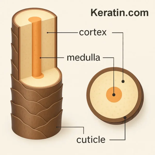
The classic description of hair structure divides the fiber into three concentric zones: the cuticle (outermost), the cortex (intermediate and dominant layer), and the medulla (central core, absent in many fine hairs). While this model remains valid, modern characterization shows that these layers are not uniform cylinders but highly engineered gradients in protein composition, cross-link density, and lipid distribution.
Hair varies widely between species and across body regions of a single individual. In humans, terminal scalp hairs can vary in diameter, ellipticity, and curl pattern depending on genetics, follicular shape, and regional signaling. The geometry of the fiber is determined by asymmetry in inner root sheath (IRS) molds, differential keratinization rates, and programmed variation in cortical cell type distribution.
The Hair Fiber Cuticle: The cuticle is a multilayered envelope of flattened, overlapping cells arranged like roof shingles. Historically described as a “single row” in the bulb, cuticle cell layers increase as differentiation proceeds upward; in the fully mature fiber, human scalp hairs generally possess six to ten layers of cuticle cells.
Cuticle cells originate in the matrix but differentiate rapidly without accumulating melanin or trichohyalin. Their keratin content reflects high proportions of hard keratins (type I and II) and KAPs that cross-link extensively, creating a mechanically robust surface. Unlike cortical cells, cuticle cells possess a robust exocuticle rich in cystine, which allows dense disulfide-bond formation. This accounts for the cuticle’s exceptional abrasion resistance.
A modern understanding of hair fiber emphasizes the four-part architecture of each cuticle cell:
Endocuticle , a lower-sulfur, softer region susceptible to chemical swelling.Exocuticle , a high-sulfur, high-cross-link layer conferring stiffness.A-layer , the outermost, extremely dense layer rich in cystine.Epicuticle , a thin hydrophobic membrane derived partly from the cell membrane complex (CMC). The CMC is now recognized as central to cuticle integrity. It consists of a lipid-protein sandwich with covalently bound 18-methyl eicosanoic acid (18-MEA) forming a protective hydrophobic boundary. Removal of 18-MEA by chemical treatments such as bleaching or perming greatly increases friction, water uptake, and surface porosity, explaining the transition from shiny, smooth hair to dull, roughened fibers.
The cuticle’s optical role is also better understood today. Its smooth, ordered structure enables specular reflection of light, producing gloss. Lifting, erosion, or loss of cuticle layers reduces surface continuity and causes diffuse reflection, lowering shine. These mechanical and optical roles confirm the cuticle’s central importance to fiber aesthetics and durability.
The Hair Cortex: The cortex is the primary structural component, comprising roughly 70–90 percent of the fiber’s volume. It determines mechanical properties, elasticity, tensile strength, and overall appearance. Modern analyses identify two major cortical cell phenotypes:
Ortho-cortical cells , with lower sulfur content and different keratin expression patterns.Para-cortical cells , with higher sulfur content and greater disulfide cross-linking. The distribution and asymmetry of these cells influence fiber shape. Straight fibers show more symmetrical arrangement, whereas curly or kinky fibers show pronounced asymmetry.
Within each cortical cell lies a hierarchical protein architecture:
Intermediate filaments (IFs) : ~7–10 nm keratin filaments, primarily type I and II keratins such as K31, K33, K35, K81, K85, and K86.Keratin-associated proteins (KAPs) : high-sulfur or ultra–high-sulfur proteins that form the matrix surrounding the IFs and control cross-link density.Macro-fibrils : bundles of IFs plus matrix material organized into longitudinal structures.Micro-fibrils and sub-fibrils : finer subdivisions observed via electron microscopy. This hierarchical assembly gives the cortex a remarkable mechanical profile: high tensile strength, appreciable elasticity, and ability to undergo reversible stretching up to approximately 30 percent extension under moderate tension.
The cortex also contains melanin pigment , which imparts color. Modern classification focuses on:
Eumelanin (brown/black) Pheomelanin (red/yellow) Mixed melanins Melanin granules vary in size, shape, and distribution patterns. Recent proteomic studies show melanosomes contribute not only color, but also affect hair stiffness and photoprotection, absorbing ultraviolet radiation and reducing structural protein degradation.
Cortical fusi , historically described as air-filled spaces, are now considered developmental remnants or voids created during keratinization. They may influence optical scattering but have minimal mechanical impact unless they are unusually numerous.
The Medulla: The medulla remains the least understood region of terminal hair fibers, largely because it is inconsistent across human hairs and often it is absent in fine scalp fibers. Modern imaging shows that when present, the medulla consists of loosely packed, vacuolated cells with air spaces that contribute to light scattering and internal reflection.
In humans, the medulla rarely plays a major biological role – as compared with other mammals, where it may regulate insulation or buoyancy. However, recent interest has focused on whether medulla patterns correlate with aging, ethnicity, or hair diameter. Findings suggest:
Coarse fibers (≥80 µm in diameter) more frequently contain continuous medullae. Fine fibers often lack a visible medulla, although polarized light microscopy can reveal fragmentary medulla-like structures. Chemical treatments and cosmetic weathering do not substantially alter medulla presence, indicating its structure is largely determined during formation. The medulla’s contribution to hair color through internal light scattering is now better understood. Air pockets create refractive interfaces that modify how light travels through the fiber, influencing perceived depth and tonal variation, especially in lighter or gray hair.
Modern Insights into Hair Fiber Chemistry and Mechanics: Advances in materials science have reframed hair as a natural composite similar in complexity to engineered polymers. Key updates include:
Cross-linking chemistry: Disulfide bonds remain central to mechanical resilience, but recent research reveals roles for: Isopeptide bonds (formed by transglutaminases); Hydrogen bonding networks influenced by humidity; Ionic interactions among keratin-associated proteins.Lipid architecture: Lipids such as 18-MEA, ceramides, and cholesterol derivatives form structured layers in the CMC and deeply influence water transport, plasticity, and chemical resistance. Damage to CMC lipids is now recognized as a primary mechanism of fiber weakening during bleaching.Water content and mechanical behaviour: Water acts as a plasticizer: increasing elasticity, reducing stiffness, and altering breakage thresholds. Modern biomechanical research uses dynamic mechanical analysis (DMA) to quantify moisture-dependent modulus changes.Fiber shape determinants: It is now established that curliness results from: Follicle asymmetry (elliptical cross sections); Differential keratinization rates across the follicle; Asymmetric distribution of para- and ortho-cortical cells. Conclusion: Our understanding of hair fiber structure has advanced substantially since early morphological descriptions. The classical view of cuticle, cortex, and medulla remains foundational, but modern biology reveals each region as a complex, highly differentiated subsystem governed by molecular gradients, chemical cross-link density, lipid architecture, and optical properties. Hair fibers represent a remarkable natural bio-material – mechanically robust, optically complex, and biochemically dynamic. A contemporary view integrates cellular origins with molecular structures and physicochemical interactions to provide a comprehensive understanding of how hair looks, behaves, and responds to environmental and chemical challenges.
Bibliography
11711645 {11711645:3WFJ6AHK},{11711645:VEE7XJRS},{11711645:TPV2SHI4},{11711645:PT4D3JF7},{11711645:H5XW7X58},{11711645:IG2S4X44},{11711645:BVTP4WDC},{11711645:TIR3JSTE},{11711645:B7TJRGP6},{11711645:92Y58CQK},{11711645:77R35QVV},{11711645:VNHLJP5A} 1 vancouver 50 date asc 2013 https://www.keratin.com/wp-content/plugins/zotpress/ %7B%22status%22%3A%22success%22%2C%22updateneeded%22%3Afalse%2C%22instance%22%3Afalse%2C%22meta%22%3A%7B%22request_last%22%3A0%2C%22request_next%22%3A0%2C%22used_cache%22%3Atrue%7D%2C%22data%22%3A%5B%7B%22key%22%3A%22B7TJRGP6%22%2C%22library%22%3A%7B%22id%22%3A11711645%7D%2C%22meta%22%3A%7B%22creatorSummary%22%3A%22Jones%20et%20al.%22%2C%22parsedDate%22%3A%221994%22%2C%22numChildren%22%3A0%7D%2C%22bib%22%3A%22%26lt%3Bdiv%20class%3D%26quot%3Bcsl-bib-body%26quot%3B%20style%3D%26quot%3Bline-height%3A%201.35%3B%20%26quot%3B%26gt%3B%5Cn%20%20%26lt%3Bdiv%20class%3D%26quot%3Bcsl-entry%26quot%3B%20style%3D%26quot%3Bclear%3A%20left%3B%20%26quot%3B%26gt%3B%5Cn%20%20%20%20%26lt%3Bdiv%20class%3D%26quot%3Bcsl-left-margin%26quot%3B%20style%3D%26quot%3Bfloat%3A%20left%3B%20padding-right%3A%200.5em%3B%20text-align%3A%20right%3B%20width%3A%201em%3B%26quot%3B%26gt%3B1.%26lt%3B%5C%2Fdiv%26gt%3B%26lt%3Bdiv%20class%3D%26quot%3Bcsl-right-inline%26quot%3B%20style%3D%26quot%3Bmargin%3A%200%20.4em%200%201.5em%3B%26quot%3B%26gt%3BJones%20LN%2C%20Horr%20TJ%2C%20Kaplin%20IJ.%20Formation%20of%20surface%20membranes%20in%20developing%20mammalian%20hair%20fibres.%20Micron.%201994%3B25%286%29%3A589%26%23x2013%3B95.%26lt%3B%5C%2Fdiv%26gt%3B%5Cn%20%20%20%26lt%3B%5C%2Fdiv%26gt%3B%5Cn%26lt%3B%5C%2Fdiv%26gt%3B%22%2C%22data%22%3A%7B%22itemType%22%3A%22journalArticle%22%2C%22title%22%3A%22Formation%20of%20surface%20membranes%20in%20developing%20mammalian%20hair%20fibres%22%2C%22creators%22%3A%5B%7B%22creatorType%22%3A%22author%22%2C%22firstName%22%3A%22L.%20N.%22%2C%22lastName%22%3A%22Jones%22%7D%2C%7B%22creatorType%22%3A%22author%22%2C%22firstName%22%3A%22T.%20J.%22%2C%22lastName%22%3A%22Horr%22%7D%2C%7B%22creatorType%22%3A%22author%22%2C%22firstName%22%3A%22I.%20J.%22%2C%22lastName%22%3A%22Kaplin%22%7D%5D%2C%22abstractNote%22%3A%22Mammalian%20hair%20fibres%20result%20from%20complex%20mechanisms%20involving%20synthesis%2C%20assembly%20and%20stabilisation%20of%20keratin%20proteins%20in%20the%20follicle.%20The%20developing%20hair%20shaft%20consists%20of%20outer%20cuticle%20cells%20surrounding%20cortical%20and%20medullary%20%28optional%29%20cell%20types.%20Presumptive%20fibre%20cuticle%20%28FC%29%20is%20contained%20by%20the%20inner%20root%20sheath%20%28IRS%29%20consisting%20of%20IRS%20cuticle%2C%20Huxley%20and%20Henle%20cells%20which%20are%20in%20turn%20enclosed%20in%20an%20outer%20root%20sheath%20%28ORS%29%20of%20epidermal-like%20cells.%20In%20the%20current%20structural%20studies%20we%20have%20used%20energy%20filtered%20transmission%20electron%20microscopy%20%28Zeiss%20902A%29%20on%20Merino%20sheep%20skin%20biopsies%20to%20examine%20the%20fine%20sequence%20of%20morphological%20changes%20involved%20in%20forming%20the%20fibre%20surface%20membrane%20and%20the%20associated%20underlying%20structural%20bands%20comprising%20the%20a-layer%20and%20exocuticle.%20Prior%20to%20the%20development%20of%20the%20exocuticle%2C%20FC%20cells%20demonstrate%20a%20typical%20plasma-membrane%20apposed%20to%20IRS%20cuticle%20plasma-membranes%20separated%20by%20an%20intercellular%20space.%20The%20formation%20of%20exocuticular%20lamellae%20is%20followed%20by%20degradation%20of%20the%20residual%20FC%20surface%20membrane%20and%20the%20appearance%20of%20intercellular%20laminae%20demonstrating%20a%20stained%20central%20band.%20As%20maturation%20continues%20cleavage%20between%20IRS%20cuticle%20and%20FC%20occurs%20along%20this%20central%20band%20liberating%20hair%20into%20the%20pilary%20canal.%20The%20mature%20surface%20consists%20of%20keratinized%20cells%20containing%20a%20well%20developed%20exocuticle%20and%20a-layer%20coated%20with%20paired%20lamina%20%28presumably%20two%20lipid%20containing%20bilayers%29%20of%20material%20approximately%2010-12%20nm%20thick%20derived%20from%20the%20intercellular%20laminae.%20The%20current%20observations%20show%20FC%20surface%20formation%20is%20similar%20to%20processes%20occurring%20in%20epidermal%20stratum%20corneum%20and%20that%20the%20cuticle%20surface%20membrane%20of%20mammalian%20fibres%20is%20not%20derived%20from%20a%20modified%20plasma-membrane%20as%20previously%20documented.%22%2C%22date%22%3A%221994%22%2C%22section%22%3A%22%22%2C%22partNumber%22%3A%22%22%2C%22partTitle%22%3A%22%22%2C%22DOI%22%3A%2210.1016%5C%2F0968-4328%2894%2990021-3%22%2C%22citationKey%22%3A%22%22%2C%22url%22%3A%22%22%2C%22PMID%22%3A%22%22%2C%22PMCID%22%3A%22%22%2C%22ISSN%22%3A%220968-4328%22%2C%22language%22%3A%22eng%22%2C%22collections%22%3A%5B%22QQGDMI57%22%5D%2C%22dateModified%22%3A%222025-11-28T15%3A16%3A05Z%22%7D%7D%2C%7B%22key%22%3A%22TPV2SHI4%22%2C%22library%22%3A%7B%22id%22%3A11711645%7D%2C%22meta%22%3A%7B%22creatorSummary%22%3A%22Nishikawa%20et%20al.%22%2C%22parsedDate%22%3A%221998-06-01%22%2C%22numChildren%22%3A0%7D%2C%22bib%22%3A%22%26lt%3Bdiv%20class%3D%26quot%3Bcsl-bib-body%26quot%3B%20style%3D%26quot%3Bline-height%3A%201.35%3B%20%26quot%3B%26gt%3B%5Cn%20%20%26lt%3Bdiv%20class%3D%26quot%3Bcsl-entry%26quot%3B%20style%3D%26quot%3Bclear%3A%20left%3B%20%26quot%3B%26gt%3B%5Cn%20%20%20%20%26lt%3Bdiv%20class%3D%26quot%3Bcsl-left-margin%26quot%3B%20style%3D%26quot%3Bfloat%3A%20left%3B%20padding-right%3A%200.5em%3B%20text-align%3A%20right%3B%20width%3A%201em%3B%26quot%3B%26gt%3B1.%26lt%3B%5C%2Fdiv%26gt%3B%26lt%3Bdiv%20class%3D%26quot%3Bcsl-right-inline%26quot%3B%20style%3D%26quot%3Bmargin%3A%200%20.4em%200%201.5em%3B%26quot%3B%26gt%3BNishikawa%20N%2C%20Tanizawa%20Y%2C%20Tanaka%20S%2C%20Horiguchi%20Y%2C%20Asakura%20T.%20Structural%20change%20of%20keratin%20protein%20in%20human%20hair%20by%20permanent%20waving%20treatment1.%20Polymer.%201998%20June%201%3B39%2816%29%3A3835%26%23x2013%3B40.%26lt%3B%5C%2Fdiv%26gt%3B%5Cn%20%20%20%26lt%3B%5C%2Fdiv%26gt%3B%5Cn%26lt%3B%5C%2Fdiv%26gt%3B%22%2C%22data%22%3A%7B%22itemType%22%3A%22journalArticle%22%2C%22title%22%3A%22Structural%20change%20of%20keratin%20protein%20in%20human%20hair%20by%20permanent%20waving%20treatment1%22%2C%22creators%22%3A%5B%7B%22creatorType%22%3A%22author%22%2C%22firstName%22%3A%22Naoki%22%2C%22lastName%22%3A%22Nishikawa%22%7D%2C%7B%22creatorType%22%3A%22author%22%2C%22firstName%22%3A%22Yoshiaki%22%2C%22lastName%22%3A%22Tanizawa%22%7D%2C%7B%22creatorType%22%3A%22author%22%2C%22firstName%22%3A%22Shoichi%22%2C%22lastName%22%3A%22Tanaka%22%7D%2C%7B%22creatorType%22%3A%22author%22%2C%22firstName%22%3A%22Yasunobu%22%2C%22lastName%22%3A%22Horiguchi%22%7D%2C%7B%22creatorType%22%3A%22author%22%2C%22firstName%22%3A%22Tetsuo%22%2C%22lastName%22%3A%22Asakura%22%7D%5D%2C%22abstractNote%22%3A%22The%20damage%20to%20human%20hair%20caused%20by%20permanent%20waving%20treatment%20was%20examined%20from%20the%20viewpoint%20of%20structure%20with%20several%20spectroscopic%20methods%2C%2013C%20CP%5C%2FMAS%20n.m.r.%2C%20wide%20angle%20X-ray%20diffraction%20%28WAXD%29%2C%20FTi.r.%20and%20Raman%20methods.%20The%20partial%20disruption%20of%20%5Cu03b1-helical%20structure%20constituting%20the%20microfibril%20in%20hair%20fibre%20was%20revealed%20by%20this%20study%2C%20although%20the%20incomplete%20re-oxidation%20of%20the%20disulphide%20bond%20has%20been%20considered%20to%20be%20the%20main%20damage%20caused%20by%20the%20treatment.%20Moreover%2C%20this%20disruption%20was%20mainly%20caused%20in%20the%20first%20reduction%20process.%20In%20particular%2C%2013C%20CP%5C%2FMAS%20n.m.r.%20gave%20the%20quantitative%20evaluation%20for%20change%20of%20%5Cu03b1-helical%20content%20in%20the%20microfibril%20with%20permanent%20waving%20treatment.%20In%20addition%2C%20FTi.r.%20and%20Raman%20spectra%20indicated%20that%20the%20%5Cu03b1-helix%20was%20partially%20changed%20to%20random%20coil%2C%20rather%20than%20to%20%5Cu03b2-sheet%20structure.%22%2C%22date%22%3A%221998-06-01%22%2C%22section%22%3A%22%22%2C%22partNumber%22%3A%22%22%2C%22partTitle%22%3A%22%22%2C%22DOI%22%3A%2210.1016%5C%2FS0032-3861%2897%2910299-3%22%2C%22citationKey%22%3A%22%22%2C%22url%22%3A%22%22%2C%22PMID%22%3A%22%22%2C%22PMCID%22%3A%22%22%2C%22ISSN%22%3A%220032-3861%22%2C%22language%22%3A%22%22%2C%22collections%22%3A%5B%22QQGDMI57%22%5D%2C%22dateModified%22%3A%222025-11-28T16%3A56%3A13Z%22%7D%7D%2C%7B%22key%22%3A%22VNHLJP5A%22%2C%22library%22%3A%7B%22id%22%3A11711645%7D%2C%22meta%22%3A%7B%22creatorSummary%22%3A%22Nagase%20et%20al.%22%2C%22parsedDate%22%3A%222002%22%2C%22numChildren%22%3A0%7D%2C%22bib%22%3A%22%26lt%3Bdiv%20class%3D%26quot%3Bcsl-bib-body%26quot%3B%20style%3D%26quot%3Bline-height%3A%201.35%3B%20%26quot%3B%26gt%3B%5Cn%20%20%26lt%3Bdiv%20class%3D%26quot%3Bcsl-entry%26quot%3B%20style%3D%26quot%3Bclear%3A%20left%3B%20%26quot%3B%26gt%3B%5Cn%20%20%20%20%26lt%3Bdiv%20class%3D%26quot%3Bcsl-left-margin%26quot%3B%20style%3D%26quot%3Bfloat%3A%20left%3B%20padding-right%3A%200.5em%3B%20text-align%3A%20right%3B%20width%3A%201em%3B%26quot%3B%26gt%3B1.%26lt%3B%5C%2Fdiv%26gt%3B%26lt%3Bdiv%20class%3D%26quot%3Bcsl-right-inline%26quot%3B%20style%3D%26quot%3Bmargin%3A%200%20.4em%200%201.5em%3B%26quot%3B%26gt%3BNagase%20S%2C%20Shibuichi%20S%2C%20Ando%20K%2C%20Kariya%20E%2C%20Satoh%20N.%20Influence%20of%20internal%20structures%20of%20hair%20fiber%20on%20hair%20appearance.%20I.%20Light%20scattering%20from%20the%20porous%20structure%20of%20the%20medulla%20of%20human%20hair.%20J%20Cosmet%20Sci.%202002%3B53%282%29%3A89%26%23x2013%3B100.%26lt%3B%5C%2Fdiv%26gt%3B%5Cn%20%20%20%26lt%3B%5C%2Fdiv%26gt%3B%5Cn%26lt%3B%5C%2Fdiv%26gt%3B%22%2C%22data%22%3A%7B%22itemType%22%3A%22journalArticle%22%2C%22title%22%3A%22Influence%20of%20internal%20structures%20of%20hair%20fiber%20on%20hair%20appearance.%20I.%20Light%20scattering%20from%20the%20porous%20structure%20of%20the%20medulla%20of%20human%20hair%22%2C%22creators%22%3A%5B%7B%22creatorType%22%3A%22author%22%2C%22firstName%22%3A%22Shinobu%22%2C%22lastName%22%3A%22Nagase%22%7D%2C%7B%22creatorType%22%3A%22author%22%2C%22firstName%22%3A%22Satoshi%22%2C%22lastName%22%3A%22Shibuichi%22%7D%2C%7B%22creatorType%22%3A%22author%22%2C%22firstName%22%3A%22Kenichi%22%2C%22lastName%22%3A%22Ando%22%7D%2C%7B%22creatorType%22%3A%22author%22%2C%22firstName%22%3A%22Emiko%22%2C%22lastName%22%3A%22Kariya%22%7D%2C%7B%22creatorType%22%3A%22author%22%2C%22firstName%22%3A%22Naoki%22%2C%22lastName%22%3A%22Satoh%22%7D%5D%2C%22abstractNote%22%3A%22In%20this%20study%20the%20influence%20of%20the%20medulla%20structure%20on%20hair%20appearance%20was%20examined.%20Hair%20with%20a%20porous%20medulla%20gave%20a%20whitish%20and%20lusterless%20appearance%20because%20of%20light%20scattering%20from%20the%20fiber%20center%2C%20whereas%20in%20the%20cases%20without%20pores%2C%20a%20clear%20and%20brilliant%20appearance%20was%20observed.%20The%20optical%20influences%20of%20the%20medulla%20pores%20were%20measured%20by%20a%20spectral%20goniophotometer%2C%20and%20obtained%20data%20were%20analyzed%20in%20terms%20of%20the%20CIE%20L%2Aa%2Ab%2A%20color%20system.%20Both%20contrasts%20in%20lightness%20and%20apparent%20color%20%28chroma%20and%20hue%29%20decreased%20in%20the%20hair%20with%20medulla%20pores%2C%20and%20the%20decreases%20in%20contrast%20caused%20a%20whitish%20and%20lusterless%20appearance.%20The%20distribution%20of%20the%20amount%20of%20medulla%20pores%20was%20investigated%20for%20Japanese%20females.%20The%20histogram%20was%20further%20analyzed%20by%20hair%20care%20behavior%20of%20individual%20panelists%2C%20and%20it%20was%20found%20that%20the%20pores%20in%20the%20medulla%20can%20be%20generated%20in%20a%20heat-drying%20process.%22%2C%22date%22%3A%222002%22%2C%22section%22%3A%22%22%2C%22partNumber%22%3A%22%22%2C%22partTitle%22%3A%22%22%2C%22DOI%22%3A%22%22%2C%22citationKey%22%3A%22%22%2C%22url%22%3A%22%22%2C%22PMID%22%3A%22%22%2C%22PMCID%22%3A%22%22%2C%22ISSN%22%3A%221525-7886%22%2C%22language%22%3A%22eng%22%2C%22collections%22%3A%5B%22QQGDMI57%22%5D%2C%22dateModified%22%3A%222025-11-28T15%3A12%3A42Z%22%7D%7D%2C%7B%22key%22%3A%22PT4D3JF7%22%2C%22library%22%3A%7B%22id%22%3A11711645%7D%2C%22meta%22%3A%7B%22creatorSummary%22%3A%22Popescu%20and%20H%5Cu00f6cker%22%2C%22parsedDate%22%3A%222009-01-01%22%2C%22numChildren%22%3A0%7D%2C%22bib%22%3A%22%26lt%3Bdiv%20class%3D%26quot%3Bcsl-bib-body%26quot%3B%20style%3D%26quot%3Bline-height%3A%201.35%3B%20%26quot%3B%26gt%3B%5Cn%20%20%26lt%3Bdiv%20class%3D%26quot%3Bcsl-entry%26quot%3B%20style%3D%26quot%3Bclear%3A%20left%3B%20%26quot%3B%26gt%3B%5Cn%20%20%20%20%26lt%3Bdiv%20class%3D%26quot%3Bcsl-left-margin%26quot%3B%20style%3D%26quot%3Bfloat%3A%20left%3B%20padding-right%3A%200.5em%3B%20text-align%3A%20right%3B%20width%3A%201em%3B%26quot%3B%26gt%3B1.%26lt%3B%5C%2Fdiv%26gt%3B%26lt%3Bdiv%20class%3D%26quot%3Bcsl-right-inline%26quot%3B%20style%3D%26quot%3Bmargin%3A%200%20.4em%200%201.5em%3B%26quot%3B%26gt%3BPopescu%20C%2C%20H%26%23xF6%3Bcker%20H.%20Chapter%204%20Cytomechanics%20of%20Hair%3A%20Basics%20of%20the%20Mechanical%20Stability.%20International%20Review%20of%20Cell%20and%20Molecular%20Biology.%202009%20Jan%201%3B277%3A137%26%23x2013%3B56.%26lt%3B%5C%2Fdiv%26gt%3B%5Cn%20%20%20%26lt%3B%5C%2Fdiv%26gt%3B%5Cn%26lt%3B%5C%2Fdiv%26gt%3B%22%2C%22data%22%3A%7B%22itemType%22%3A%22journalArticle%22%2C%22title%22%3A%22Chapter%204%20Cytomechanics%20of%20Hair%3A%20Basics%20of%20the%20Mechanical%20Stability%22%2C%22creators%22%3A%5B%7B%22creatorType%22%3A%22author%22%2C%22firstName%22%3A%22Crisan%22%2C%22lastName%22%3A%22Popescu%22%7D%2C%7B%22creatorType%22%3A%22author%22%2C%22firstName%22%3A%22Hartwig%22%2C%22lastName%22%3A%22H%5Cu00f6cker%22%7D%5D%2C%22abstractNote%22%3A%22Hair%20is%20a%20complex%20%5Cu201ccornified%5Cu201d%20multicellular%20tissue%20composed%20of%20cuticle%20and%20cortex%20cells%20mechanically%20acting%20as%20a%20whole.%20The%20cuticle%20cells%20overlap%20and%20%5Cu2026%22%2C%22date%22%3A%222009%5C%2F01%5C%2F01%22%2C%22section%22%3A%22%22%2C%22partNumber%22%3A%22%22%2C%22partTitle%22%3A%22%22%2C%22DOI%22%3A%22%22%2C%22citationKey%22%3A%22%22%2C%22url%22%3A%22%22%2C%22PMID%22%3A%22%22%2C%22PMCID%22%3A%22%22%2C%22ISSN%22%3A%22%22%2C%22language%22%3A%22en-US%22%2C%22collections%22%3A%5B%22QQGDMI57%22%5D%2C%22dateModified%22%3A%222025-11-28T17%3A02%3A22Z%22%7D%7D%2C%7B%22key%22%3A%2277R35QVV%22%2C%22library%22%3A%7B%22id%22%3A11711645%7D%2C%22meta%22%3A%7B%22creatorSummary%22%3A%22Yang%20et%20al.%22%2C%22parsedDate%22%3A%222014%22%2C%22numChildren%22%3A0%7D%2C%22bib%22%3A%22%26lt%3Bdiv%20class%3D%26quot%3Bcsl-bib-body%26quot%3B%20style%3D%26quot%3Bline-height%3A%201.35%3B%20%26quot%3B%26gt%3B%5Cn%20%20%26lt%3Bdiv%20class%3D%26quot%3Bcsl-entry%26quot%3B%20style%3D%26quot%3Bclear%3A%20left%3B%20%26quot%3B%26gt%3B%5Cn%20%20%20%20%26lt%3Bdiv%20class%3D%26quot%3Bcsl-left-margin%26quot%3B%20style%3D%26quot%3Bfloat%3A%20left%3B%20padding-right%3A%200.5em%3B%20text-align%3A%20right%3B%20width%3A%201em%3B%26quot%3B%26gt%3B1.%26lt%3B%5C%2Fdiv%26gt%3B%26lt%3Bdiv%20class%3D%26quot%3Bcsl-right-inline%26quot%3B%20style%3D%26quot%3Bmargin%3A%200%20.4em%200%201.5em%3B%26quot%3B%26gt%3BYang%20FC%2C%20Zhang%20Y%2C%20Rheinst%26%23xE4%3Bdter%20MC.%20The%20structure%20of%20people%26%23x2019%3Bs%20hair.%20PeerJ.%202014%3B2%3Ae619.%26lt%3B%5C%2Fdiv%26gt%3B%5Cn%20%20%20%26lt%3B%5C%2Fdiv%26gt%3B%5Cn%26lt%3B%5C%2Fdiv%26gt%3B%22%2C%22data%22%3A%7B%22itemType%22%3A%22journalArticle%22%2C%22title%22%3A%22The%20structure%20of%20people%27s%20hair%22%2C%22creators%22%3A%5B%7B%22creatorType%22%3A%22author%22%2C%22firstName%22%3A%22Fei-Chi%22%2C%22lastName%22%3A%22Yang%22%7D%2C%7B%22creatorType%22%3A%22author%22%2C%22firstName%22%3A%22Yuchen%22%2C%22lastName%22%3A%22Zhang%22%7D%2C%7B%22creatorType%22%3A%22author%22%2C%22firstName%22%3A%22Maikel%20C.%22%2C%22lastName%22%3A%22Rheinst%5Cu00e4dter%22%7D%5D%2C%22abstractNote%22%3A%22Hair%20is%20a%20filamentous%20biomaterial%20consisting%20mainly%20of%20proteins%20in%20particular%20keratin.%20The%20structure%20of%20human%20hair%20is%20well%20known%3A%20the%20medulla%20is%20a%20loosely%20packed%2C%20disordered%20region%20near%20the%20centre%20of%20the%20hair%20surrounded%20by%20the%20cortex%2C%20which%20contains%20the%20major%20part%20of%20the%20fibre%20mass%2C%20mainly%20consisting%20of%20keratin%20proteins%20and%20structural%20lipids.%20The%20cortex%20is%20surrounded%20by%20the%20cuticle%2C%20a%20layer%20of%20dead%2C%20overlapping%20cells%20forming%20a%20protective%20layer%20around%20the%20hair.%20The%20corresponding%20structures%20have%20been%20studied%20extensively%20using%20a%20variety%20of%20different%20techniques%2C%20such%20as%20light%2C%20electron%20and%20atomic%20force%20microscopes%2C%20and%20also%20X-ray%20diffraction.%20We%20were%20interested%20in%20the%20question%20how%20much%20the%20molecular%20hair%20structure%20differs%20from%20person%20to%20person%2C%20between%20male%20and%20female%20hair%2C%20hair%20of%20different%20appearances%20such%20as%20colour%20and%20waviness.%20We%20included%20hair%20from%20parent%20and%20child%2C%20identical%20and%20fraternal%20twins%20in%20the%20study%20to%20see%20if%20genetically%20similar%20hair%20would%20show%20similar%20structural%20features.%20The%20molecular%20structure%20of%20the%20hair%20samples%20was%20studied%20using%20high-resolution%20X-ray%20diffraction%2C%20which%20covers%20length%20scales%20from%20molecules%20up%20to%20the%20organization%20of%20secondary%20structures.%20Signals%20due%20to%20the%20coiled-coil%20phase%20of%20%5Cu03b1-helical%20keratin%20proteins%2C%20intermediate%20keratin%20filaments%20in%20the%20cortex%20and%20from%20the%20lipid%20layers%20in%20the%20cell%20membrane%20complex%20were%20observed%20in%20the%20specimen%20of%20all%20individuals%2C%20with%20very%20small%20deviations.%20Despite%20the%20relatively%20small%20number%20of%20individuals%20%2812%29%20included%20in%20this%20study%2C%20some%20conclusions%20can%20be%20drawn.%20While%20the%20general%20features%20were%20observed%20in%20all%20individuals%20and%20the%20corresponding%20molecular%20structures%20were%20almost%20identical%2C%20additional%20signals%20were%20observed%20in%20some%20specimen%20and%20assigned%20to%20different%20types%20of%20lipids%20in%20the%20cell%20membrane%20complex.%20Genetics%20seem%20to%20play%20a%20role%20in%20this%20composition%20as%20identical%20patterns%20were%20observed%20in%20hair%20from%20father%20and%20daughter%20and%20identical%20twins%2C%20however%2C%20not%20for%20fraternal%20twins.%20Identification%20and%20characterization%20of%20these%20features%20is%20an%20important%20step%20towards%20the%20detection%20of%20abnormalities%20in%20the%20molecular%20structure%20of%20hair%20as%20a%20potential%20diagnostic%20tool%20for%20certain%20diseases.%22%2C%22date%22%3A%222014%22%2C%22section%22%3A%22%22%2C%22partNumber%22%3A%22%22%2C%22partTitle%22%3A%22%22%2C%22DOI%22%3A%2210.7717%5C%2Fpeerj.619%22%2C%22citationKey%22%3A%22%22%2C%22url%22%3A%22%22%2C%22PMID%22%3A%22%22%2C%22PMCID%22%3A%22%22%2C%22ISSN%22%3A%222167-8359%22%2C%22language%22%3A%22eng%22%2C%22collections%22%3A%5B%22QQGDMI57%22%5D%2C%22dateModified%22%3A%222025-11-28T15%3A13%3A46Z%22%7D%7D%2C%7B%22key%22%3A%22VEE7XJRS%22%2C%22library%22%3A%7B%22id%22%3A11711645%7D%2C%22meta%22%3A%7B%22creatorSummary%22%3A%22Erdo%5Cu011fan%22%2C%22parsedDate%22%3A%222017-05-03%22%2C%22numChildren%22%3A0%7D%2C%22bib%22%3A%22%26lt%3Bdiv%20class%3D%26quot%3Bcsl-bib-body%26quot%3B%20style%3D%26quot%3Bline-height%3A%201.35%3B%20%26quot%3B%26gt%3B%5Cn%20%20%26lt%3Bdiv%20class%3D%26quot%3Bcsl-entry%26quot%3B%20style%3D%26quot%3Bclear%3A%20left%3B%20%26quot%3B%26gt%3B%5Cn%20%20%20%20%26lt%3Bdiv%20class%3D%26quot%3Bcsl-left-margin%26quot%3B%20style%3D%26quot%3Bfloat%3A%20left%3B%20padding-right%3A%200.5em%3B%20text-align%3A%20right%3B%20width%3A%201em%3B%26quot%3B%26gt%3B1.%26lt%3B%5C%2Fdiv%26gt%3B%26lt%3Bdiv%20class%3D%26quot%3Bcsl-right-inline%26quot%3B%20style%3D%26quot%3Bmargin%3A%200%20.4em%200%201.5em%3B%26quot%3B%26gt%3BErdo%26%23x11F%3Ban%20B.%20Anatomy%20and%20Physiology%20of%20Hair.%20In%3A%20Hair%20and%20Scalp%20Disorders.%20IntechOpen%3B%202017.%26lt%3B%5C%2Fdiv%26gt%3B%5Cn%20%20%20%26lt%3B%5C%2Fdiv%26gt%3B%5Cn%26lt%3B%5C%2Fdiv%26gt%3B%22%2C%22data%22%3A%7B%22itemType%22%3A%22bookSection%22%2C%22title%22%3A%22Anatomy%20and%20Physiology%20of%20Hair%22%2C%22creators%22%3A%5B%7B%22creatorType%22%3A%22author%22%2C%22firstName%22%3A%22Bilgen%22%2C%22lastName%22%3A%22Erdo%5Cu011fan%22%7D%5D%2C%22abstractNote%22%3A%22Hair%20is%20one%20of%20the%20characteristic%20features%20of%20mammals%20and%20has%20various%20functions%20such%20as%20protection%20against%20external%20factors%3B%20producing%20sebum%2C%20apocrine%20sweat%20and%20pheromones%3B%20impact%20on%20social%20and%20sexual%20interactions%3B%20thermoregulation%20and%20being%20a%20resource%20for%20stem%20cells.%20Hair%20is%20a%20derivative%20of%20the%20epidermis%20and%20consists%20of%20two%20distinct%20parts%3A%20the%20follicle%20and%20the%20hair%20shaft.%20The%20follicle%20is%20the%20essential%20unit%20for%20the%20generation%20of%20hair.%20The%20hair%20shaft%20consists%20of%20a%20cortex%20and%20cuticle%20cells%2C%20and%20a%20medulla%20for%20some%20types%20of%20hairs.%20Hair%20follicle%20has%20a%20continuous%20growth%20and%20rest%20sequence%20named%20hair%20cycle.%20The%20duration%20of%20growth%20and%20rest%20cycles%20is%20coordinated%20by%20many%20endocrine%2C%20vascular%20and%20neural%20stimuli%20and%20depends%20not%20only%20on%20localization%20of%20the%20hair%20but%20also%20on%20various%20factors%2C%20like%20age%20and%20nutritional%20habits.%20Distinctive%20anatomy%20and%20physiology%20of%20hair%20follicle%20are%20presented%20in%20this%20chapter.%20Extensive%20knowledge%20on%20anatomical%20and%20physiological%20aspects%20of%20hair%20can%20contribute%20to%20understand%20and%20heal%20different%20hair%20disorders.%22%2C%22bookTitle%22%3A%22Hair%20and%20Scalp%20Disorders%22%2C%22date%22%3A%222017%5C%2F05%5C%2F03%22%2C%22originalDate%22%3A%22%22%2C%22originalPublisher%22%3A%22%22%2C%22originalPlace%22%3A%22%22%2C%22format%22%3A%22%22%2C%22ISBN%22%3A%229789535130987%22%2C%22DOI%22%3A%22%22%2C%22citationKey%22%3A%22%22%2C%22url%22%3A%22%22%2C%22ISSN%22%3A%22%22%2C%22language%22%3A%22en%22%2C%22collections%22%3A%5B%22QQGDMI57%22%5D%2C%22dateModified%22%3A%222025-11-28T16%3A57%3A32Z%22%7D%7D%2C%7B%22key%22%3A%2292Y58CQK%22%2C%22library%22%3A%7B%22id%22%3A11711645%7D%2C%22meta%22%3A%7B%22creatorSummary%22%3A%22Plowman%20and%20Harland%22%2C%22parsedDate%22%3A%222018%22%2C%22numChildren%22%3A0%7D%2C%22bib%22%3A%22%26lt%3Bdiv%20class%3D%26quot%3Bcsl-bib-body%26quot%3B%20style%3D%26quot%3Bline-height%3A%201.35%3B%20%26quot%3B%26gt%3B%5Cn%20%20%26lt%3Bdiv%20class%3D%26quot%3Bcsl-entry%26quot%3B%20style%3D%26quot%3Bclear%3A%20left%3B%20%26quot%3B%26gt%3B%5Cn%20%20%20%20%26lt%3Bdiv%20class%3D%26quot%3Bcsl-left-margin%26quot%3B%20style%3D%26quot%3Bfloat%3A%20left%3B%20padding-right%3A%200.5em%3B%20text-align%3A%20right%3B%20width%3A%201em%3B%26quot%3B%26gt%3B1.%26lt%3B%5C%2Fdiv%26gt%3B%26lt%3Bdiv%20class%3D%26quot%3Bcsl-right-inline%26quot%3B%20style%3D%26quot%3Bmargin%3A%200%20.4em%200%201.5em%3B%26quot%3B%26gt%3BPlowman%20JE%2C%20Harland%20DP.%20Fibre%20Ultrastructure.%20Adv%20Exp%20Med%20Biol.%202018%3B1054%3A3%26%23x2013%3B13.%26lt%3B%5C%2Fdiv%26gt%3B%5Cn%20%20%20%26lt%3B%5C%2Fdiv%26gt%3B%5Cn%26lt%3B%5C%2Fdiv%26gt%3B%22%2C%22data%22%3A%7B%22itemType%22%3A%22journalArticle%22%2C%22title%22%3A%22Fibre%20Ultrastructure%22%2C%22creators%22%3A%5B%7B%22creatorType%22%3A%22author%22%2C%22firstName%22%3A%22Jeffrey%20E.%22%2C%22lastName%22%3A%22Plowman%22%7D%2C%7B%22creatorType%22%3A%22author%22%2C%22firstName%22%3A%22Duane%20P.%22%2C%22lastName%22%3A%22Harland%22%7D%5D%2C%22abstractNote%22%3A%22Mammalian%20hair%20fibres%20can%20be%20structurally%20divided%20into%20three%20main%20components%3A%20a%20cuticle%2C%20cortex%20and%20sometimes%20a%20medulla.%20The%20cuticle%20consists%20of%20a%20thin%20layer%20of%20overlapping%20cells%20on%20the%20surface%20of%20the%20fibre%2C%20constituting%20around%2010%25%20of%20the%20total%20fibre%20weight.%20The%20cortex%20makes%20up%20the%20remaining%2086-90%25%20and%20is%20made%20up%20of%20axially%20aligned%20spindle-shaped%20cells%20of%20which%20three%20major%20types%20have%20been%20recognised%20in%20wool%3A%20ortho%2C%20meso%20and%20para.%20Cortical%20cells%20are%20packed%20full%20of%20macrofibril%20bundles%2C%20which%20are%20a%20composite%20of%20aligned%20intermediate%20filaments%20embedded%20in%20an%20amorphous%20matrix.%20The%20spacing%20and%20three-dimensional%20arrangement%20of%20the%20intermediate%20filaments%20vary%20with%20cell%20type.%20The%20medulla%20consists%20of%20a%20continuous%20or%20discontinuous%20column%20of%20horizontal%20spaces%20in%20the%20centre%20of%20the%20cortex%20that%20becomes%20more%20prevalent%20as%20the%20fibre%20diameter%20increases.%22%2C%22date%22%3A%222018%22%2C%22section%22%3A%22%22%2C%22partNumber%22%3A%22%22%2C%22partTitle%22%3A%22%22%2C%22DOI%22%3A%2210.1007%5C%2F978-981-10-8195-8_1%22%2C%22citationKey%22%3A%22%22%2C%22url%22%3A%22%22%2C%22PMID%22%3A%22%22%2C%22PMCID%22%3A%22%22%2C%22ISSN%22%3A%220065-2598%22%2C%22language%22%3A%22eng%22%2C%22collections%22%3A%5B%22QQGDMI57%22%5D%2C%22dateModified%22%3A%222025-11-28T15%3A14%3A52Z%22%7D%7D%2C%7B%22key%22%3A%22BVTP4WDC%22%2C%22library%22%3A%7B%22id%22%3A11711645%7D%2C%22meta%22%3A%7B%22creatorSummary%22%3A%22Cloete%20et%20al.%22%2C%22parsedDate%22%3A%222019%22%2C%22numChildren%22%3A0%7D%2C%22bib%22%3A%22%26lt%3Bdiv%20class%3D%26quot%3Bcsl-bib-body%26quot%3B%20style%3D%26quot%3Bline-height%3A%201.35%3B%20%26quot%3B%26gt%3B%5Cn%20%20%26lt%3Bdiv%20class%3D%26quot%3Bcsl-entry%26quot%3B%20style%3D%26quot%3Bclear%3A%20left%3B%20%26quot%3B%26gt%3B%5Cn%20%20%20%20%26lt%3Bdiv%20class%3D%26quot%3Bcsl-left-margin%26quot%3B%20style%3D%26quot%3Bfloat%3A%20left%3B%20padding-right%3A%200.5em%3B%20text-align%3A%20right%3B%20width%3A%201em%3B%26quot%3B%26gt%3B1.%26lt%3B%5C%2Fdiv%26gt%3B%26lt%3Bdiv%20class%3D%26quot%3Bcsl-right-inline%26quot%3B%20style%3D%26quot%3Bmargin%3A%200%20.4em%200%201.5em%3B%26quot%3B%26gt%3BCloete%20E%2C%20Khumalo%20NP%2C%20Van%20Wyk%20JC%2C%20Ngoepe%20MN.%20Systems%20Approach%20to%20Human%20Hair%20Fibers%3A%20Interdependence%20Between%20Physical%2C%20Mechanical%2C%20Biochemical%20and%20Geometric%20Properties%20of%20Natural%20Healthy%20Hair.%20Front%20Physiol.%202019%3B10%3A112.%26lt%3B%5C%2Fdiv%26gt%3B%5Cn%20%20%20%26lt%3B%5C%2Fdiv%26gt%3B%5Cn%26lt%3B%5C%2Fdiv%26gt%3B%22%2C%22data%22%3A%7B%22itemType%22%3A%22journalArticle%22%2C%22title%22%3A%22Systems%20Approach%20to%20Human%20Hair%20Fibers%3A%20Interdependence%20Between%20Physical%2C%20Mechanical%2C%20Biochemical%20and%20Geometric%20Properties%20of%20Natural%20Healthy%20Hair%22%2C%22creators%22%3A%5B%7B%22creatorType%22%3A%22author%22%2C%22firstName%22%3A%22Elsabe%22%2C%22lastName%22%3A%22Cloete%22%7D%2C%7B%22creatorType%22%3A%22author%22%2C%22firstName%22%3A%22Nonhlanhla%20P.%22%2C%22lastName%22%3A%22Khumalo%22%7D%2C%7B%22creatorType%22%3A%22author%22%2C%22firstName%22%3A%22Jennifer%20C.%22%2C%22lastName%22%3A%22Van%20Wyk%22%7D%2C%7B%22creatorType%22%3A%22author%22%2C%22firstName%22%3A%22Malebogo%20N.%22%2C%22lastName%22%3A%22Ngoepe%22%7D%5D%2C%22abstractNote%22%3A%22Contextual%20interpretation%20of%20hair%20fiber%20data%20is%20often%20blind%20to%20the%20effects%20of%20the%20dynamic%20complexity%20between%20different%20fiber%20properties.%20This%20intrinsic%20complexity%20requires%20systems%20thinking%20to%20decipher%20hair%20fiber%20accurately.%20Hair%20research%2C%20studied%20by%20various%20disciplines%2C%20follows%20a%20reductionist%20research%20approach%2C%20where%20elements%20of%20interest%20are%20studied%20from%20a%20local%20context%20with%20a%20certain%20amount%20of%20detachment%20from%20other%20elements%20or%20contexts.%20Following%20a%20systems%20approach%2C%20the%20authors%20are%20currently%20developing%20a%20cross-disciplinary%20taxonomy%20to%20provide%20a%20holistic%20view%20of%20fiber%20constituents%20and%20their%20interactions%20within%20large-scale%20dynamics.%20Based%20on%20the%20development%20process%2C%20this%20paper%20presents%20a%20review%20that%20explores%20the%20associated%20features%2C%20interrelationships%20and%20interactive%20complexities%20between%20physical%2C%20mechanical%2C%20biochemical%20and%20geometric%20features%20of%20natural%2C%20healthy%20hair%20fibers.%20Through%20the%20review%2C%20the%20importance%20of%20an%20appropriate%20taxonomy%20for%20interpreting%20hair%20fiber%20data%20across%20different%20disciplines%20is%20revealed.%20The%20review%20also%20demonstrates%20how%20seemingly%20unrelated%20fiber%20constituents%20are%20indeed%20interdependent%20and%20that%20these%20interdependencies%20may%20affect%20the%20behavior%20of%20the%20fiber.%20Finally%2C%20the%20review%20highlights%20how%20a%20non-integrative%20approach%20may%20have%20a%20negative%20impact%20on%20the%20reliability%20of%20hair%20data%20interpretation.%22%2C%22date%22%3A%222019%22%2C%22section%22%3A%22%22%2C%22partNumber%22%3A%22%22%2C%22partTitle%22%3A%22%22%2C%22DOI%22%3A%2210.3389%5C%2Ffphys.2019.00112%22%2C%22citationKey%22%3A%22%22%2C%22url%22%3A%22%22%2C%22PMID%22%3A%22%22%2C%22PMCID%22%3A%22%22%2C%22ISSN%22%3A%221664-042X%22%2C%22language%22%3A%22eng%22%2C%22collections%22%3A%5B%22QQGDMI57%22%5D%2C%22dateModified%22%3A%222025-11-28T15%3A16%3A56Z%22%7D%7D%2C%7B%22key%22%3A%22H5XW7X58%22%2C%22library%22%3A%7B%22id%22%3A11711645%7D%2C%22meta%22%3A%7B%22creatorSummary%22%3A%22Rogers%22%2C%22parsedDate%22%3A%222019-05-09%22%2C%22numChildren%22%3A0%7D%2C%22bib%22%3A%22%26lt%3Bdiv%20class%3D%26quot%3Bcsl-bib-body%26quot%3B%20style%3D%26quot%3Bline-height%3A%201.35%3B%20%26quot%3B%26gt%3B%5Cn%20%20%26lt%3Bdiv%20class%3D%26quot%3Bcsl-entry%26quot%3B%20style%3D%26quot%3Bclear%3A%20left%3B%20%26quot%3B%26gt%3B%5Cn%20%20%20%20%26lt%3Bdiv%20class%3D%26quot%3Bcsl-left-margin%26quot%3B%20style%3D%26quot%3Bfloat%3A%20left%3B%20padding-right%3A%200.5em%3B%20text-align%3A%20right%3B%20width%3A%201em%3B%26quot%3B%26gt%3B1.%26lt%3B%5C%2Fdiv%26gt%3B%26lt%3Bdiv%20class%3D%26quot%3Bcsl-right-inline%26quot%3B%20style%3D%26quot%3Bmargin%3A%200%20.4em%200%201.5em%3B%26quot%3B%26gt%3BRogers%20GE.%20Known%20and%20Unknown%20Features%20of%20Hair%20Cuticle%20Structure%3A%20A%20Brief%20Review.%20Cosmetics.%202019%20May%209%3B6%282%29%3A32.%26lt%3B%5C%2Fdiv%26gt%3B%5Cn%20%20%20%26lt%3B%5C%2Fdiv%26gt%3B%5Cn%26lt%3B%5C%2Fdiv%26gt%3B%22%2C%22data%22%3A%7B%22itemType%22%3A%22journalArticle%22%2C%22title%22%3A%22Known%20and%20Unknown%20Features%20of%20Hair%20Cuticle%20Structure%3A%20A%20Brief%20Review%22%2C%22creators%22%3A%5B%7B%22creatorType%22%3A%22author%22%2C%22firstName%22%3A%22George%20E.%22%2C%22lastName%22%3A%22Rogers%22%7D%5D%2C%22abstractNote%22%3A%22The%20cuticle%20is%20the%20outermost%20layer%20of%20overlapping%20flattened%20cells%20of%20hair%20and%20has%20been%20subjected%20to%20many%20years%20of%20study%20to%20understand%20its%20structure%20and%20how%20it%20develops%20in%20the%20follicle.%20The%20essential%20function%20of%20the%20cuticle%20with%20its%20tough%20inelastic%20protein%20content%20is%20to%20protect%20the%20inner%20cortex%20that%20provides%20the%20elastic%20properties%20of%20hair.%20Progress%20in%20our%20knowledge%20of%20hair%20came%20from%20studies%20with%20the%20electron%20microscope%2C%20initially%20transmission%20electron%20microscopy%20%28TEM%29%20for%20internal%20structure%20and%20later%20the%20scanning%20electron%20microscope%20%28SEM%29%20for%20cuticle%20surface%20shape%20and%20for%20investigating%20changes%20caused%20by%20various%20environmental%20influences%20such%20as%20cosmetic%20treatments%20and%20industrial%20processing%20of%20wool.%20Other%20physical%20techniques%20have%20been%20successfully%20applied%20in%20conjunction%20with%20proteomics.%20The%20outstanding%20internal%20features%20of%20the%20cuticle%20cells%20are%20the%20internal%20layers%20consisting%20of%20keratin%20filament%20proteins%20and%20the%20keratin-associated%20proteins.%20The%20stability%20and%20physical%20toughness%20of%20the%20cuticle%20cell%20is%20partly%20accounted%20for%20by%20the%20high%20content%20of%20disulphide%20crosslinking.%20The%20material%20between%20the%20cells%20that%20holds%20them%20tightly%20together%2C%20the%20cell%20membrane%20complex%2C%20consists%20of%20a%20layer%20of%20lipid%20on%20both%20sides%20of%20a%20central%20protein%20layer.%20The%20lipid%20contains%2018-methyleicosanoic%20acid%20that%20is%20part%20of%20the%20hydrophobic%20lipid%20surface%20of%20hair.%20For%20the%20past%20decade%20there%20have%20been%20aspects%20that%20remained%20unanswered%20because%20they%20are%20difficult%20to%20study.%20Some%20of%20these%20are%20discussed%20in%20this%20brief%20review%20with%20suggestions%20for%20experimental%20approaches%20to%20shed%20more%20light.%22%2C%22date%22%3A%222019-05-09%22%2C%22section%22%3A%22%22%2C%22partNumber%22%3A%22%22%2C%22partTitle%22%3A%22%22%2C%22DOI%22%3A%2210.3390%5C%2Fcosmetics6020032%22%2C%22citationKey%22%3A%22%22%2C%22url%22%3A%22%22%2C%22PMID%22%3A%22%22%2C%22PMCID%22%3A%22%22%2C%22ISSN%22%3A%222079-9284%22%2C%22language%22%3A%22en%22%2C%22collections%22%3A%5B%22QQGDMI57%22%5D%2C%22dateModified%22%3A%222025-11-28T15%3A19%3A29Z%22%7D%7D%2C%7B%22key%22%3A%22IG2S4X44%22%2C%22library%22%3A%7B%22id%22%3A11711645%7D%2C%22meta%22%3A%7B%22creatorSummary%22%3A%22Cloete%20et%20al.%22%2C%22parsedDate%22%3A%222019-11%22%2C%22numChildren%22%3A0%7D%2C%22bib%22%3A%22%26lt%3Bdiv%20class%3D%26quot%3Bcsl-bib-body%26quot%3B%20style%3D%26quot%3Bline-height%3A%201.35%3B%20%26quot%3B%26gt%3B%5Cn%20%20%26lt%3Bdiv%20class%3D%26quot%3Bcsl-entry%26quot%3B%20style%3D%26quot%3Bclear%3A%20left%3B%20%26quot%3B%26gt%3B%5Cn%20%20%20%20%26lt%3Bdiv%20class%3D%26quot%3Bcsl-left-margin%26quot%3B%20style%3D%26quot%3Bfloat%3A%20left%3B%20padding-right%3A%200.5em%3B%20text-align%3A%20right%3B%20width%3A%201em%3B%26quot%3B%26gt%3B1.%26lt%3B%5C%2Fdiv%26gt%3B%26lt%3Bdiv%20class%3D%26quot%3Bcsl-right-inline%26quot%3B%20style%3D%26quot%3Bmargin%3A%200%20.4em%200%201.5em%3B%26quot%3B%26gt%3BCloete%20E%2C%20Khumalo%20NP%2C%20Ngoepe%20MN.%20The%20what%2C%20why%20and%20how%20of%20curly%20hair%3A%20a%20review.%20Proc%20Math%20Phys%20Eng%20Sci.%202019%20Nov%3B475%282231%29%3A20190516.%26lt%3B%5C%2Fdiv%26gt%3B%5Cn%20%20%20%26lt%3B%5C%2Fdiv%26gt%3B%5Cn%26lt%3B%5C%2Fdiv%26gt%3B%22%2C%22data%22%3A%7B%22itemType%22%3A%22journalArticle%22%2C%22title%22%3A%22The%20what%2C%20why%20and%20how%20of%20curly%20hair%3A%20a%20review%22%2C%22creators%22%3A%5B%7B%22creatorType%22%3A%22author%22%2C%22firstName%22%3A%22Elsabe%22%2C%22lastName%22%3A%22Cloete%22%7D%2C%7B%22creatorType%22%3A%22author%22%2C%22firstName%22%3A%22Nonhlanhla%20P.%22%2C%22lastName%22%3A%22Khumalo%22%7D%2C%7B%22creatorType%22%3A%22author%22%2C%22firstName%22%3A%22Malebogo%20N.%22%2C%22lastName%22%3A%22Ngoepe%22%7D%5D%2C%22abstractNote%22%3A%22An%20attempt%20to%20understand%20and%20explain%20a%20peculiarity%20that%20was%20observed%20for%20curly%20fibres%20during%20experimentation%20revealed%20disparate%20literature%20reporting%20on%20several%20key%20issues.%20The%20phenotypical%20nature%20of%20curly%20fibres%20is%20only%20accurately%20understood%20within%20the%20larger%20scope%20of%20hair%20fibres%2C%20which%20are%20highly%20complex%20biological%20structures.%20A%20brief%20literature%20search%20produced%20thousands%20of%20research%20items.%20Besides%20the%20large%20amount%20of%20information%20on%20the%20topic%2C%20there%20was%20also%20great%20variability%20in%20research%20focus.%20From%20our%20review%2C%20it%20appeared%20that%20the%20complexity%20of%20hair%20biology%2C%20combined%20with%20the%20variety%20of%20research%20subtopics%2C%20often%20results%20in%20uncertainty%20when%20relating%20different%20aspects%20of%20investigation.%20During%20the%20literature%20investigation%2C%20we%20systematically%20categorized%20elements%20of%20curly%20hair%20research%20into%20three%20basic%20topics%3A%20essentially%20asking%20why%20fibres%20curl%2C%20what%20the%20curly%20fibre%20looks%20like%20and%20how%20the%20curly%20fibre%20behaves.%20These%20categories%20were%20subsequently%20formalized%20into%20a%20curvature%20fibre%20model%20that%20is%20composed%20of%20successive%20but%20distinctive%20tiers%20comprising%20the%20elements%20in%20curly%20hair%20research.%20The%20purpose%20of%20this%20paper%20is%20twofold%3A%20namely%20to%20present%20%28i%29%20a%20literature%20review%20that%20explores%20the%20different%20aspects%20of%20curly%20human%20scalp%20hair%20and%20%28ii%29%20the%20curvature%20fibre%20model%20as%20a%20systemized%20approach%20to%20investigating%20curly%20hair.%22%2C%22date%22%3A%222019-11%22%2C%22section%22%3A%22%22%2C%22partNumber%22%3A%22%22%2C%22partTitle%22%3A%22%22%2C%22DOI%22%3A%2210.1098%5C%2Frspa.2019.0516%22%2C%22citationKey%22%3A%22%22%2C%22url%22%3A%22%22%2C%22PMID%22%3A%22%22%2C%22PMCID%22%3A%22%22%2C%22ISSN%22%3A%221364-5021%22%2C%22language%22%3A%22eng%22%2C%22collections%22%3A%5B%22QQGDMI57%22%5D%2C%22dateModified%22%3A%222025-11-28T15%3A17%3A58Z%22%7D%7D%2C%7B%22key%22%3A%22TIR3JSTE%22%2C%22library%22%3A%7B%22id%22%3A11711645%7D%2C%22meta%22%3A%7B%22creatorSummary%22%3A%22Yamamoto%20et%20al.%22%2C%22parsedDate%22%3A%222021-05%22%2C%22numChildren%22%3A0%7D%2C%22bib%22%3A%22%26lt%3Bdiv%20class%3D%26quot%3Bcsl-bib-body%26quot%3B%20style%3D%26quot%3Bline-height%3A%201.35%3B%20%26quot%3B%26gt%3B%5Cn%20%20%26lt%3Bdiv%20class%3D%26quot%3Bcsl-entry%26quot%3B%20style%3D%26quot%3Bclear%3A%20left%3B%20%26quot%3B%26gt%3B%5Cn%20%20%20%20%26lt%3Bdiv%20class%3D%26quot%3Bcsl-left-margin%26quot%3B%20style%3D%26quot%3Bfloat%3A%20left%3B%20padding-right%3A%200.5em%3B%20text-align%3A%20right%3B%20width%3A%201em%3B%26quot%3B%26gt%3B1.%26lt%3B%5C%2Fdiv%26gt%3B%26lt%3Bdiv%20class%3D%26quot%3Bcsl-right-inline%26quot%3B%20style%3D%26quot%3Bmargin%3A%200%20.4em%200%201.5em%3B%26quot%3B%26gt%3BYamamoto%20M%2C%20Sakamoto%20Y%2C%20Honda%20Y%2C%20Koike%20K%2C%20Nakamura%20H%2C%20Matsumoto%20T%2C%20et%20al.%20De%20novo%20filament%20formation%20by%20human%20hair%20keratins%20K85%20and%20K35%20follows%20a%20filament%20development%20pattern%20distinct%20from%20cytokeratin%20filament%20networks.%20FEBS%20Open%20Bio.%202021%20May%3B11%285%29%3A1299%26%23x2013%3B312.%26lt%3B%5C%2Fdiv%26gt%3B%5Cn%20%20%20%26lt%3B%5C%2Fdiv%26gt%3B%5Cn%26lt%3B%5C%2Fdiv%26gt%3B%22%2C%22data%22%3A%7B%22itemType%22%3A%22journalArticle%22%2C%22title%22%3A%22De%20novo%20filament%20formation%20by%20human%20hair%20keratins%20K85%20and%20K35%20follows%20a%20filament%20development%20pattern%20distinct%20from%20cytokeratin%20filament%20networks%22%2C%22creators%22%3A%5B%7B%22creatorType%22%3A%22author%22%2C%22firstName%22%3A%22Masaki%22%2C%22lastName%22%3A%22Yamamoto%22%7D%2C%7B%22creatorType%22%3A%22author%22%2C%22firstName%22%3A%22Yasuko%22%2C%22lastName%22%3A%22Sakamoto%22%7D%2C%7B%22creatorType%22%3A%22author%22%2C%22firstName%22%3A%22Yuko%22%2C%22lastName%22%3A%22Honda%22%7D%2C%7B%22creatorType%22%3A%22author%22%2C%22firstName%22%3A%22Kenzo%22%2C%22lastName%22%3A%22Koike%22%7D%2C%7B%22creatorType%22%3A%22author%22%2C%22firstName%22%3A%22Hideaki%22%2C%22lastName%22%3A%22Nakamura%22%7D%2C%7B%22creatorType%22%3A%22author%22%2C%22firstName%22%3A%22Toshihiko%22%2C%22lastName%22%3A%22Matsumoto%22%7D%2C%7B%22creatorType%22%3A%22author%22%2C%22firstName%22%3A%22Shoji%22%2C%22lastName%22%3A%22Ando%22%7D%5D%2C%22abstractNote%22%3A%22In%20human%20hair%20follicles%2C%20the%20hair-forming%20cells%20express%2016%20hair%20keratin%20genes%20depending%20on%20the%20differentiation%20stages.%20K85%20and%20K35%20are%20the%20first%20hair%20keratins%20expressed%20in%20cortical%20cells%20at%20the%20early%20stage%20of%20the%20differentiation.%20Two%20types%20of%20mutations%20in%20the%20gene%20encoding%20K85%20are%20associated%20with%20ectodermal%20dysplasia%20of%20hair%20and%20nail%20type.%20Here%2C%20we%20transfected%20cultured%20SW-13%20cells%20with%20human%20K85%20and%20K35%20genes%20and%20characterized%20filament%20formation.%20The%20K85-K35%20pair%20formed%20short%20filaments%20in%20the%20cytoplasm%2C%20which%20gradually%20elongated%20and%20became%20thicker%20and%20entangled%20around%20the%20nucleus%2C%20indicating%20that%20K85-K35%20promotes%20lateral%20association%20of%20short%20intermediate%20filaments%20%28IFs%29%20into%20bundles%20but%20cannot%20form%20IF%20networks%20in%20the%20cytoplasm.%20Of%20the%20K85%20mutations%20related%20to%20ectodermal%20dysplasia%20of%20hair%20and%20nail%20type%2C%20a%20two-nucleotide%20%28C1448%20T1449%20%29%20deletion%20%28delCT%29%20in%20the%20protein%20tail%20domain%20of%20K85%20interfered%20with%20the%20K85-K35%20filament%20formation%20and%20gave%20only%20aggregates%2C%20whereas%20a%20missense%20mutation%20%28233A%26gt%3BG%29%20that%20replaces%20Arg78%20with%20His%20%28R78H%29%20in%20the%20head%20domain%20of%20K85%20did%20not%20interfere%20with%20the%20filament%20formation.%20Transfection%20of%20cultured%20MCF-7%20cells%20with%20all%20the%20hair%20keratin%20gene%20combinations%2C%20K85-K35%2C%20K85%28R78H%29-K35%20and%20K85%28delCT%29-K35%2C%20as%20well%20as%20the%20individual%20hair%20keratin%20genes%2C%20formed%20well-developed%20cytoplasmic%20IF%20networks%2C%20probably%20by%20incorporating%20into%20the%20endogenous%20cytokeratin%20IF%20networks.%20Thus%2C%20the%20unique%20de%5Cu00a0novo%20assembly%20properties%20of%20the%20K85-K35%20pair%20might%20play%20a%20key%20role%20in%20the%20early%20stage%20of%20hair%20formation.%22%2C%22date%22%3A%222021-05%22%2C%22section%22%3A%22%22%2C%22partNumber%22%3A%22%22%2C%22partTitle%22%3A%22%22%2C%22DOI%22%3A%2210.1002%5C%2F2211-5463.13126%22%2C%22citationKey%22%3A%22%22%2C%22url%22%3A%22%22%2C%22PMID%22%3A%22%22%2C%22PMCID%22%3A%22%22%2C%22ISSN%22%3A%222211-5463%22%2C%22language%22%3A%22eng%22%2C%22collections%22%3A%5B%22QQGDMI57%22%5D%2C%22dateModified%22%3A%222025-11-28T15%3A16%3A25Z%22%7D%7D%2C%7B%22key%22%3A%223WFJ6AHK%22%2C%22library%22%3A%7B%22id%22%3A11711645%7D%2C%22meta%22%3A%7B%22creatorSummary%22%3A%22Pelissier-Alicot%22%2C%22parsedDate%22%3A%222023-12-18%22%2C%22numChildren%22%3A0%7D%2C%22bib%22%3A%22%26lt%3Bdiv%20class%3D%26quot%3Bcsl-bib-body%26quot%3B%20style%3D%26quot%3Bline-height%3A%201.35%3B%20%26quot%3B%26gt%3B%5Cn%20%20%26lt%3Bdiv%20class%3D%26quot%3Bcsl-entry%26quot%3B%20style%3D%26quot%3Bclear%3A%20left%3B%20%26quot%3B%26gt%3B%5Cn%20%20%20%20%26lt%3Bdiv%20class%3D%26quot%3Bcsl-left-margin%26quot%3B%20style%3D%26quot%3Bfloat%3A%20left%3B%20padding-right%3A%200.5em%3B%20text-align%3A%20right%3B%20width%3A%201em%3B%26quot%3B%26gt%3B1.%26lt%3B%5C%2Fdiv%26gt%3B%26lt%3Bdiv%20class%3D%26quot%3Bcsl-right-inline%26quot%3B%20style%3D%26quot%3Bmargin%3A%200%20.4em%200%201.5em%3B%26quot%3B%26gt%3BPelissier-Alicot%20AL.%20Anatomy%20and%20Biology%20of%20Hair%20at%20Different%20Ages.%20In%3A%20Kintz%20P%2C%20Salomone%20A%2C%20Vincenti%20M%2C%20editors.%20Perspectives%20and%20Challenges%20of%20Hair%20Analysis.%20Royal%20Society%20of%20Chemistry%3B%202023.%20p.%201%26%23x2013%3B18.%26lt%3B%5C%2Fdiv%26gt%3B%5Cn%20%20%20%26lt%3B%5C%2Fdiv%26gt%3B%5Cn%26lt%3B%5C%2Fdiv%26gt%3B%22%2C%22data%22%3A%7B%22itemType%22%3A%22bookSection%22%2C%22title%22%3A%22Anatomy%20and%20Biology%20of%20Hair%20at%20Different%20Ages%22%2C%22creators%22%3A%5B%7B%22creatorType%22%3A%22editor%22%2C%22firstName%22%3A%22Pascal%22%2C%22lastName%22%3A%22Kintz%22%7D%2C%7B%22creatorType%22%3A%22editor%22%2C%22firstName%22%3A%22Alberto%22%2C%22lastName%22%3A%22Salomone%22%7D%2C%7B%22creatorType%22%3A%22editor%22%2C%22firstName%22%3A%22Marco%22%2C%22lastName%22%3A%22Vincenti%22%7D%2C%7B%22creatorType%22%3A%22author%22%2C%22firstName%22%3A%22A.%20L.%22%2C%22lastName%22%3A%22Pelissier-Alicot%22%7D%5D%2C%22abstractNote%22%3A%22Often%20considered%20as%20a%20mini-organ%2C%20human%20hair%20displays%20complex%20functions.%20Adult%20hair%20is%20divided%20into%20two%20parts%3A%20the%20hair%20shaft%2C%20composed%20of%20dead%2C%20fully%20keratinized%20epithelial%20cells%20visible%20on%20the%20surface%20of%20the%20scalp%2C%20and%20the%20root%2C%20which%20includes%20the%20hair%20follicle%20and%20its%20appendages%2C%20the%20sweat%20and%20sebaceous%20glands%20as%20well%20as%20the%20arrector%20muscle%2C%20to%20form%20the%20pilosebaceous%20unit.%20The%20follicle%20presents%20a%20continuous%20cycle%20of%20growth%20and%20regression%2C%20controlled%20by%20an%20environment%20requiring%20surrounding%20niches%20for%20hair%20follicle%20stem%20cells%20and%20various%20signaling%20pathways.%20To%20achieve%20such%20a%20complex%20organization%20between%20hair%20follicles%20and%20the%20surrounding%20environment%2C%20sophisticated%20morphogenesis%20is%20required%20during%20embryonic%20development.%20Indeed%2C%20hair%20development%20begins%20around%20the%20eighth%20week%20of%20fetal%20development%20and%20consists%20of%20three%20phases%2C%20induction%2C%20organogenesis%2C%20and%20cytodifferentiation.%20This%20process%20requires%20close%20interaction%20between%20the%20ectoderm%20and%20the%20mesoderm%20via%20growth%20factors%2C%20cytokines%2C%20neuropeptides%2C%20neurotransmitters%2C%20and%20hormones.%20The%20first%20hair%20emerges%20in%20successive%20waves%20and%20presents%20different%20morphological%20and%20growth%20characteristics%20from%20the%20terminal%20hair%2C%20which%20appears%20between%2012%20and%2018%20months.%20Comprehension%20of%20these%20phenomena%20is%20essential%20to%20understand%20the%20mechanisms%20of%20drug%20incorporation%20into%20hair%2C%20as%20well%20as%20the%20difficulties%20of%20interpretation%20of%20the%20concentrations%2C%20particularly%20in%20early%20childhood.%22%2C%22bookTitle%22%3A%22Perspectives%20and%20Challenges%20of%20Hair%20Analysis%22%2C%22date%22%3A%222023-12-18%22%2C%22originalDate%22%3A%22%22%2C%22originalPublisher%22%3A%22%22%2C%22originalPlace%22%3A%22%22%2C%22format%22%3A%22%22%2C%22ISBN%22%3A%229781839167263%209781837671953%209781837671946%22%2C%22DOI%22%3A%22%22%2C%22citationKey%22%3A%22%22%2C%22url%22%3A%22%22%2C%22ISSN%22%3A%22%22%2C%22language%22%3A%22en%22%2C%22collections%22%3A%5B%22QQGDMI57%22%5D%2C%22dateModified%22%3A%222025-11-28T16%3A58%3A31Z%22%7D%7D%5D%7D 1.
Jones LN, Horr TJ, Kaplin IJ. Formation of surface membranes in developing mammalian hair fibres. Micron. 1994;25(6):589–95.
1.
Nishikawa N, Tanizawa Y, Tanaka S, Horiguchi Y, Asakura T. Structural change of keratin protein in human hair by permanent waving treatment1. Polymer. 1998 June 1;39(16):3835–40.
1.
Nagase S, Shibuichi S, Ando K, Kariya E, Satoh N. Influence of internal structures of hair fiber on hair appearance. I. Light scattering from the porous structure of the medulla of human hair. J Cosmet Sci. 2002;53(2):89–100.
1.
Popescu C, Höcker H. Chapter 4 Cytomechanics of Hair: Basics of the Mechanical Stability. International Review of Cell and Molecular Biology. 2009 Jan 1;277:137–56.
1.
Yang FC, Zhang Y, Rheinstädter MC. The structure of people’s hair. PeerJ. 2014;2:e619.
1.
Erdoğan B. Anatomy and Physiology of Hair. In: Hair and Scalp Disorders. IntechOpen; 2017.
1.
Plowman JE, Harland DP. Fibre Ultrastructure. Adv Exp Med Biol. 2018;1054:3–13.
1.
Cloete E, Khumalo NP, Van Wyk JC, Ngoepe MN. Systems Approach to Human Hair Fibers: Interdependence Between Physical, Mechanical, Biochemical and Geometric Properties of Natural Healthy Hair. Front Physiol. 2019;10:112.
1.
Rogers GE. Known and Unknown Features of Hair Cuticle Structure: A Brief Review. Cosmetics. 2019 May 9;6(2):32.
1.
Cloete E, Khumalo NP, Ngoepe MN. The what, why and how of curly hair: a review. Proc Math Phys Eng Sci. 2019 Nov;475(2231):20190516.
1.
Yamamoto M, Sakamoto Y, Honda Y, Koike K, Nakamura H, Matsumoto T, et al. De novo filament formation by human hair keratins K85 and K35 follows a filament development pattern distinct from cytokeratin filament networks. FEBS Open Bio. 2021 May;11(5):1299–312.
1.
Pelissier-Alicot AL. Anatomy and Biology of Hair at Different Ages. In: Kintz P, Salomone A, Vincenti M, editors. Perspectives and Challenges of Hair Analysis. Royal Society of Chemistry; 2023. p. 1–18.