Introduction: Alopecia areata is an autoimmune condition characterized by non-scarring hair loss, affecting individuals of all ages, genders, and ethnicities. It is marked by the sudden onset of hair loss, often in circular patches, and can impact the scalp, face, and body. The severity of the condition varies widely, ranging from small, isolated patches to complete loss of scalp and body hair. While the cause of alopecia areata remains unclear, genetic, environmental, and immunological factors are known to play crucial roles in its development. Significant advancements have been made in recent years in understanding the disease’s immunological basis, genetic predispositions, and potential treatment approaches. While it is not a life threatening condition, alopecia areata can have a profound impact on individuals’ psychological well-being and quality of life. This article aims to provide a comprehensive overview of alopecia areata, focusing on its epidemiology, pathophysiology, clinical presentation, diagnosis, and treatment modalities. Check out other pages about alopecia areata on keratin.com here.
Epidemiology: Alopecia areata is experienced by approximately 1-2% of the global population at some point in their lives. At any one moment in time it affects approximately 0.1-0.2% of the global population. Until recently it was thought that there was no predilection for gender or race, however, recent studies suggest people of African and Asian ancestry may have a higher risk of developing AA as compared to people of White European descent. Some studies suggest AA might be more common in women than men, but the difference is slight and other studies have presented conflicting results. The condition typically manifests before the age of 30, with a peak incidence between the second and third decades of life. However, it can occur at any age, even in babies. The lifetime risk of developing alopecia areata is estimated to be around 2.1%, indicating that it is relatively common.
Genetics: Alopecia areata has a strong genetic component. Approximately 10-20% of patients have a family history of the disorder, and first-degree relatives have a significantly increased risk. The condition is also associated with other autoimmune diseases, including vitiligo, thyroiditis, and type 1 diabetes mellitus, further highlighting its autoimmune nature. While it may be possible that having AA can trigger another form of autoimmune disease, it is more likely that particular genes that make someone susceptible to AA also make them susceptible to developing other autoimmune conditions.
Research has identified several genetic loci associated with an increased risk of alopecia areata, particularly those involved in the regulation of the immune system, such as genes in the major histocompatibility complex (MHC) region, particularly HLA-DR and HLA-DQ. Additionally, polymorphisms in genes related to the cytotoxic T-lymphocyte antigen 4 (CTLA-4) and interleukin-2 receptor (IL2RA) have been implicated. Some of these genes that have been linked to AA also show up in research into other autoimmune conditions.
Pathophysiology: Alopecia areata is primarily an autoimmune disorder in which the immune system mistakenly targets hair follicles, leading to hair loss. The hair follicle is typically “immune-privileged”, meaning it is protected from the immune system. However, in alopecia areata, this privilege is compromised, leading to an autoimmune response.
Environmental factors, including stress, infections, and possibly dietary components, may act as triggers in genetically predisposed individuals. The exact mechanism by which these factors contribute to the onset of the disease is still under investigation, but they are thought to trigger the autoimmune response that leads to hair follicle destruction.
The immune response in alopecia areata is primarily mediated by T lymphocytes, particularly CD8+ cytotoxic T cells. These cells infiltrate the hair follicle, targeting and destroying the anagen (active growth phase) hair follicles. This destruction is facilitated by the release of pro-inflammatory cytokines, such as interferon-gamma (IFN-γ), tumor necrosis factor-alpha (TNF-α), and interleukin-15 (IL-15), which exacerbate the immune response. Regulatory T cells, which typically act to suppress autoimmune responses, are also dysregulated in alopecia areata, further contributing to the disease’s progression.
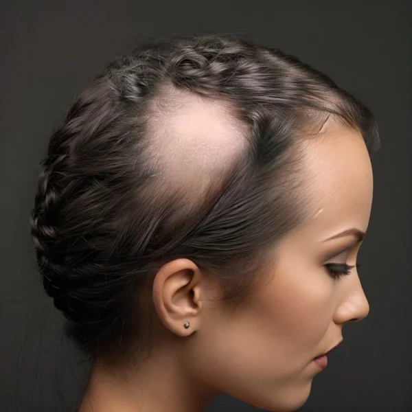
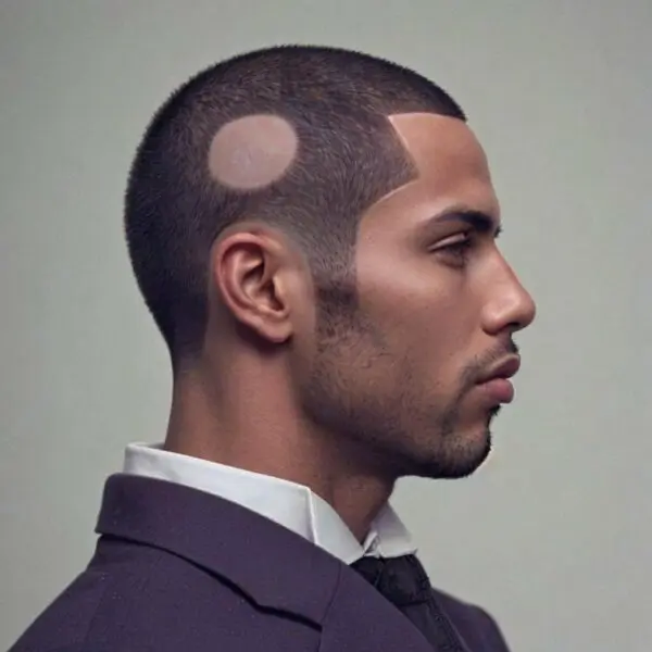
Clinical Presentation: Alopecia areata presents with distinctive clinical features that make it recognizable, though the pattern and extent of hair loss can vary. The most common presentation is the sudden appearance of one or more well-circumscribed, round or oval patches of hair loss on the scalp. The affected areas are typically smooth and without signs of inflammation or scarring, although some patients may experience mild itching or a tingling sensation. The disease course is often unpredictable, with periods of spontaneous remission and relapse.
Patchy Alopecia Areata is the most common form, characterized by discrete patches of hair loss. In some cases (about 30% of people who get AA), alopecia areata may progress beyond patchy hair loss. Alopecia Totalis involves the complete loss of all scalp hair. Alopecia Universalis is the most extensive form, characterized by the complete loss of hair on the scalp and body, including eyebrows, eyelashes, and other body hair. Other variants of alopecia areata include Ophiasis where hair loss occurs in a band-like pattern around the sides and lower back of the scalp. Diffuse Alopecia Areata involves diffuse thinning of hair across the scalp without distinct patches. Alopecia Barbae involves hair loss confined to the beard area.
Nail changes are seen in about 10-20% of patients and can include pitting, ridging, and brittleness, which may be a marker of more severe disease.
Diagnosis: The diagnosis of alopecia areata is primarily clinical, based on the characteristic appearance of the hair loss. Dermoscopy can aid in the diagnosis by revealing specific features such as “exclamation mark” hairs (broken hairs that are narrower at the base), yellow dots (representing sebaceous gland hyperplasia), and black dots (broken hairs within follicles).
In cases where the diagnosis is uncertain, or if there is suspicion of another underlying condition, a scalp biopsy may be performed. Histopathological examination typically shows a peribulbar lymphocytic infiltrate around the affected hair follicles, often described as a “swarm of bees” appearance. Other findings include miniaturization of hair follicles and increased numbers of catagen and telogen follicles, indicating a shift from the anagen phase.
Differential diagnoses to consider include tinea capitis, trichotillomania, androgenetic alopecia, and telogen effluvium, among others.
Psychological Impact: The psychological impact of alopecia areata can be profound, given that hair is often closely tied to a person’s identity and self-esteem. Patients may experience significant emotional distress, anxiety, depression, and social withdrawal due to the unpredictable nature of the disease and its visible impact. The condition can severely affect quality of life, particularly in cases of extensive hair loss such as alopecia totalis or universalis.
Depression and anxiety are common comorbidities, and addressing the mental health aspects of the condition is a critical component of patient care. Supportive care, including counseling and support groups, plays a crucial role in the management of the psychological aspects of alopecia areata. Health care providers should be aware of these potential issues and offer appropriate referrals to mental health professionals when necessary.
Treatment: The treatment of alopecia areata is challenging, as there is no cure, and the course of the disease is unpredictable. Management strategies aim to induce hair regrowth and prevent further hair loss, but treatment responses vary widely among patients.
Topical Therapies:
Corticosteroids: Topical corticosteroids, such as clobetasol propionate, are commonly used as a first-line treatment, particularly for limited disease. They help reduce inflammation and suppress the immune response around hair follicles.
Minoxidil: Topical minoxidil is often used in conjunction with corticosteroids to promote hair regrowth. While it does not address the underlying autoimmune process, it can stimulate hair follicles to enter the anagen phase.
Anthralin: This is a topical immunomodulator that can be used in more resistant cases. It causes a mild irritant reaction, which is thought to modify the local immune response.
Intralesional Therapies:
Intralesional Corticosteroids: Triamcinolone acetonide is commonly injected into affected areas of the scalp to promote hair regrowth in localized alopecia areata. This approach is typically reserved for smaller, stable patches of hair loss.
Systemic Therapies:
Oral Corticosteroids: Systemic corticosteroids may be used in cases of rapid progression or extensive disease, though their use is limited by potential side effects such as weight gain, hypertension, and osteoporosis.
Immunosuppressants: Agents like methotrexate, cyclosporine, and azathioprine have been used with varying success in severe or recalcitrant cases of alopecia areata. However, these treatments are not typical and are not used in most clinics.
JAK Inhibitors: Janus kinase (JAK) inhibitors, such as tofacitinib and ruxolitinib, represent a newer class of systemic therapies that have shown promise in clinical trials. These agents target specific pathways involved in the immune response and have been associated with significant hair regrowth in some patients, although they are not yet universally approved for this indication.
Contact Sensitizing Agents and Irritants:
PUVA (Psoralen + UVA) and NBUVB (Narrowband UVB): Phototherapy has been used with some success, particularly in patients with extensive or resistant disease. It works by modulating the immune response and promoting hair regrowth, although the exact mechanism is not fully understood.
DPCP (Diphencyprone): DPCP is a contact sensitizer that induces a mild allergic reaction in the scalp, thereby redirecting the immune response away from the hair follicles. It has been shown to promote hair regrowth in some patients, particularly those with more extensive patchy alopecia areata.
SADBE (Squaric Acid Dibutyl Ester): Similar to DPCP, SADBE is another topical sensitizing agent used to induce a controlled allergic reaction, which may help in stimulating hair regrowth. It is often considered for patients who do not respond to other treatments.
Anthralin: This is a topical irritant that causes mild inflammation, intended to disrupt the autoimmune process in the scalp. Although primarily used in psoriasis, it is sometimes employed in alopecia areata to promote hair regrowth by inducing an irritant dermatitis.
Prognosis: The prognosis of alopecia areata is highly variable and unpredictable. Some patients experience spontaneous regrowth of hair, particularly in cases of limited disease, while others may develop chronic, relapsing, or more severe forms of the disease. Factors associated with a poorer prognosis include early onset (especially in young children), extensive hair loss at the initial presentation, a family history of alopecia areata, and the presence of nail changes.
Long-term management often involves a combination of treatments, patient education, and psychological support. For some patients, particularly those with extensive or treatment-resistant disease, the focus may shift to cosmetic solutions, such as wigs, hairpieces, and scalp micropigmentation.
Future Directions and Research: Research into alopecia areata continues to evolve, with ongoing studies exploring the genetic and immunological underpinnings of the disease. Advances in genomics and immunotherapy hold promise for the development of more targeted and effective treatments. Additionally, the identification of biomarkers may lead to more personalized approaches to therapy, improving outcomes for patients. The role of environmental triggers, gut microbiota, and lifestyle factors in the development and progression of alopecia areata is also an area of active investigation. Understanding these factors may provide new insights into disease prevention and management.
Conclusion: Alopecia areata is a complex autoimmune disorder with a significant impact on affected individuals. While the exact cause remains unclear, advances in research have highlighted the role of genetic, immunological, and environmental factors in its pathogenesis. The clinical presentation is varied, and the disease course is unpredictable, making management challenging. Current treatments aim to suppress the immune response and promote hair regrowth, but they are often met with varying degrees of success and may not be effective for all patients. The psychological burden of the condition underscores the need for comprehensive care that includes both medical and emotional support. As research progresses, there is hope for more targeted and effective therapies that could offer better outcomes for those affected by this condition, ultimately improving their quality of life. Continued exploration into the underlying mechanisms and potential therapeutic targets remains essential in the pursuit of more effective and personalized treatments for alopecia areata.
Lu W, Shapiro J, Yu M, Barekatain A, Lo B, Finner A, et al. Alopecia areata: pathogenesis and potential for therapy. Expert Rev Mol Med. 2006 June 20;8(14):1–19.
1.
Alkhalifah A, Alsantali A, Wang E, McElwee KJ, Shapiro J. Alopecia areata update: part I. Clinical picture, histopathology, and pathogenesis. J Am Acad Dermatol. 2010 Feb;62(2):177–88, quiz 189–90.
1.
Alkhalifah A, Alsantali A, Wang E, McElwee KJ, Shapiro J. Alopecia areata update: part II. Treatment. J Am Acad Dermatol. 2010 Feb;62(2):191–202, quiz 203–4.
1.
Wang E, McElwee KJ. Etiopathogenesis of alopecia areata: Why do our patients get it? Dermatol Ther. 2011;24(3):337–47.
1.
McElwee KJ, Gilhar A, Tobin DJ, Ramot Y, Sundberg JP, Nakamura M, et al. What causes alopecia areata? Exp Dermatol. 2013 Sept;22(9):609–26.
1.
Guo H, Cheng Y, Shapiro J, McElwee K. The role of lymphocytes in the development and treatment of alopecia areata. Expert Rev Clin Immunol. 2015;11(12):1335–51.
Alopecia areata (AA) is a prevalent autoimmune condition, characterized by non-scarring hair loss. Many studies have been done on the epidemiology of this disorder, which…
Introduction: Alopecia areata (AA) is an autoimmune disorder most often characterized by patchy hair loss that can significantly impact the quality of life for those…
Manage Cookie Consent
We use technologies like cookies to store and/or access device information. We do this to improve browsing experience and to show (non-) personalized ads. Consenting to these technologies will allow us to process data such as browsing behavior or unique IDs on this site. Not consenting or withdrawing consent, may adversely affect certain features and functions.
Functional Always active
The technical storage or access is strictly necessary for the legitimate purpose of enabling the use of a specific service explicitly requested by the subscriber or user, or for the sole purpose of carrying out the transmission of a communication over an electronic communications network.
Preferences
The technical storage or access is necessary for the legitimate purpose of storing preferences that are not requested by the subscriber or user.
Statistics
The technical storage or access that is used exclusively for statistical purposes.The technical storage or access that is used exclusively for anonymous statistical purposes. Without a subpoena, voluntary compliance on the part of your Internet Service Provider, or additional records from a third party, information stored or retrieved for this purpose alone cannot usually be used to identify you.
Marketing
The technical storage or access is required to create user profiles to send advertising, or to track the user on a website or across several websites for similar marketing purposes.