Introduction: Kerion Celsi is a severe, inflammatory form of tinea capitis, primarily affecting children. It is characterized by a boggy, purulent, and sometimes painful lesion on the scalp. The condition arises from a hypersensitivity reaction to dermatophytes (fungi that live on the skin), often leading to misdiagnosis as a bacterial infection due to its atypical presentation. This article aims to demystify Kerion Celsi, exploring its causes, symptoms, treatment, and the importance of early diagnosis to prevent lasting effects.
What are dermatophytes : Dermatophytes are a group of fungi that have the ability to infect and survive on and in keratinized tissues, including the skin, hair, and nails. These fungi are the causative agents of various forms of tinea capitis (ringworm) infections in humans and animals. They are characterized by their ability to utilize keratin as a nutrient source, leading to the degradation of keratinized tissue, the proliferation of the fungi, and subsequent infection. Dermatophyte infections are generally superficial, affecting the outer layers of the skin, and are known for their circular, ring-like lesions, which is why they are commonly referred to as “ringworm”.
The infections can be transmitted through direct contact with infected individuals or animals, or indirectly through contact with contaminated objects or surfaces. Common species of dermatophytes include Trichophyton, Microsporum, and Epidermophyton. The most common fungi that causes scalp ringworm varies across different regions of the world, and it has also been seen to change over time. However, in the last 50 years or so, Trichophyton tonsurans has become the most common culprit in North America.
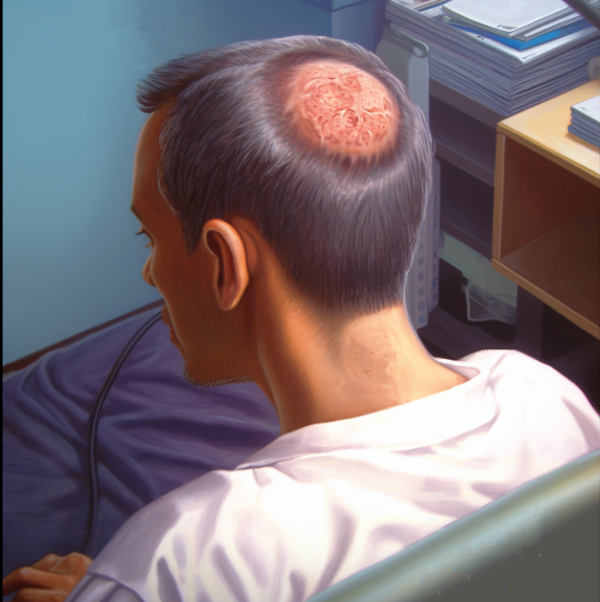
What is Kerion Celsi: A kerion is not an infectious agent in itself rather a kerion is a severe, inflammatory skin lesion that develops when an infectious agent that normally causes scalp ringworm (tinea capitis) becomes more aggressive. Deep boggy red areas characterized by a severe acute inflammatory infiltrate with pustule formation are termed kerions or kerion celsi. Normally, scalp ringworm inducing agents cause circular patches of red crusty skin. Although not pleasant, the problem is relatively mild and reversible with proper treatment. However, if the infection gets out of hand a kerion may develop. It results from an exaggerated immune response to the fungus, leading to severe inflammation.
Who Gets Kerion Celsi: Primarily affecting children, Kerion Celsi doesn’t discriminate based on gender or ethnicity. Children between 3 and 7 years are most susceptible, possibly due to their developing immune systems and close physical contact in settings like schools and playgrounds. However, adults, especially those with compromised immune systems, can also develop this condition.
How Does Kerion Celsi Occur: Transmission of the fungal dermatophytes causing Kerion Celsi usually happens through direct contact with infected individuals or animals. The fungi thrive in warm, moist environments, making children’s scalps an ideal breeding ground. Sharing combs, hats, or pillows can also facilitate the spread of these fungi. Kerion Celsi are more likely to occur in hot humid parts of the world, but they can be found in people the world over.
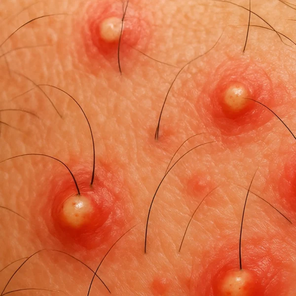
Kerion Symptoms : When a kerion develops it first starts out as a more typical presentation of tinea capitis with a flaky, crusty patch of skin on the scalp. This can quickly deteriorate into a boggy, puritic mass of inflamed tissue. This is a kerion and it may be accompanied by hair loss and tenderness. The kerion can deteriorate to a nasty, deep abscess if it is not treated correctly. This has the potential to elicit scarring and permanent alopecia. When a kerion develops with severe inflammation it is fairly common for the regional lymph nodes in the neck to become enlarged (a condition called cervical lymphadenopathy). Some people can become quite ill and may develop a widespread rash and fever.
Suppuration and kerion formation are more commonly are typically associated with Trichophyton tonsurans infection. Kerion formation with other infectious agents that cause tinea capitis are less likely, but still possible. For example, a fungus found on the skin of dogs and cats called Microsporum canis, can cause a kerion if it transfers from the pet to humans in the home.
Kerion Diagnosis : Kerion Celsi presents as a swollen, red, and sometimes oozing lesion on the scalp which you might think would be a fairly distinctive condition. However, misdiagnosis is common, as it can resemble bacterial infections or other scalp conditions. A healthcare provider will typically diagnose it through clinical examination and sometimes the use of a “Wood’s lamp” (basically a blacklight).
This type of light shines UV light onto the skin. If there is an infection on the area where the Wood’s lamp is illuminating, the area will fluoresce (usually a green-yellow color). It does not allow for a clear distinction to be made between a fungal infection of a bacterial infection, but it does help to identify if an infectious agents is present. Sometimes the Wood’s lamp test give a false negative result – the lesion has deteriorated to the extent that the inflammation can hide the fungal infection. Further, some types of fungi do not fluoresce, so the Wood’s lamp is quick and easy to do, but it is not a definitive test.
For a clear diagnosis of fungal infection, a dermatologist will take skin scrapings which can then be sent for culture in a laboratory. Sometimes as a kerion deteriorates, bacterial infection also takes hold. Consequently, dermatologists may also send samples to the lab for bacterial culture as well as fungal culture. More modern PCR tests are also available, but these tend not to be popular, probably because they are relatively expensive to perform.
The dermatologist may also do some tests in their own clinic (it depends on their training and experience). For those suitably trained, they may do examinations of skin and hair samples under a microscope (such as Chicago sky blue staining, Periodic acid–Schiff staining and/or other similar sample processing). “Old-School” dermatologists will do a potassium hydroxide (KOH) test where KOH is used to digest the skin or hair – but fungi are resistant so they can be clearly seen under the microscope.
Kerion differential diagnosis : Sometimes Kerion Celsi is confused with other conditions due to a lack of diagnostic testing. The most common confusion seems to be between kerion and impetigo which can look somewhat similar. In one report, some patients were hospitalized with the diagnosis of Staphylococcal abscess while a microbiological diagnostic test would have shown the cause was due to Trichophyton verrucosum infection resulting in a kerion. Occasionally, a kerion can look much like some forms of scarring alopecia such as dissecting cellulitis or erosive pustular dermatosis.
Because of this apparent similarity in presentation, the doctor needs to be very careful in their investigation and knowledgeable about the potential for confusing kerions with other diagnoses. The patient who suspects they have a kerion caused by an infectious agent or something similar needs to find an experienced dermatologist to improve the chances of getting a correct diagnosis. The average general practitioner is probably not in a position to make a kerion diagnosis with complete confidence.
Treatment and Hair Loss Management: Early initiation of treatment is extremely important with kerions. The sooner treatment is started the less likely the kerion will promote a permanent scarring alopecia. Unfortunately, some reports suggest that rapid diagnosis and treatment of kerions only occurs in a minority of cases or that kerions are often misdiagnosed at first. There seems to be a certain lack of knowledge about tinea capitis and kerions among some general practitioners and this can lead to a delay in referral to a dermatologist and then receiving proper treatment. The longer kerions persist the more damaging they become.
Treatment of Kerion Celsi involves oral antifungal medications, such as griseofulvin, terbinafine, or itraconazole. The course of treatment can last several weeks to ensure complete eradication of the fungus. Topical treatments are less effective due to the depth of hair follicle involvement. Many patients with kerions and a primary diagnosis of tinea capitis also have a secondary bacterial infection of the kerion. Anti-fungals are not good for treating bacterial or yeast infections so other anti-bacterial treatments may be given along with the anti-fungal drug. Sometimes oral corticosteroids are also given in addition to the anti-fungal agent, although the few published studies comparing treatments with and without corticosteroids have shown little added benefit. However in principle, oral corticosteroids could help reduce the inflammation in the kerion. Topical corticosteroids are not used as this can complicate treatment of the local fungal infection.
Early and appropriate treatment is crucial to prevent permanent hair loss or scarring. Usually the hair does grow back after completion of anti-fungal treatment, though it can take some weeks for the hair follicles to get their act together and start growing hair again. If the hair loss is permanent due to scar tissue formation, the only practical option may be a small hair transplant.
Conclusion: Kerion Celsi, while alarming in appearance, is effectively treatable with timely medical intervention. Awareness and understanding of this condition are vital in preventing misdiagnosis and ensuring prompt treatment. Parents should be vigilant for symptoms in children and seek medical advice early. By doing so, the long-term effects of Kerion Celsi, such as scarring and hair loss, can be minimized.
Bibliography
11711645 {11711645:PTZT7QBU},{11711645:SVK4WCDH},{11711645:MDPISVDK},{11711645:HHWUTSMQ},{11711645:G9JDM9QC},{11711645:6V2J2RBF},{11711645:IG78WGDV},{11711645:D6BJZB4B},{11711645:735QNSX3},{11711645:CZQ35R3X},{11711645:P3766X9W},{11711645:7VUGWCP8},{11711645:XXH8FRJX},{11711645:WD39DPZD},{11711645:6JJ54RK6},{11711645:Q5HPWXD6},{11711645:KCBMPM47} 1 vancouver 50 date asc 703 https://www.keratin.com/wp-content/plugins/zotpress/ %7B%22status%22%3A%22success%22%2C%22updateneeded%22%3Afalse%2C%22instance%22%3Afalse%2C%22meta%22%3A%7B%22request_last%22%3A0%2C%22request_next%22%3A0%2C%22used_cache%22%3Atrue%7D%2C%22data%22%3A%5B%7B%22key%22%3A%22HHWUTSMQ%22%2C%22library%22%3A%7B%22id%22%3A11711645%7D%2C%22meta%22%3A%7B%22creatorSummary%22%3A%22Fitch%22%2C%22parsedDate%22%3A%221879-11%22%2C%22numChildren%22%3A0%7D%2C%22bib%22%3A%22%26lt%3Bdiv%20class%3D%26quot%3Bcsl-bib-body%26quot%3B%20style%3D%26quot%3Bline-height%3A%201.35%3B%20%26quot%3B%26gt%3B%5Cn%20%20%26lt%3Bdiv%20class%3D%26quot%3Bcsl-entry%26quot%3B%20style%3D%26quot%3Bclear%3A%20left%3B%20%26quot%3B%26gt%3B%5Cn%20%20%20%20%26lt%3Bdiv%20class%3D%26quot%3Bcsl-left-margin%26quot%3B%20style%3D%26quot%3Bfloat%3A%20left%3B%20padding-right%3A%200.5em%3B%20text-align%3A%20right%3B%20width%3A%201em%3B%26quot%3B%26gt%3B1.%26lt%3B%5C%2Fdiv%26gt%3B%26lt%3Bdiv%20class%3D%26quot%3Bcsl-right-inline%26quot%3B%20style%3D%26quot%3Bmargin%3A%200%20.4em%200%201.5em%3B%26quot%3B%26gt%3BFitch%20WH.%20Tinea%20Kerion.%20Chic%20Med%20J%20Exam.%201879%20Nov%3B39%285%29%3A478%26%23x2013%3B81.%26lt%3B%5C%2Fdiv%26gt%3B%5Cn%20%20%20%26lt%3B%5C%2Fdiv%26gt%3B%5Cn%26lt%3B%5C%2Fdiv%26gt%3B%22%2C%22data%22%3A%7B%22itemType%22%3A%22journalArticle%22%2C%22title%22%3A%22Tinea%20Kerion%22%2C%22creators%22%3A%5B%7B%22creatorType%22%3A%22author%22%2C%22firstName%22%3A%22Wm%20H.%22%2C%22lastName%22%3A%22Fitch%22%7D%5D%2C%22abstractNote%22%3A%22%22%2C%22date%22%3A%221879-11%22%2C%22section%22%3A%22%22%2C%22partNumber%22%3A%22%22%2C%22partTitle%22%3A%22%22%2C%22DOI%22%3A%22%22%2C%22citationKey%22%3A%22%22%2C%22url%22%3A%22%22%2C%22PMID%22%3A%22%22%2C%22PMCID%22%3A%22%22%2C%22ISSN%22%3A%22%22%2C%22language%22%3A%22eng%22%2C%22collections%22%3A%5B%227M63KT3E%22%5D%2C%22dateModified%22%3A%222023-12-26T15%3A36%3A36Z%22%7D%7D%2C%7B%22key%22%3A%22G9JDM9QC%22%2C%22library%22%3A%7B%22id%22%3A11711645%7D%2C%22meta%22%3A%7B%22creatorSummary%22%3A%22Ginsburg%20et%20al.%22%2C%22parsedDate%22%3A%221987-12%22%2C%22numChildren%22%3A0%7D%2C%22bib%22%3A%22%26lt%3Bdiv%20class%3D%26quot%3Bcsl-bib-body%26quot%3B%20style%3D%26quot%3Bline-height%3A%201.35%3B%20%26quot%3B%26gt%3B%5Cn%20%20%26lt%3Bdiv%20class%3D%26quot%3Bcsl-entry%26quot%3B%20style%3D%26quot%3Bclear%3A%20left%3B%20%26quot%3B%26gt%3B%5Cn%20%20%20%20%26lt%3Bdiv%20class%3D%26quot%3Bcsl-left-margin%26quot%3B%20style%3D%26quot%3Bfloat%3A%20left%3B%20padding-right%3A%200.5em%3B%20text-align%3A%20right%3B%20width%3A%201em%3B%26quot%3B%26gt%3B1.%26lt%3B%5C%2Fdiv%26gt%3B%26lt%3Bdiv%20class%3D%26quot%3Bcsl-right-inline%26quot%3B%20style%3D%26quot%3Bmargin%3A%200%20.4em%200%201.5em%3B%26quot%3B%26gt%3BGinsburg%20CM%2C%20Gan%20VN%2C%20Petruska%20M.%20Randomized%20controlled%20trial%20of%20intralesional%20corticosteroid%20and%20griseofulvin%20vs.%20griseofulvin%20alone%20for%20treatment%20of%20kerion.%20Pediatr%20Infect%20Dis%20J.%201987%20Dec%3B6%2812%29%3A1084%26%23x2013%3B7.%26lt%3B%5C%2Fdiv%26gt%3B%5Cn%20%20%20%26lt%3B%5C%2Fdiv%26gt%3B%5Cn%26lt%3B%5C%2Fdiv%26gt%3B%22%2C%22data%22%3A%7B%22itemType%22%3A%22journalArticle%22%2C%22title%22%3A%22Randomized%20controlled%20trial%20of%20intralesional%20corticosteroid%20and%20griseofulvin%20vs.%20griseofulvin%20alone%20for%20treatment%20of%20kerion%22%2C%22creators%22%3A%5B%7B%22creatorType%22%3A%22author%22%2C%22firstName%22%3A%22C.%20M.%22%2C%22lastName%22%3A%22Ginsburg%22%7D%2C%7B%22creatorType%22%3A%22author%22%2C%22firstName%22%3A%22V.%20N.%22%2C%22lastName%22%3A%22Gan%22%7D%2C%7B%22creatorType%22%3A%22author%22%2C%22firstName%22%3A%22M.%22%2C%22lastName%22%3A%22Petruska%22%7D%5D%2C%22abstractNote%22%3A%22We%20studied%2030%20children%20with%20tinea%20capitis%20and%20kerion%20to%20define%20the%20epidemiology%20of%20the%20disease%20and%20to%20compare%20the%20efficacy%20of%20intralesional%20steroid%20injection%20and%20griseofulvin%20to%20griseofulvin%20alone%20for%20treatment%20of%20this%20disorder.%20Patients%20ranged%20in%20age%20from%201%20year%20to%2012%20years%2C%201%20month%20%28mean%2C%205%20years%2C%207%20months%29.%20All%20patients%20were%20black%20and%2023%20%2877%25%29%20were%20female.%20%28The%20racial%20composition%20of%20our%20clinic%20population%20is%2045%25%20black%2C%2018%25%20Latin%20American%2C%2034%25%20Caucasian%20and%203%25%20Asian.%29%20Fungus%20cultures%20were%20positive%20in%20all%20but%20one%20patient%20and%20Trichophyton%20tonsurans%20was%20isolated%20from%2026%20of%2030%20%2887%25%29%20of%20the%20pretreatment%20hair%20cultures.%20Direct%20microscopic%20examinations%20of%20KOH-treated%20hair%20samples%20were%20negative%20in%2013%20of%2029%20%2843%25%29%20culture-positive%20patients.%20Patients%20were%20randomly%20assigned%20to%20receive%20intralesional%20steroid%20injection%20%282.5%20mg%29%20and%20griseofulvin%20%2814%20patients%29%20or%20griseofulvin%20only%20%2816%20patients%29.%20The%20treatment%20groups%20were%20comparable%20with%20regard%20to%20age%2C%20sex%2C%20duration%20of%20lesions%20before%20treatment%20and%20type%20of%20lesions.%20There%20were%20no%20significant%20differences%20between%20the%20two%20treatment%20groups%20in%20the%20time%20to%20negative%20culture%2C%20time%20of%20onset%20of%20new%20hair%20growth%2C%20complete%20regrowth%20of%20hair%20and%20time%20to%20scalp%20clearing.%20We%20conclude%20that%20intralesional%20injection%20of%20corticosteroid%20is%20an%20unnecessary%20adjunct%20to%20therapy%20for%20patients%20with%20tinea%20capitis%20with%20kerion.%22%2C%22date%22%3A%221987-12%22%2C%22section%22%3A%22%22%2C%22partNumber%22%3A%22%22%2C%22partTitle%22%3A%22%22%2C%22DOI%22%3A%22%22%2C%22citationKey%22%3A%22%22%2C%22url%22%3A%22%22%2C%22PMID%22%3A%22%22%2C%22PMCID%22%3A%22%22%2C%22ISSN%22%3A%220891-3668%22%2C%22language%22%3A%22eng%22%2C%22collections%22%3A%5B%227M63KT3E%22%5D%2C%22dateModified%22%3A%222023-12-26T15%3A19%3A03Z%22%7D%7D%2C%7B%22key%22%3A%22WD39DPZD%22%2C%22library%22%3A%7B%22id%22%3A11711645%7D%2C%22meta%22%3A%7B%22creatorSummary%22%3A%22Sperling%22%2C%22parsedDate%22%3A%221991-03%22%2C%22numChildren%22%3A0%7D%2C%22bib%22%3A%22%26lt%3Bdiv%20class%3D%26quot%3Bcsl-bib-body%26quot%3B%20style%3D%26quot%3Bline-height%3A%201.35%3B%20%26quot%3B%26gt%3B%5Cn%20%20%26lt%3Bdiv%20class%3D%26quot%3Bcsl-entry%26quot%3B%20style%3D%26quot%3Bclear%3A%20left%3B%20%26quot%3B%26gt%3B%5Cn%20%20%20%20%26lt%3Bdiv%20class%3D%26quot%3Bcsl-left-margin%26quot%3B%20style%3D%26quot%3Bfloat%3A%20left%3B%20padding-right%3A%200.5em%3B%20text-align%3A%20right%3B%20width%3A%201em%3B%26quot%3B%26gt%3B1.%26lt%3B%5C%2Fdiv%26gt%3B%26lt%3Bdiv%20class%3D%26quot%3Bcsl-right-inline%26quot%3B%20style%3D%26quot%3Bmargin%3A%200%20.4em%200%201.5em%3B%26quot%3B%26gt%3BSperling%20LC.%20Inflammatory%20tinea%20capitis%20%28kerion%29%20mimicking%20dissecting%20cellulitis.%20Occurrence%20in%20two%20adolescents.%20Int%20J%20Dermatol.%201991%20Mar%3B30%283%29%3A190%26%23x2013%3B2.%26lt%3B%5C%2Fdiv%26gt%3B%5Cn%20%20%20%26lt%3B%5C%2Fdiv%26gt%3B%5Cn%26lt%3B%5C%2Fdiv%26gt%3B%22%2C%22data%22%3A%7B%22itemType%22%3A%22journalArticle%22%2C%22title%22%3A%22Inflammatory%20tinea%20capitis%20%28kerion%29%20mimicking%20dissecting%20cellulitis.%20Occurrence%20in%20two%20adolescents%22%2C%22creators%22%3A%5B%7B%22creatorType%22%3A%22author%22%2C%22firstName%22%3A%22L.%20C.%22%2C%22lastName%22%3A%22Sperling%22%7D%5D%2C%22abstractNote%22%3A%22Tinea%20capitis%20is%20unusual%20in%20postpubertal%20individuals%20and%20is%20frequently%20misdiagnosed.%20In%20cases%20of%20inflammatory%20disease%2C%20prompt%20initiation%20of%20therapy%20is%20essential%20to%20prevent%20scarring%20and%20permanent%20hair%20loss.%20Two%20examples%20are%20presented%20to%20illustrate%20principles%20of%20evaluation%20and%20treatment.%22%2C%22date%22%3A%221991-03%22%2C%22section%22%3A%22%22%2C%22partNumber%22%3A%22%22%2C%22partTitle%22%3A%22%22%2C%22DOI%22%3A%2210.1111%5C%2Fj.1365-4362.1991.tb03849.x%22%2C%22citationKey%22%3A%22%22%2C%22url%22%3A%22%22%2C%22PMID%22%3A%22%22%2C%22PMCID%22%3A%22%22%2C%22ISSN%22%3A%220011-9059%22%2C%22language%22%3A%22eng%22%2C%22collections%22%3A%5B%227M63KT3E%22%5D%2C%22dateModified%22%3A%222023-12-26T15%3A22%3A50Z%22%7D%7D%2C%7B%22key%22%3A%22735QNSX3%22%2C%22library%22%3A%7B%22id%22%3A11711645%7D%2C%22meta%22%3A%7B%22creatorSummary%22%3A%22McDonagh%20and%20Bleehen%22%2C%22parsedDate%22%3A%221991-05%22%2C%22numChildren%22%3A0%7D%2C%22bib%22%3A%22%26lt%3Bdiv%20class%3D%26quot%3Bcsl-bib-body%26quot%3B%20style%3D%26quot%3Bline-height%3A%201.35%3B%20%26quot%3B%26gt%3B%5Cn%20%20%26lt%3Bdiv%20class%3D%26quot%3Bcsl-entry%26quot%3B%20style%3D%26quot%3Bclear%3A%20left%3B%20%26quot%3B%26gt%3B%5Cn%20%20%20%20%26lt%3Bdiv%20class%3D%26quot%3Bcsl-left-margin%26quot%3B%20style%3D%26quot%3Bfloat%3A%20left%3B%20padding-right%3A%200.5em%3B%20text-align%3A%20right%3B%20width%3A%201em%3B%26quot%3B%26gt%3B1.%26lt%3B%5C%2Fdiv%26gt%3B%26lt%3Bdiv%20class%3D%26quot%3Bcsl-right-inline%26quot%3B%20style%3D%26quot%3Bmargin%3A%200%20.4em%200%201.5em%3B%26quot%3B%26gt%3BMcDonagh%20AJ%2C%20Bleehen%20SS.%20Kerion%20masquerading%20as%20erosive%20pustular%20dermatosis%20of%20the%20scalp.%20Br%20J%20Dermatol.%201991%20May%3B124%285%29%3A507%26%23x2013%3B8.%26lt%3B%5C%2Fdiv%26gt%3B%5Cn%20%20%20%26lt%3B%5C%2Fdiv%26gt%3B%5Cn%26lt%3B%5C%2Fdiv%26gt%3B%22%2C%22data%22%3A%7B%22itemType%22%3A%22journalArticle%22%2C%22title%22%3A%22Kerion%20masquerading%20as%20erosive%20pustular%20dermatosis%20of%20the%20scalp%22%2C%22creators%22%3A%5B%7B%22creatorType%22%3A%22author%22%2C%22firstName%22%3A%22A.%20J.%22%2C%22lastName%22%3A%22McDonagh%22%7D%2C%7B%22creatorType%22%3A%22author%22%2C%22firstName%22%3A%22S.%20S.%22%2C%22lastName%22%3A%22Bleehen%22%7D%5D%2C%22abstractNote%22%3A%22%22%2C%22date%22%3A%221991-05%22%2C%22section%22%3A%22%22%2C%22partNumber%22%3A%22%22%2C%22partTitle%22%3A%22%22%2C%22DOI%22%3A%2210.1111%5C%2Fj.1365-2133.1991.tb00640.x%22%2C%22citationKey%22%3A%22%22%2C%22url%22%3A%22%22%2C%22PMID%22%3A%22%22%2C%22PMCID%22%3A%22%22%2C%22ISSN%22%3A%220007-0963%22%2C%22language%22%3A%22eng%22%2C%22collections%22%3A%5B%227M63KT3E%22%5D%2C%22dateModified%22%3A%222023-12-26T15%3A21%3A46Z%22%7D%7D%2C%7B%22key%22%3A%226V2J2RBF%22%2C%22library%22%3A%7B%22id%22%3A11711645%7D%2C%22meta%22%3A%7B%22creatorSummary%22%3A%22Honig%20et%20al.%22%2C%22parsedDate%22%3A%221993-09%22%2C%22numChildren%22%3A0%7D%2C%22bib%22%3A%22%26lt%3Bdiv%20class%3D%26quot%3Bcsl-bib-body%26quot%3B%20style%3D%26quot%3Bline-height%3A%201.35%3B%20%26quot%3B%26gt%3B%5Cn%20%20%26lt%3Bdiv%20class%3D%26quot%3Bcsl-entry%26quot%3B%20style%3D%26quot%3Bclear%3A%20left%3B%20%26quot%3B%26gt%3B%5Cn%20%20%20%20%26lt%3Bdiv%20class%3D%26quot%3Bcsl-left-margin%26quot%3B%20style%3D%26quot%3Bfloat%3A%20left%3B%20padding-right%3A%200.5em%3B%20text-align%3A%20right%3B%20width%3A%201em%3B%26quot%3B%26gt%3B1.%26lt%3B%5C%2Fdiv%26gt%3B%26lt%3Bdiv%20class%3D%26quot%3Bcsl-right-inline%26quot%3B%20style%3D%26quot%3Bmargin%3A%200%20.4em%200%201.5em%3B%26quot%3B%26gt%3BHonig%20PJ%2C%20Caputo%20GL%2C%20Leyden%20JJ%2C%20McGinley%20K%2C%20Selbst%20SM%2C%20McGravey%20AR.%20Microbiology%20of%20kerions.%20J%20Pediatr.%201993%20Sept%3B123%283%29%3A422%26%23x2013%3B4.%26lt%3B%5C%2Fdiv%26gt%3B%5Cn%20%20%20%26lt%3B%5C%2Fdiv%26gt%3B%5Cn%26lt%3B%5C%2Fdiv%26gt%3B%22%2C%22data%22%3A%7B%22itemType%22%3A%22journalArticle%22%2C%22title%22%3A%22Microbiology%20of%20kerions%22%2C%22creators%22%3A%5B%7B%22creatorType%22%3A%22author%22%2C%22firstName%22%3A%22P.%20J.%22%2C%22lastName%22%3A%22Honig%22%7D%2C%7B%22creatorType%22%3A%22author%22%2C%22firstName%22%3A%22G.%20L.%22%2C%22lastName%22%3A%22Caputo%22%7D%2C%7B%22creatorType%22%3A%22author%22%2C%22firstName%22%3A%22J.%20J.%22%2C%22lastName%22%3A%22Leyden%22%7D%2C%7B%22creatorType%22%3A%22author%22%2C%22firstName%22%3A%22K.%22%2C%22lastName%22%3A%22McGinley%22%7D%2C%7B%22creatorType%22%3A%22author%22%2C%22firstName%22%3A%22S.%20M.%22%2C%22lastName%22%3A%22Selbst%22%7D%2C%7B%22creatorType%22%3A%22author%22%2C%22firstName%22%3A%22A.%20R.%22%2C%22lastName%22%3A%22McGravey%22%7D%5D%2C%22abstractNote%22%3A%22Qualitative%20and%20semiquantitative%20bacterial%20culture%20specimens%20were%20obtained%20from%2044%20patients%20with%20kerions.%20Staphylococcus%20aureus%20was%20isolated%20from%20the%20scalp%20surface%20overlying%20the%20kerion%20in%2029%25%20of%20patients%20and%20from%20the%20pus%20within%20the%20kerion%20in%2048%25%20of%20patients.%20Gram-negative%20bacteria%20were%20found%20in%20the%20same%20locations%20in%2011.3%25%20and%2018%25%20of%20patients%2C%20respectively.%20The%20data%20indicate%20that%20bacteria%20are%20frequently%20cultured%20from%20kerions.%22%2C%22date%22%3A%221993-09%22%2C%22section%22%3A%22%22%2C%22partNumber%22%3A%22%22%2C%22partTitle%22%3A%22%22%2C%22DOI%22%3A%2210.1016%5C%2Fs0022-3476%2805%2981749-4%22%2C%22citationKey%22%3A%22%22%2C%22url%22%3A%22%22%2C%22PMID%22%3A%22%22%2C%22PMCID%22%3A%22%22%2C%22ISSN%22%3A%220022-3476%22%2C%22language%22%3A%22eng%22%2C%22collections%22%3A%5B%227M63KT3E%22%5D%2C%22dateModified%22%3A%222023-12-26T15%3A21%3A07Z%22%7D%7D%2C%7B%22key%22%3A%22P3766X9W%22%2C%22library%22%3A%7B%22id%22%3A11711645%7D%2C%22meta%22%3A%7B%22creatorSummary%22%3A%22Pomeranz%20and%20Fairley%22%2C%22parsedDate%22%3A%221994-06%22%2C%22numChildren%22%3A0%7D%2C%22bib%22%3A%22%26lt%3Bdiv%20class%3D%26quot%3Bcsl-bib-body%26quot%3B%20style%3D%26quot%3Bline-height%3A%201.35%3B%20%26quot%3B%26gt%3B%5Cn%20%20%26lt%3Bdiv%20class%3D%26quot%3Bcsl-entry%26quot%3B%20style%3D%26quot%3Bclear%3A%20left%3B%20%26quot%3B%26gt%3B%5Cn%20%20%20%20%26lt%3Bdiv%20class%3D%26quot%3Bcsl-left-margin%26quot%3B%20style%3D%26quot%3Bfloat%3A%20left%3B%20padding-right%3A%200.5em%3B%20text-align%3A%20right%3B%20width%3A%201em%3B%26quot%3B%26gt%3B1.%26lt%3B%5C%2Fdiv%26gt%3B%26lt%3Bdiv%20class%3D%26quot%3Bcsl-right-inline%26quot%3B%20style%3D%26quot%3Bmargin%3A%200%20.4em%200%201.5em%3B%26quot%3B%26gt%3BPomeranz%20AJ%2C%20Fairley%20JA.%20Management%20errors%20leading%20to%20unnecessary%20hospitalization%20for%20kerion.%20Pediatrics.%201994%20June%3B93%286%20Pt%201%29%3A986%26%23x2013%3B8.%26lt%3B%5C%2Fdiv%26gt%3B%5Cn%20%20%20%26lt%3B%5C%2Fdiv%26gt%3B%5Cn%26lt%3B%5C%2Fdiv%26gt%3B%22%2C%22data%22%3A%7B%22itemType%22%3A%22journalArticle%22%2C%22title%22%3A%22Management%20errors%20leading%20to%20unnecessary%20hospitalization%20for%20kerion%22%2C%22creators%22%3A%5B%7B%22creatorType%22%3A%22author%22%2C%22firstName%22%3A%22A.%20J.%22%2C%22lastName%22%3A%22Pomeranz%22%7D%2C%7B%22creatorType%22%3A%22author%22%2C%22firstName%22%3A%22J.%20A.%22%2C%22lastName%22%3A%22Fairley%22%7D%5D%2C%22abstractNote%22%3A%22OBJECTIVE%3A%20Tinea%20capitis%20is%20a%20common%20problem%20in%20the%20inner%20city%2C%20outpatient%20population.%20It%20is%20known%20that%20some%20children%20were%20being%20admitted%20for%20treatment%20of%20the%20kerion%20type%20of%20tinea%20capitis.%20The%20purpose%20of%20this%20study%20was%20to%20determine%20why%20these%20children%20were%20being%20admitted%20and%20whether%20hospitalization%20was%20justified.%5CnDESIGN%3A%20A%20retrospective%20study%20of%20all%20children%20hospitalized%20at%20Children%26%23039%3Bs%20Hospital%20of%20Wisconsin%20between%20January%201990%20and%20December%201992%20with%20the%20diagnosis%20of%20tinea%20capitis%20was%20performed.%5CnRESULTS%3A%20Twenty-nine%20children%20who%20were%20hospitalized%20with%20tinea%20capitis%20were%20identified.%20In%2010%20cases%2C%20tinea%20capitis%20was%20the%20primary%20reason%20for%20admission%2C%20in%2019%20patients%20it%20was%20a%20secondary%20diagnosis.%20All%20of%20the%20patients%20with%20a%20primary%20diagnosis%20of%20tinea%20capitis%20were%20presumed%20to%20have%20a%20secondary%20bacterial%20infection%20of%20the%20kerion%3B%20however%2C%20this%20was%20not%20verified%20by%20the%20workup%20of%20any%20patient.%20Numerous%20deviations%20from%20optimal%20treatment%20were%20noted%20in%20both%20groups%20of%20patients.%20Although%20tinea%20capitis%20had%20been%20correctly%20diagnosed%20before%20admission%20in%20seven%20primary%20tinea%20capitis%20patients%2C%20only%20two%20of%20these%20patients%20received%20oral%20griseofulvin.%20The%20patients%20in%20whom%20tinea%20capitis%20was%20a%20secondary%20diagnosis%20also%20had%20a%20number%20of%20therapeutic%20deficiencies%20including%20failure%20to%20prescribe%20griseofulvin%20%2816%25%29%2C%20failure%20to%20administer%20griseofulvin%20with%20a%20fatty%20food%20or%20meal%20%2858%25%29%2C%20and%20failure%20to%20prescribe%20a%20sporicidal%20shampoo%20%2863%25%29.%5CnCONCLUSIONS%3A%20Although%20all%20patients%20were%20assumed%20to%20have%20a%20secondary%20bacterial%20infection%2C%20this%20could%20not%20be%20documented.%20With%20the%20possible%20exception%20of%20one%20patient%20all%20the%20symptoms%20described%20by%20the%20patients%20could%20be%20explained%20by%20the%20highly%20inflammatory%20nature%20of%20a%20kerion.%20A%20better%20understanding%20of%20the%20degree%20of%20inflammation%20that%20may%20accompany%20a%20kerion%2C%20as%20well%20as%20its%20proper%20treatment%2C%20may%20prevent%20unnecessary%20hospitalization%20of%20children.%22%2C%22date%22%3A%221994-06%22%2C%22section%22%3A%22%22%2C%22partNumber%22%3A%22%22%2C%22partTitle%22%3A%22%22%2C%22DOI%22%3A%22%22%2C%22citationKey%22%3A%22%22%2C%22url%22%3A%22%22%2C%22PMID%22%3A%22%22%2C%22PMCID%22%3A%22%22%2C%22ISSN%22%3A%220031-4005%22%2C%22language%22%3A%22eng%22%2C%22collections%22%3A%5B%227M63KT3E%22%5D%2C%22dateModified%22%3A%222023-12-26T15%3A18%3A34Z%22%7D%7D%2C%7B%22key%22%3A%22KCBMPM47%22%2C%22library%22%3A%7B%22id%22%3A11711645%7D%2C%22meta%22%3A%7B%22creatorSummary%22%3A%22Zaror%20et%20al.%22%2C%22parsedDate%22%3A%221995-08%22%2C%22numChildren%22%3A0%7D%2C%22bib%22%3A%22%26lt%3Bdiv%20class%3D%26quot%3Bcsl-bib-body%26quot%3B%20style%3D%26quot%3Bline-height%3A%201.35%3B%20%26quot%3B%26gt%3B%5Cn%20%20%26lt%3Bdiv%20class%3D%26quot%3Bcsl-entry%26quot%3B%20style%3D%26quot%3Bclear%3A%20left%3B%20%26quot%3B%26gt%3B%5Cn%20%20%20%20%26lt%3Bdiv%20class%3D%26quot%3Bcsl-left-margin%26quot%3B%20style%3D%26quot%3Bfloat%3A%20left%3B%20padding-right%3A%200.5em%3B%20text-align%3A%20right%3B%20width%3A%201em%3B%26quot%3B%26gt%3B1.%26lt%3B%5C%2Fdiv%26gt%3B%26lt%3Bdiv%20class%3D%26quot%3Bcsl-right-inline%26quot%3B%20style%3D%26quot%3Bmargin%3A%200%20.4em%200%201.5em%3B%26quot%3B%26gt%3BZaror%20L%2C%20Hering%20M%2C%20Moreno%20MI%2C%20Navarrete%20M.%20%5BKerion%20Celsi.%20A%20diagnostic%20problem%3F%20Experience%20with%206%20cases%5D.%20Rev%20Med%20Chil.%201995%20Aug%3B123%288%29%3A1006%26%23x2013%3B8.%26lt%3B%5C%2Fdiv%26gt%3B%5Cn%20%20%20%26lt%3B%5C%2Fdiv%26gt%3B%5Cn%26lt%3B%5C%2Fdiv%26gt%3B%22%2C%22data%22%3A%7B%22itemType%22%3A%22journalArticle%22%2C%22title%22%3A%22%5BKerion%20Celsi.%20A%20diagnostic%20problem%3F%20Experience%20with%206%20cases%5D%22%2C%22creators%22%3A%5B%7B%22creatorType%22%3A%22author%22%2C%22firstName%22%3A%22L.%22%2C%22lastName%22%3A%22Zaror%22%7D%2C%7B%22creatorType%22%3A%22author%22%2C%22firstName%22%3A%22M.%22%2C%22lastName%22%3A%22Hering%22%7D%2C%7B%22creatorType%22%3A%22author%22%2C%22firstName%22%3A%22M.%20I.%22%2C%22lastName%22%3A%22Moreno%22%7D%2C%7B%22creatorType%22%3A%22author%22%2C%22firstName%22%3A%22M.%22%2C%22lastName%22%3A%22Navarrete%22%7D%5D%2C%22abstractNote%22%3A%22We%20report%20six%20patients%20with%20Kerion%20Celsi%20due%20to%20Trichophyton%20verrucosum.%20Five%20of%20the%20patients%20were%20hospitalized%20with%20the%20diagnosis%20of%20Staphylococcal%20abscess.%20This%20confusion%20is%20due%20to%20that%20highly%20suppurative%20and%20inflammatory%20nature%20of%20the%20infection.%20Griseofulvin%20is%20the%20antimicrobial%20of%20choice%20for%20treatment%2C%20associated%20with%20imidazolics%20and%20corticosteroids%20to%20prevent%20alopecia.%20The%20authors%20suggest%20that%20an%20adequate%20use%20of%20simple%20microbiological%20diagnostic%20tests%20in%20the%20diagnosis%20of%20pyodermitis%20in%20rural%20children%2C%20may%20prevent%20unnecessary%20hospitalizations%20and%20permanent%20hair%20loss.%22%2C%22date%22%3A%221995-08%22%2C%22section%22%3A%22%22%2C%22partNumber%22%3A%22%22%2C%22partTitle%22%3A%22%22%2C%22DOI%22%3A%22%22%2C%22citationKey%22%3A%22%22%2C%22url%22%3A%22%22%2C%22PMID%22%3A%22%22%2C%22PMCID%22%3A%22%22%2C%22ISSN%22%3A%220034-9887%22%2C%22language%22%3A%22spa%22%2C%22collections%22%3A%5B%227M63KT3E%22%5D%2C%22dateModified%22%3A%222023-12-26T14%3A58%3A11Z%22%7D%7D%2C%7B%22key%22%3A%22PTZT7QBU%22%2C%22library%22%3A%7B%22id%22%3A11711645%7D%2C%22meta%22%3A%7B%22creatorSummary%22%3A%22Aste%20et%20al.%22%2C%22parsedDate%22%3A%221998%22%2C%22numChildren%22%3A0%7D%2C%22bib%22%3A%22%26lt%3Bdiv%20class%3D%26quot%3Bcsl-bib-body%26quot%3B%20style%3D%26quot%3Bline-height%3A%201.35%3B%20%26quot%3B%26gt%3B%5Cn%20%20%26lt%3Bdiv%20class%3D%26quot%3Bcsl-entry%26quot%3B%20style%3D%26quot%3Bclear%3A%20left%3B%20%26quot%3B%26gt%3B%5Cn%20%20%20%20%26lt%3Bdiv%20class%3D%26quot%3Bcsl-left-margin%26quot%3B%20style%3D%26quot%3Bfloat%3A%20left%3B%20padding-right%3A%200.5em%3B%20text-align%3A%20right%3B%20width%3A%201em%3B%26quot%3B%26gt%3B1.%26lt%3B%5C%2Fdiv%26gt%3B%26lt%3Bdiv%20class%3D%26quot%3Bcsl-right-inline%26quot%3B%20style%3D%26quot%3Bmargin%3A%200%20.4em%200%201.5em%3B%26quot%3B%26gt%3BAste%20N%2C%20Pau%20M%2C%20Biggio%20P.%20Kerion%20Celsi%3A%20a%20clinical%20epidemiological%20study.%20Mycoses.%201998%3B41%283%26%23x2013%3B4%29%3A169%26%23x2013%3B73.%26lt%3B%5C%2Fdiv%26gt%3B%5Cn%20%20%20%26lt%3B%5C%2Fdiv%26gt%3B%5Cn%26lt%3B%5C%2Fdiv%26gt%3B%22%2C%22data%22%3A%7B%22itemType%22%3A%22journalArticle%22%2C%22title%22%3A%22Kerion%20Celsi%3A%20a%20clinical%20epidemiological%20study%22%2C%22creators%22%3A%5B%7B%22creatorType%22%3A%22author%22%2C%22firstName%22%3A%22N.%22%2C%22lastName%22%3A%22Aste%22%7D%2C%7B%22creatorType%22%3A%22author%22%2C%22firstName%22%3A%22M.%22%2C%22lastName%22%3A%22Pau%22%7D%2C%7B%22creatorType%22%3A%22author%22%2C%22firstName%22%3A%22P.%22%2C%22lastName%22%3A%22Biggio%22%7D%5D%2C%22abstractNote%22%3A%22We%20present%20a%20case%20record%20of%20patients%20with%20kerion%20Celsi%20observed%20in%20the%20Clinic%20of%20Dermatology%2C%20University%20of%20Cagliari%20%28Italy%29%20from%201981%20to%201995.%20Altogether%2C%2031%20children%20%2818%20male%20and%2013%20female%29%20with%20an%20age%20range%20of%2040%20days%20to%2012%20years%20were%20studied.%20In%20all%20cases%20the%20lesions%20were%20confined%20to%20the%20scalp.%20In%2010%20cases%20the%20disease%20developed%20as%20such%2C%20while%20in%2021%20patients%20it%20represented%20the%20inflammatory%20evolution%20of%20tinea%20capitis%20secondary%20to%20incongruous%20topical%20treatment.%20Microsporum%20canis%20was%20isolated%20in%2022%20cases%20and%20Trichophyton%20mentagrophytes%20in%20nine%20cases.%20Systemic%20treatment%20was%20carried%20out%20in%2020%20patients%20with%20griseofulvin%2C%20in%20six%20with%20ketoconazole%20and%20in%20five%20with%20terbinafine.%20Complete%20recovery%20was%20obtained%20in%2025-30%20days.%20No%20cases%20of%20relapse%20were%20observed.%20The%20authors%20stress%20the%20importance%20of%20an%20early%20diagnosis%20followed%20by%20timely%20and%20effective%20treatment%20to%20prevent%20scarring%20and%20permanent%20hair%20loss.%22%2C%22date%22%3A%221998%22%2C%22section%22%3A%22%22%2C%22partNumber%22%3A%22%22%2C%22partTitle%22%3A%22%22%2C%22DOI%22%3A%2210.1111%5C%2Fj.1439-0507.1998.tb00319.x%22%2C%22citationKey%22%3A%22%22%2C%22url%22%3A%22%22%2C%22PMID%22%3A%22%22%2C%22PMCID%22%3A%22%22%2C%22ISSN%22%3A%220933-7407%22%2C%22language%22%3A%22eng%22%2C%22collections%22%3A%5B%227M63KT3E%22%5D%2C%22dateModified%22%3A%222023-12-26T15%3A17%3A40Z%22%7D%7D%2C%7B%22key%22%3A%22Q5HPWXD6%22%2C%22library%22%3A%7B%22id%22%3A11711645%7D%2C%22meta%22%3A%7B%22creatorSummary%22%3A%22Tanuma%20et%20al.%22%2C%22parsedDate%22%3A%221999%22%2C%22numChildren%22%3A0%7D%2C%22bib%22%3A%22%26lt%3Bdiv%20class%3D%26quot%3Bcsl-bib-body%26quot%3B%20style%3D%26quot%3Bline-height%3A%201.35%3B%20%26quot%3B%26gt%3B%5Cn%20%20%26lt%3Bdiv%20class%3D%26quot%3Bcsl-entry%26quot%3B%20style%3D%26quot%3Bclear%3A%20left%3B%20%26quot%3B%26gt%3B%5Cn%20%20%20%20%26lt%3Bdiv%20class%3D%26quot%3Bcsl-left-margin%26quot%3B%20style%3D%26quot%3Bfloat%3A%20left%3B%20padding-right%3A%200.5em%3B%20text-align%3A%20right%3B%20width%3A%201em%3B%26quot%3B%26gt%3B1.%26lt%3B%5C%2Fdiv%26gt%3B%26lt%3Bdiv%20class%3D%26quot%3Bcsl-right-inline%26quot%3B%20style%3D%26quot%3Bmargin%3A%200%20.4em%200%201.5em%3B%26quot%3B%26gt%3BTanuma%20H%2C%20Doi%20M%2C%20Abe%20M%2C%20Kume%20H%2C%20Nishiyama%20S%2C%20Katsuoka%20K.%20Case%20report.%20Kerion%20Celsi%20effectively%20treated%20with%20terbinafine.%20Characteristics%20of%20kerion%20Celsi%20in%20the%20elderly%20in%20Japan.%20Mycoses.%201999%3B42%289%26%23x2013%3B10%29%3A581%26%23x2013%3B5.%26lt%3B%5C%2Fdiv%26gt%3B%5Cn%20%20%20%26lt%3B%5C%2Fdiv%26gt%3B%5Cn%26lt%3B%5C%2Fdiv%26gt%3B%22%2C%22data%22%3A%7B%22itemType%22%3A%22journalArticle%22%2C%22title%22%3A%22Case%20report.%20Kerion%20Celsi%20effectively%20treated%20with%20terbinafine.%20Characteristics%20of%20kerion%20Celsi%20in%20the%20elderly%20in%20Japan%22%2C%22creators%22%3A%5B%7B%22creatorType%22%3A%22author%22%2C%22firstName%22%3A%22H.%22%2C%22lastName%22%3A%22Tanuma%22%7D%2C%7B%22creatorType%22%3A%22author%22%2C%22firstName%22%3A%22M.%22%2C%22lastName%22%3A%22Doi%22%7D%2C%7B%22creatorType%22%3A%22author%22%2C%22firstName%22%3A%22M.%22%2C%22lastName%22%3A%22Abe%22%7D%2C%7B%22creatorType%22%3A%22author%22%2C%22firstName%22%3A%22H.%22%2C%22lastName%22%3A%22Kume%22%7D%2C%7B%22creatorType%22%3A%22author%22%2C%22firstName%22%3A%22S.%22%2C%22lastName%22%3A%22Nishiyama%22%7D%2C%7B%22creatorType%22%3A%22author%22%2C%22firstName%22%3A%22K.%22%2C%22lastName%22%3A%22Katsuoka%22%7D%5D%2C%22abstractNote%22%3A%22A%2075-year-old%20non-working%20male%20living%20in%20Sagamihara%2C%20Kanagawa%20Prefecture%2C%20had%20erythematous%20plaques%20with%20scales%20associated%20with%20follicular%20pustules%20in%20the%20head%20area%20extending%20from%20the%20occipital%20to%20right%20temporal%20regions%20about%201%20month%20prior%20to%20his%20initial%20visit%2C%20when%20hair%20loss%20increased.%20The%20diagnosis%20was%20kerion%20Celsi.%20Trichophyton%20rubrum%20was%20isolated%20from%20scales%20and%20tissues%20taken%20from%20lesions%20in%20the%20head.%20Histopathological%20examinations%20showed%20irregular%20epidermal%20thickening%20with%20dense%20cell%20infiltration%20from%20the%20dermis%20to%20subcutaneous%20adipose%20tissues.%20Granulomatous%20reactions%20involving%20neutrophils%2C%20histiocytes%20and%20giant%20cells%20were%20seen%20mainly%20in%20the%20hair%20follicles.%20Periodic%20acid-Schiff%20%28PAS%29%20and%20Grocott-positive%20microbial%20elements%20were%20detected%20in%20the%20horny%20layer%2C%20and%20inside%20and%20outside%20the%20hair%20follicles.%20Pustules%20disappeared%201%20week%20after%20starting%20the%20oral%20treatment%20with%20terbinafine%20%28125%20mg%20day-1%29.%20A%20cure%20was%20achieved%202%20weeks%20after%20starting%20the%20treatment%2C%20with%20only%20slight%20scales%20remaining.%20No%20recurrence%20has%20been%20observed%20to%20date.%20Terbinafine%20was%20thought%20to%20be%20very%20effective%20and%20safe%20for%20kerion%20Celsi.%20We%20reviewed%2027%20cases%20of%20kerion%20Celsi%20reported%20in%20patients%2C%20aged%20at%20least%2070%20years%2C%20in%20Japan%20and%20found%20that%20the%20major%20characteristics%20of%20this%20disease%20in%20Japan%20include%20the%20following%3A%20%281%29%20female%20cases%20outnumber%20male%20cases%3B%20%282%29%20the%20causative%20organism%20was%20T.%20rubrum%20in%2014%20of%2027%20patients%20%2851.9%25%29%3B%20and%20%283%29%20topical%20application%20of%20steroids%20often%20induces%20this%20disease%20in%20patients%20with%20superficial%20tinea%20capitis.%22%2C%22date%22%3A%221999%22%2C%22section%22%3A%22%22%2C%22partNumber%22%3A%22%22%2C%22partTitle%22%3A%22%22%2C%22DOI%22%3A%2210.1046%5C%2Fj.1439-0507.1999.00488.x%22%2C%22citationKey%22%3A%22%22%2C%22url%22%3A%22%22%2C%22PMID%22%3A%22%22%2C%22PMCID%22%3A%22%22%2C%22ISSN%22%3A%220933-7407%22%2C%22language%22%3A%22eng%22%2C%22collections%22%3A%5B%227M63KT3E%22%5D%2C%22dateModified%22%3A%222023-12-26T14%3A59%3A04Z%22%7D%7D%2C%7B%22key%22%3A%22IG78WGDV%22%2C%22library%22%3A%7B%22id%22%3A11711645%7D%2C%22meta%22%3A%7B%22creatorSummary%22%3A%22Hussain%20et%20al.%22%2C%22parsedDate%22%3A%221999-04%22%2C%22numChildren%22%3A0%7D%2C%22bib%22%3A%22%26lt%3Bdiv%20class%3D%26quot%3Bcsl-bib-body%26quot%3B%20style%3D%26quot%3Bline-height%3A%201.35%3B%20%26quot%3B%26gt%3B%5Cn%20%20%26lt%3Bdiv%20class%3D%26quot%3Bcsl-entry%26quot%3B%20style%3D%26quot%3Bclear%3A%20left%3B%20%26quot%3B%26gt%3B%5Cn%20%20%20%20%26lt%3Bdiv%20class%3D%26quot%3Bcsl-left-margin%26quot%3B%20style%3D%26quot%3Bfloat%3A%20left%3B%20padding-right%3A%200.5em%3B%20text-align%3A%20right%3B%20width%3A%201em%3B%26quot%3B%26gt%3B1.%26lt%3B%5C%2Fdiv%26gt%3B%26lt%3Bdiv%20class%3D%26quot%3Bcsl-right-inline%26quot%3B%20style%3D%26quot%3Bmargin%3A%200%20.4em%200%201.5em%3B%26quot%3B%26gt%3BHussain%20I%2C%20Muzaffar%20F%2C%20Rashid%20T%2C%20Ahmad%20TJ%2C%20Jahangir%20M%2C%20Haroon%20TS.%20A%20randomized%2C%20comparative%20trial%20of%20treatment%20of%20kerion%20celsi%20with%20griseofulvin%20plus%20oral%20prednisolone%20vs.%20griseofulvin%20alone.%20Med%20Mycol.%201999%20Apr%3B37%282%29%3A97%26%23x2013%3B9.%26lt%3B%5C%2Fdiv%26gt%3B%5Cn%20%20%20%26lt%3B%5C%2Fdiv%26gt%3B%5Cn%26lt%3B%5C%2Fdiv%26gt%3B%22%2C%22data%22%3A%7B%22itemType%22%3A%22journalArticle%22%2C%22title%22%3A%22A%20randomized%2C%20comparative%20trial%20of%20treatment%20of%20kerion%20celsi%20with%20griseofulvin%20plus%20oral%20prednisolone%20vs.%20griseofulvin%20alone%22%2C%22creators%22%3A%5B%7B%22creatorType%22%3A%22author%22%2C%22firstName%22%3A%22I.%22%2C%22lastName%22%3A%22Hussain%22%7D%2C%7B%22creatorType%22%3A%22author%22%2C%22firstName%22%3A%22F.%22%2C%22lastName%22%3A%22Muzaffar%22%7D%2C%7B%22creatorType%22%3A%22author%22%2C%22firstName%22%3A%22T.%22%2C%22lastName%22%3A%22Rashid%22%7D%2C%7B%22creatorType%22%3A%22author%22%2C%22firstName%22%3A%22T.%20J.%22%2C%22lastName%22%3A%22Ahmad%22%7D%2C%7B%22creatorType%22%3A%22author%22%2C%22firstName%22%3A%22M.%22%2C%22lastName%22%3A%22Jahangir%22%7D%2C%7B%22creatorType%22%3A%22author%22%2C%22firstName%22%3A%22T.%20S.%22%2C%22lastName%22%3A%22Haroon%22%7D%5D%2C%22abstractNote%22%3A%22Glucocorticoids%20are%20often%20recommended%20along%20with%20oral%20antifungals%20in%20the%20treatment%20of%20kerion%20celsi.%20In%20this%20randomized%20study%2C%20the%20efficacy%20of%20combination%20therapy%20with%20oral%20griseofulvin%20and%20oral%20prednisolone%20%28n%20%3D17%29%20was%20compared%20to%20oral%20griseofulvin%20alone%20%28n%3D13%29%20in%20the%20treatment%20of%20kerion%20celsi.%20Both%20groups%20were%20treated%20with%20oral%20griseofulvin%20for%208%20weeks%20whereas%20oral%20prednisolone%20was%20given%20in%20tapering%20doses%20for%203-4%20weeks%20to%20the%20first%20group%20only.%20The%20final%20evaluation%20at%20week%2012%20showed%20a%20cure%20rate%20of%20100%25%20in%20both%20groups%20without%20any%20significant%20difference%20in%20terms%20of%20clinical%20or%20mycological%20cure%20%28P%26gt%3B0.05%29.%20No%20adverse%20events%20were%20noted%20in%20either%20group.%20In%20our%20opinion%20the%20combination%20of%20oral%20prednisolone%20with%20griseofulvin%20does%20not%20result%20in%20additional%20objective%20or%20subjective%20improvement%20compared%20to%20griseofulvin%20alone%20in%20cases%20with%20kerion%20celsi.%22%2C%22date%22%3A%221999-04%22%2C%22section%22%3A%22%22%2C%22partNumber%22%3A%22%22%2C%22partTitle%22%3A%22%22%2C%22DOI%22%3A%22%22%2C%22citationKey%22%3A%22%22%2C%22url%22%3A%22%22%2C%22PMID%22%3A%22%22%2C%22PMCID%22%3A%22%22%2C%22ISSN%22%3A%221369-3786%22%2C%22language%22%3A%22eng%22%2C%22collections%22%3A%5B%227M63KT3E%22%5D%2C%22dateModified%22%3A%222023-12-26T15%3A19%3A43Z%22%7D%7D%2C%7B%22key%22%3A%22XXH8FRJX%22%2C%22library%22%3A%7B%22id%22%3A11711645%7D%2C%22meta%22%3A%7B%22creatorSummary%22%3A%22Sarabi%20and%20Khachemoune%22%2C%22parsedDate%22%3A%222007-12%22%2C%22numChildren%22%3A0%7D%2C%22bib%22%3A%22%26lt%3Bdiv%20class%3D%26quot%3Bcsl-bib-body%26quot%3B%20style%3D%26quot%3Bline-height%3A%201.35%3B%20%26quot%3B%26gt%3B%5Cn%20%20%26lt%3Bdiv%20class%3D%26quot%3Bcsl-entry%26quot%3B%20style%3D%26quot%3Bclear%3A%20left%3B%20%26quot%3B%26gt%3B%5Cn%20%20%20%20%26lt%3Bdiv%20class%3D%26quot%3Bcsl-left-margin%26quot%3B%20style%3D%26quot%3Bfloat%3A%20left%3B%20padding-right%3A%200.5em%3B%20text-align%3A%20right%3B%20width%3A%201em%3B%26quot%3B%26gt%3B1.%26lt%3B%5C%2Fdiv%26gt%3B%26lt%3Bdiv%20class%3D%26quot%3Bcsl-right-inline%26quot%3B%20style%3D%26quot%3Bmargin%3A%200%20.4em%200%201.5em%3B%26quot%3B%26gt%3BSarabi%20K%2C%20Khachemoune%20A.%20Tinea%20capitis%3A%20a%20review.%20Dermatol%20Nurs.%202007%20Dec%3B19%286%29%3A525%26%23x2013%3B9%3B%20quiz%20530.%26lt%3B%5C%2Fdiv%26gt%3B%5Cn%20%20%20%26lt%3B%5C%2Fdiv%26gt%3B%5Cn%26lt%3B%5C%2Fdiv%26gt%3B%22%2C%22data%22%3A%7B%22itemType%22%3A%22journalArticle%22%2C%22title%22%3A%22Tinea%20capitis%3A%20a%20review%22%2C%22creators%22%3A%5B%7B%22creatorType%22%3A%22author%22%2C%22firstName%22%3A%22Khashayar%22%2C%22lastName%22%3A%22Sarabi%22%7D%2C%7B%22creatorType%22%3A%22author%22%2C%22firstName%22%3A%22Amor%22%2C%22lastName%22%3A%22Khachemoune%22%7D%5D%2C%22abstractNote%22%3A%22Tinea%20capitis%20causes%20hair%20loss%2C%20scaling%2C%20erythema%2C%20and%20impetigo-like%20lesions.%20It%20is%20the%20most%20common%20dermatophyte%20infection%20found%20in%20children%20under%20the%20age%20of%2012%2C%20especially%20in%20African%20Americans.%20A%20good%20knowledge%20and%20understanding%20of%20the%20dosages%2C%20duration%2C%20and%20potential%20side%20effects%20of%20different%20antifungals%20is%20important%20for%20managing%20tinea%20capitis.%22%2C%22date%22%3A%222007-12%22%2C%22section%22%3A%22%22%2C%22partNumber%22%3A%22%22%2C%22partTitle%22%3A%22%22%2C%22DOI%22%3A%22%22%2C%22citationKey%22%3A%22%22%2C%22url%22%3A%22%22%2C%22PMID%22%3A%22%22%2C%22PMCID%22%3A%22%22%2C%22ISSN%22%3A%221060-3441%22%2C%22language%22%3A%22eng%22%2C%22collections%22%3A%5B%227M63KT3E%22%5D%2C%22dateModified%22%3A%222023-12-26T15%3A02%3A33Z%22%7D%7D%2C%7B%22key%22%3A%22CZQ35R3X%22%2C%22library%22%3A%7B%22id%22%3A11711645%7D%2C%22meta%22%3A%7B%22creatorSummary%22%3A%22Patel%20and%20Schwartz%22%2C%22parsedDate%22%3A%222011-05%22%2C%22numChildren%22%3A0%7D%2C%22bib%22%3A%22%26lt%3Bdiv%20class%3D%26quot%3Bcsl-bib-body%26quot%3B%20style%3D%26quot%3Bline-height%3A%201.35%3B%20%26quot%3B%26gt%3B%5Cn%20%20%26lt%3Bdiv%20class%3D%26quot%3Bcsl-entry%26quot%3B%20style%3D%26quot%3Bclear%3A%20left%3B%20%26quot%3B%26gt%3B%5Cn%20%20%20%20%26lt%3Bdiv%20class%3D%26quot%3Bcsl-left-margin%26quot%3B%20style%3D%26quot%3Bfloat%3A%20left%3B%20padding-right%3A%200.5em%3B%20text-align%3A%20right%3B%20width%3A%201em%3B%26quot%3B%26gt%3B1.%26lt%3B%5C%2Fdiv%26gt%3B%26lt%3Bdiv%20class%3D%26quot%3Bcsl-right-inline%26quot%3B%20style%3D%26quot%3Bmargin%3A%200%20.4em%200%201.5em%3B%26quot%3B%26gt%3BPatel%20GA%2C%20Schwartz%20RA.%20Tinea%20capitis%3A%20still%20an%20unsolved%20problem%3F%20Mycoses.%202011%20May%3B54%283%29%3A183%26%23x2013%3B8.%26lt%3B%5C%2Fdiv%26gt%3B%5Cn%20%20%20%26lt%3B%5C%2Fdiv%26gt%3B%5Cn%26lt%3B%5C%2Fdiv%26gt%3B%22%2C%22data%22%3A%7B%22itemType%22%3A%22journalArticle%22%2C%22title%22%3A%22Tinea%20capitis%3A%20still%20an%20unsolved%20problem%3F%22%2C%22creators%22%3A%5B%7B%22creatorType%22%3A%22author%22%2C%22firstName%22%3A%22Gopal%20A.%22%2C%22lastName%22%3A%22Patel%22%7D%2C%7B%22creatorType%22%3A%22author%22%2C%22firstName%22%3A%22Robert%20A.%22%2C%22lastName%22%3A%22Schwartz%22%7D%5D%2C%22abstractNote%22%3A%22Tinea%20capitis%20is%20a%20fungal%20infection%20specifically%20involving%20the%20scalp%20and%20hair.%20It%20is%20the%20most%20common%20dermatophyte%20infection%20in%20children%20under%2012%20years%20of%20age%2C%20with%20a%20predominance%20in%20those%20of%20sub-Saharan%20African%20descent.%20Common%20signs%20include%20hair%20loss%2C%20scaling%2C%20erythema%20and%20impetigo-like%20plaques.%20Adults%20may%20also%20be%20affected%2C%20but%20to%20a%20lesser%20degree.%20The%20causative%20species%20are%20from%20the%20Microsporum%20and%20Trichophyton%20genera.%20Limited%20treatment%20options%20and%20diverse%20modes%20of%20transmission%20complicate%20the%20clinician%26%23039%3Bs%20ability%20to%20address%20this%20disease%20adequately.%20Although%20dermatophytes%20are%20ubiquitous%20in%20our%20environment%20and%20tinea%20capitis%20is%20common%2C%20therapeutic%20options%20can%20be%20utilised%20to%20reduce%20morbidity.%22%2C%22date%22%3A%222011-05%22%2C%22section%22%3A%22%22%2C%22partNumber%22%3A%22%22%2C%22partTitle%22%3A%22%22%2C%22DOI%22%3A%2210.1111%5C%2Fj.1439-0507.2009.01819.x%22%2C%22citationKey%22%3A%22%22%2C%22url%22%3A%22%22%2C%22PMID%22%3A%22%22%2C%22PMCID%22%3A%22%22%2C%22ISSN%22%3A%221439-0507%22%2C%22language%22%3A%22eng%22%2C%22collections%22%3A%5B%227M63KT3E%22%5D%2C%22dateModified%22%3A%222023-12-26T15%3A03%3A17Z%22%7D%7D%2C%7B%22key%22%3A%227VUGWCP8%22%2C%22library%22%3A%7B%22id%22%3A11711645%7D%2C%22meta%22%3A%7B%22creatorSummary%22%3A%22Sacchidanand%20et%20al.%22%2C%22parsedDate%22%3A%222012-01%22%2C%22numChildren%22%3A0%7D%2C%22bib%22%3A%22%26lt%3Bdiv%20class%3D%26quot%3Bcsl-bib-body%26quot%3B%20style%3D%26quot%3Bline-height%3A%201.35%3B%20%26quot%3B%26gt%3B%5Cn%20%20%26lt%3Bdiv%20class%3D%26quot%3Bcsl-entry%26quot%3B%20style%3D%26quot%3Bclear%3A%20left%3B%20%26quot%3B%26gt%3B%5Cn%20%20%20%20%26lt%3Bdiv%20class%3D%26quot%3Bcsl-left-margin%26quot%3B%20style%3D%26quot%3Bfloat%3A%20left%3B%20padding-right%3A%200.5em%3B%20text-align%3A%20right%3B%20width%3A%201em%3B%26quot%3B%26gt%3B1.%26lt%3B%5C%2Fdiv%26gt%3B%26lt%3Bdiv%20class%3D%26quot%3Bcsl-right-inline%26quot%3B%20style%3D%26quot%3Bmargin%3A%200%20.4em%200%201.5em%3B%26quot%3B%26gt%3BSacchidanand%20S%2C%20Savitha%20A%2C%20Aparna%20A%2C%20Shilpa%20K.%20Significance%20of%20scraping%20scalp%20lesions%20in%20adults.%20Int%20J%20Trichology.%202012%20Jan%3B4%281%29%3A48%26%23x2013%3B9.%26lt%3B%5C%2Fdiv%26gt%3B%5Cn%20%20%20%26lt%3B%5C%2Fdiv%26gt%3B%5Cn%26lt%3B%5C%2Fdiv%26gt%3B%22%2C%22data%22%3A%7B%22itemType%22%3A%22journalArticle%22%2C%22title%22%3A%22Significance%20of%20scraping%20scalp%20lesions%20in%20adults%22%2C%22creators%22%3A%5B%7B%22creatorType%22%3A%22author%22%2C%22firstName%22%3A%22S.%22%2C%22lastName%22%3A%22Sacchidanand%22%7D%2C%7B%22creatorType%22%3A%22author%22%2C%22firstName%22%3A%22As%22%2C%22lastName%22%3A%22Savitha%22%7D%2C%7B%22creatorType%22%3A%22author%22%2C%22firstName%22%3A%22Ad%22%2C%22lastName%22%3A%22Aparna%22%7D%2C%7B%22creatorType%22%3A%22author%22%2C%22firstName%22%3A%22K.%22%2C%22lastName%22%3A%22Shilpa%22%7D%5D%2C%22abstractNote%22%3A%22%22%2C%22date%22%3A%222012-01%22%2C%22section%22%3A%22%22%2C%22partNumber%22%3A%22%22%2C%22partTitle%22%3A%22%22%2C%22DOI%22%3A%2210.4103%5C%2F0974-7753.96099%22%2C%22citationKey%22%3A%22%22%2C%22url%22%3A%22%22%2C%22PMID%22%3A%22%22%2C%22PMCID%22%3A%22%22%2C%22ISSN%22%3A%220974-9241%22%2C%22language%22%3A%22eng%22%2C%22collections%22%3A%5B%227M63KT3E%22%5D%2C%22dateModified%22%3A%222023-12-26T15%3A24%3A32Z%22%7D%7D%2C%7B%22key%22%3A%22SVK4WCDH%22%2C%22library%22%3A%7B%22id%22%3A11711645%7D%2C%22meta%22%3A%7B%22creatorSummary%22%3A%22Chia%20and%20Dahl%22%2C%22parsedDate%22%3A%222013-02%22%2C%22numChildren%22%3A0%7D%2C%22bib%22%3A%22%26lt%3Bdiv%20class%3D%26quot%3Bcsl-bib-body%26quot%3B%20style%3D%26quot%3Bline-height%3A%201.35%3B%20%26quot%3B%26gt%3B%5Cn%20%20%26lt%3Bdiv%20class%3D%26quot%3Bcsl-entry%26quot%3B%20style%3D%26quot%3Bclear%3A%20left%3B%20%26quot%3B%26gt%3B%5Cn%20%20%20%20%26lt%3Bdiv%20class%3D%26quot%3Bcsl-left-margin%26quot%3B%20style%3D%26quot%3Bfloat%3A%20left%3B%20padding-right%3A%200.5em%3B%20text-align%3A%20right%3B%20width%3A%201em%3B%26quot%3B%26gt%3B1.%26lt%3B%5C%2Fdiv%26gt%3B%26lt%3Bdiv%20class%3D%26quot%3Bcsl-right-inline%26quot%3B%20style%3D%26quot%3Bmargin%3A%200%20.4em%200%201.5em%3B%26quot%3B%26gt%3BChia%20C%2C%20Dahl%20MV.%20Kerion%20mimicking%20erosive%20pustular%20dermatosis%20in%20elderly%20patients.%20Cutis.%202013%20Feb%3B91%282%29%3A73%26%23x2013%3B7.%26lt%3B%5C%2Fdiv%26gt%3B%5Cn%20%20%20%26lt%3B%5C%2Fdiv%26gt%3B%5Cn%26lt%3B%5C%2Fdiv%26gt%3B%22%2C%22data%22%3A%7B%22itemType%22%3A%22journalArticle%22%2C%22title%22%3A%22Kerion%20mimicking%20erosive%20pustular%20dermatosis%20in%20elderly%20patients%22%2C%22creators%22%3A%5B%7B%22creatorType%22%3A%22author%22%2C%22firstName%22%3A%22Charles%22%2C%22lastName%22%3A%22Chia%22%7D%2C%7B%22creatorType%22%3A%22author%22%2C%22firstName%22%3A%22Mark%20V.%22%2C%22lastName%22%3A%22Dahl%22%7D%5D%2C%22abstractNote%22%3A%22Erosive%20pustular%20dermatosis%20of%20the%20scalp%20typically%20occurs%20in%20elderly%20patients%3B%20a%20diagnosis%20of%20fungal%20kerion%20infection%20in%20this%20patient%20population%20may%20seem%20unlikely.%20We%20present%203%20elderly%20patients%20who%20developed%20pustular%20eruptions%20on%20the%20scalp%20that%20were%20suggestive%20of%20erosive%20pustular%20dermatosis.%20Culture%20and%5C%2For%20biopsy%20findings%20initially%20excluded%20kerion%20fungal%20infections.%20Later%2C%20cultures%20isolated%20Trichophyton%20species%20from%201%20patient%20and%20Microsporum%20species%20from%202%20patients%2C%20and%20the%20correct%20diagnosis%20of%20kerion%20was%20made.%20All%20patients%20were%20treated%20successfully%20with%20oral%20terbinafine%20hydrochloride.%20Fungal%20infection%20can%20be%20suspected%20in%20some%20elderly%20patients%20with%20erosive%20pustular%20dermatoses%20of%20the%20scalp.%20Repeated%20cultures%20and%20biopsies%20of%20hair-bearing%20skin%2C%20scale%2C%20and%20cut%20hair%20samples%20may%20be%20required%20to%20establish%20the%20correct%20diagnosis%20of%20kerion.%22%2C%22date%22%3A%222013-02%22%2C%22section%22%3A%22%22%2C%22partNumber%22%3A%22%22%2C%22partTitle%22%3A%22%22%2C%22DOI%22%3A%22%22%2C%22citationKey%22%3A%22%22%2C%22url%22%3A%22%22%2C%22PMID%22%3A%22%22%2C%22PMCID%22%3A%22%22%2C%22ISSN%22%3A%220011-4162%22%2C%22language%22%3A%22eng%22%2C%22collections%22%3A%5B%227M63KT3E%22%5D%2C%22dateModified%22%3A%222023-12-26T15%3A22%3A14Z%22%7D%7D%2C%7B%22key%22%3A%226JJ54RK6%22%2C%22library%22%3A%7B%22id%22%3A11711645%7D%2C%22meta%22%3A%7B%22creatorSummary%22%3A%22Stein%20et%20al.%22%2C%22parsedDate%22%3A%222013-09%22%2C%22numChildren%22%3A0%7D%2C%22bib%22%3A%22%26lt%3Bdiv%20class%3D%26quot%3Bcsl-bib-body%26quot%3B%20style%3D%26quot%3Bline-height%3A%201.35%3B%20%26quot%3B%26gt%3B%5Cn%20%20%26lt%3Bdiv%20class%3D%26quot%3Bcsl-entry%26quot%3B%20style%3D%26quot%3Bclear%3A%20left%3B%20%26quot%3B%26gt%3B%5Cn%20%20%20%20%26lt%3Bdiv%20class%3D%26quot%3Bcsl-left-margin%26quot%3B%20style%3D%26quot%3Bfloat%3A%20left%3B%20padding-right%3A%200.5em%3B%20text-align%3A%20right%3B%20width%3A%201em%3B%26quot%3B%26gt%3B1.%26lt%3B%5C%2Fdiv%26gt%3B%26lt%3Bdiv%20class%3D%26quot%3Bcsl-right-inline%26quot%3B%20style%3D%26quot%3Bmargin%3A%200%20.4em%200%201.5em%3B%26quot%3B%26gt%3BStein%20LL%2C%20Adams%20EG%2C%20Holcomb%20KZ.%20Inflammatory%20tinea%20capitis%20mimicking%20dissecting%20cellulitis%20in%20a%20postpubertal%20male%3A%20a%20case%20report%20and%20review%20of%20the%20literature.%20Mycoses.%202013%20Sept%3B56%285%29%3A596%26%23x2013%3B600.%26lt%3B%5C%2Fdiv%26gt%3B%5Cn%20%20%20%26lt%3B%5C%2Fdiv%26gt%3B%5Cn%26lt%3B%5C%2Fdiv%26gt%3B%22%2C%22data%22%3A%7B%22itemType%22%3A%22journalArticle%22%2C%22title%22%3A%22Inflammatory%20tinea%20capitis%20mimicking%20dissecting%20cellulitis%20in%20a%20postpubertal%20male%3A%20a%20case%20report%20and%20review%20of%20the%20literature%22%2C%22creators%22%3A%5B%7B%22creatorType%22%3A%22author%22%2C%22firstName%22%3A%22Loretta%20L.%22%2C%22lastName%22%3A%22Stein%22%7D%2C%7B%22creatorType%22%3A%22author%22%2C%22firstName%22%3A%22Erin%20G.%22%2C%22lastName%22%3A%22Adams%22%7D%2C%7B%22creatorType%22%3A%22author%22%2C%22firstName%22%3A%22Katherine%20Z.%22%2C%22lastName%22%3A%22Holcomb%22%7D%5D%2C%22abstractNote%22%3A%22Tinea%20capitis%20in%20postpubertal%20patients%20is%20unusual%20and%20may%20be%20misdiagnosed%20as%20dissecting%20cellulitis.%20We%20report%20a%20case%20of%20a%20healthy%2019-year-old%20Hispanic%20male%20presenting%20with%20a%202-month%20history%20of%20a%20large%2C%20painful%20subcutaneous%20boggy%20plaque%20on%20the%20scalp%20with%20patchy%20alopecia%2C%20erythematous%20papules%2C%20cysts%20and%20pustules.%20Although%20initially%20diagnosed%20as%20dissecting%20cellulitis%2C%20potassium%20hydroxide%20evaluation%20%28KOH%20preparation%29%20of%20the%20hair%20from%20the%20affected%20region%20was%20positive.%20A%20punch%20biopsy%20of%20the%20scalp%20demonstrated%20endothrix%20consistent%20with%20tinea%20capitis%2C%20but%20with%20a%20brisk%2C%20deep%20mixed%20inflammatory%20infiltrate%20as%20can%20be%20seen%20with%20chronic%20dissecting%20cellulitis.%20Fungal%20culture%20revealed%20Trichophyton%20tonsurans%2C%20and%20a%20diagnosis%20of%20inflammatory%20tinea%20capitis%20was%20made.%20The%20patient%20was%20treated%20over%20the%20course%20of%2017%5Cu00a0months%20with%20multiple%20systemic%20and%20topical%20antifungal%20medications%2C%20with%20slow%2C%20but%20demonstrable%20clinical%20and%20histopathological%20improvement.%20A%20rare%20diagnosis%20in%20adults%2C%20clinicians%20should%20have%20a%20high%20index%20of%20suspicion%20for%20this%20condition%20in%20an%20adult%20with%20an%20inflammatory%20scalp%20disorder%20not%20classic%20for%20dissecting%20cellulitis%20or%20with%20a%20recalcitrant%20dissecting%20cellulitis.%20Prompt%2C%20appropriate%20diagnosis%20and%20treatment%20is%20necessary%20to%20prevent%20the%20long-term%20complications%20of%20scarring%20alopecia.%22%2C%22date%22%3A%222013-09%22%2C%22section%22%3A%22%22%2C%22partNumber%22%3A%22%22%2C%22partTitle%22%3A%22%22%2C%22DOI%22%3A%2210.1111%5C%2Fmyc.12082%22%2C%22citationKey%22%3A%22%22%2C%22url%22%3A%22%22%2C%22PMID%22%3A%22%22%2C%22PMCID%22%3A%22%22%2C%22ISSN%22%3A%221439-0507%22%2C%22language%22%3A%22eng%22%2C%22collections%22%3A%5B%227M63KT3E%22%5D%2C%22dateModified%22%3A%222023-12-26T15%3A25%3A18Z%22%7D%7D%2C%7B%22key%22%3A%22D6BJZB4B%22%2C%22library%22%3A%7B%22id%22%3A11711645%7D%2C%22meta%22%3A%7B%22creatorSummary%22%3A%22John%20et%20al.%22%2C%22parsedDate%22%3A%222018-01%22%2C%22numChildren%22%3A0%7D%2C%22bib%22%3A%22%26lt%3Bdiv%20class%3D%26quot%3Bcsl-bib-body%26quot%3B%20style%3D%26quot%3Bline-height%3A%201.35%3B%20%26quot%3B%26gt%3B%5Cn%20%20%26lt%3Bdiv%20class%3D%26quot%3Bcsl-entry%26quot%3B%20style%3D%26quot%3Bclear%3A%20left%3B%20%26quot%3B%26gt%3B%5Cn%20%20%20%20%26lt%3Bdiv%20class%3D%26quot%3Bcsl-left-margin%26quot%3B%20style%3D%26quot%3Bfloat%3A%20left%3B%20padding-right%3A%200.5em%3B%20text-align%3A%20right%3B%20width%3A%201em%3B%26quot%3B%26gt%3B1.%26lt%3B%5C%2Fdiv%26gt%3B%26lt%3Bdiv%20class%3D%26quot%3Bcsl-right-inline%26quot%3B%20style%3D%26quot%3Bmargin%3A%200%20.4em%200%201.5em%3B%26quot%3B%26gt%3BJohn%20AM%2C%20Schwartz%20RA%2C%20Janniger%20CK.%20The%20kerion%3A%20an%20angry%20tinea%20capitis.%20Int%20J%20Dermatol.%202018%20Jan%3B57%281%29%3A3%26%23x2013%3B9.%26lt%3B%5C%2Fdiv%26gt%3B%5Cn%20%20%20%26lt%3B%5C%2Fdiv%26gt%3B%5Cn%26lt%3B%5C%2Fdiv%26gt%3B%22%2C%22data%22%3A%7B%22itemType%22%3A%22journalArticle%22%2C%22title%22%3A%22The%20kerion%3A%20an%20angry%20tinea%20capitis%22%2C%22creators%22%3A%5B%7B%22creatorType%22%3A%22author%22%2C%22firstName%22%3A%22Ann%20M.%22%2C%22lastName%22%3A%22John%22%7D%2C%7B%22creatorType%22%3A%22author%22%2C%22firstName%22%3A%22Robert%20A.%22%2C%22lastName%22%3A%22Schwartz%22%7D%2C%7B%22creatorType%22%3A%22author%22%2C%22firstName%22%3A%22Camila%20K.%22%2C%22lastName%22%3A%22Janniger%22%7D%5D%2C%22abstractNote%22%3A%22Tinea%20capitis%20has%20a%20high%20incidence%20with%20a%20global%20changing%20pathogen%20distribution%2C%20making%20this%20condition%20a%20public%20health%20concern%20around%20the%20world.%20As%20the%20infection%20is%20initially%20asymptomatic%2C%20it%20is%20easily%20spread.%20Moreover%2C%20it%20is%20present%20in%20many%20fomites%2C%20including%20hairbrushes%2C%20pillows%2C%20and%20bedding.%20Prompt%20recognition%20and%20treatment%20is%20necessary%20for%20kerion%2C%20an%20inflammatory%20subtype%20characterized%20by%20tender%20boggy%20plaques%20with%20purulent%20drainage.%20Kerion%20is%20usually%20associated%20with%20infection%20by%20zoophilic%20dermatophytes%2C%20although%20other%20sources%20have%20been%20described.%20Treatment%20for%20this%20severe%20form%20of%20dermatophytic%20infection%20can%20be%20challenging.%20In%20addition%20to%20the%20use%20of%20topical%20treatments%2C%20oral%20administration%20of%20griseofulvin%2C%20terbinafine%2C%20itraconazole%2C%20or%20fluconazole%20is%20often%20required.%20Griseofulvin%2C%20the%20first-line%20treatment%2C%20may%20not%20completely%20eradicate%20pathogen%20colonization%20of%20the%20host%20and%20may%20contribute%20to%20reinfection%20and%20prevalence%20of%20infective%20but%20asymptomatic%20carriers.%20This%20review%20highlights%20new%20agents%20that%20are%20being%20evaluated%20for%20the%20treatment%20of%20kerion%20and%20typical%20tinea%20capitis%2C%20enhanced%20diagnostic%20criteria%2C%20and%20a%20grading%20system%20for%20kerion%20evaluation.%22%2C%22date%22%3A%222018-01%22%2C%22section%22%3A%22%22%2C%22partNumber%22%3A%22%22%2C%22partTitle%22%3A%22%22%2C%22DOI%22%3A%2210.1111%5C%2Fijd.13423%22%2C%22citationKey%22%3A%22%22%2C%22url%22%3A%22%22%2C%22PMID%22%3A%22%22%2C%22PMCID%22%3A%22%22%2C%22ISSN%22%3A%221365-4632%22%2C%22language%22%3A%22eng%22%2C%22collections%22%3A%5B%227M63KT3E%22%5D%2C%22dateModified%22%3A%222023-12-26T14%3A57%3A54Z%22%7D%7D%2C%7B%22key%22%3A%22MDPISVDK%22%2C%22library%22%3A%7B%22id%22%3A11711645%7D%2C%22meta%22%3A%7B%22creatorSummary%22%3A%22Chiriac%20et%20al.%22%2C%22parsedDate%22%3A%222023-11-20%22%2C%22numChildren%22%3A0%7D%2C%22bib%22%3A%22%26lt%3Bdiv%20class%3D%26quot%3Bcsl-bib-body%26quot%3B%20style%3D%26quot%3Bline-height%3A%201.35%3B%20%26quot%3B%26gt%3B%5Cn%20%20%26lt%3Bdiv%20class%3D%26quot%3Bcsl-entry%26quot%3B%20style%3D%26quot%3Bclear%3A%20left%3B%20%26quot%3B%26gt%3B%5Cn%20%20%20%20%26lt%3Bdiv%20class%3D%26quot%3Bcsl-left-margin%26quot%3B%20style%3D%26quot%3Bfloat%3A%20left%3B%20padding-right%3A%200.5em%3B%20text-align%3A%20right%3B%20width%3A%201em%3B%26quot%3B%26gt%3B1.%26lt%3B%5C%2Fdiv%26gt%3B%26lt%3Bdiv%20class%3D%26quot%3Bcsl-right-inline%26quot%3B%20style%3D%26quot%3Bmargin%3A%200%20.4em%200%201.5em%3B%26quot%3B%26gt%3BChiriac%20A%2C%20Diaconeasa%20A%2C%20Voicu%20C%2C%20Ivaniciuc%20M%2C%20Miulescu%20R%2C%20Chiriac%20AE%2C%20et%20al.%20Kerion%20Celsi%20in%20infants%20and%20children-A%20narrative%20review%202010-2023.%20Mycoses.%202023%20Nov%2020%3B%26lt%3B%5C%2Fdiv%26gt%3B%5Cn%20%20%20%26lt%3B%5C%2Fdiv%26gt%3B%5Cn%26lt%3B%5C%2Fdiv%26gt%3B%22%2C%22data%22%3A%7B%22itemType%22%3A%22journalArticle%22%2C%22title%22%3A%22Kerion%20Celsi%20in%20infants%20and%20children-A%20narrative%20review%202010-2023%22%2C%22creators%22%3A%5B%7B%22creatorType%22%3A%22author%22%2C%22firstName%22%3A%22Anca%22%2C%22lastName%22%3A%22Chiriac%22%7D%2C%7B%22creatorType%22%3A%22author%22%2C%22firstName%22%3A%22Adriana%22%2C%22lastName%22%3A%22Diaconeasa%22%7D%2C%7B%22creatorType%22%3A%22author%22%2C%22firstName%22%3A%22Cristiana%22%2C%22lastName%22%3A%22Voicu%22%7D%2C%7B%22creatorType%22%3A%22author%22%2C%22firstName%22%3A%22Mihaela%22%2C%22lastName%22%3A%22Ivaniciuc%22%7D%2C%7B%22creatorType%22%3A%22author%22%2C%22firstName%22%3A%22Raluca%22%2C%22lastName%22%3A%22Miulescu%22%7D%2C%7B%22creatorType%22%3A%22author%22%2C%22firstName%22%3A%22Anca%20E.%22%2C%22lastName%22%3A%22Chiriac%22%7D%2C%7B%22creatorType%22%3A%22author%22%2C%22firstName%22%3A%22Pietro%22%2C%22lastName%22%3A%22Nenoff%22%7D%2C%7B%22creatorType%22%3A%22author%22%2C%22firstName%22%3A%22Uwe%22%2C%22lastName%22%3A%22Wollina%22%7D%5D%2C%22abstractNote%22%3A%22Kerion%20Celsi%20is%20an%20inflammatory%2C%20deep%20fungal%20infection%20of%20the%20scalp.%20It%20is%20rare%20in%20neonates%20but%20gets%20more%20common%20in%20children%20about%203%5Cu2009years%20and%20older.%20It%20represents%20with%20swelling%2C%20boggy%20lesions%2C%20pain%2C%20alopecia%20and%20purulent%20secretions.%20Secondary%20bacterial%20infection%20is%20not%20unusual%20after%20maceration.%20Extracutaneous%20manifestations%20include%20regional%20lymphadenopathy%2C%20fever%20and%20very%20rare%20fungemia.%20Id-reactions%20can%20occur.%20Diagnosis%20is%20based%20on%20clinical%20suspicion%2C%20clinical%20examination%20and%20medical%20history.%20Diagnosis%20should%20be%20confirmed%20by%20microscopy%2C%20fungal%20culture%20and%20molecular%20procedures.%20The%20most%20common%20isolated%20fungal%20species%20are%20anthropophilic%20Trichophyton%20%28T.%29%20tonsurans%20and%20zoophilic%20Microsporum%20%28M.%29%20canis%2C%20while%20geophilic%20species%20and%20moulds%20rarely%20cause%20Kerion%20Celsi.%20Treatment%20is%20medical%20with%20systemic%20and%20topical%20antifungals%20supplemented%20by%20systemic%20antibiotics%20when%20necessary%2C%20while%20surgery%20needs%20to%20be%20avoided.%20Early%20and%20sufficient%20treatment%20prevents%20scarring%20alopecia.%20The%20most%20important%20differential%20diagnosis%20is%20bacterial%20skin%20and%20soft%20tissue%20infections.%22%2C%22date%22%3A%222023-11-20%22%2C%22section%22%3A%22%22%2C%22partNumber%22%3A%22%22%2C%22partTitle%22%3A%22%22%2C%22DOI%22%3A%2210.1111%5C%2Fmyc.13675%22%2C%22citationKey%22%3A%22%22%2C%22url%22%3A%22%22%2C%22PMID%22%3A%22%22%2C%22PMCID%22%3A%22%22%2C%22ISSN%22%3A%221439-0507%22%2C%22language%22%3A%22eng%22%2C%22collections%22%3A%5B%227M63KT3E%22%5D%2C%22dateModified%22%3A%222023-12-26T14%3A59%3A40Z%22%7D%7D%5D%7D 1.
Fitch WH. Tinea Kerion. Chic Med J Exam. 1879 Nov;39(5):478–81.
1.
Ginsburg CM, Gan VN, Petruska M. Randomized controlled trial of intralesional corticosteroid and griseofulvin vs. griseofulvin alone for treatment of kerion. Pediatr Infect Dis J. 1987 Dec;6(12):1084–7.
1.
Sperling LC. Inflammatory tinea capitis (kerion) mimicking dissecting cellulitis. Occurrence in two adolescents. Int J Dermatol. 1991 Mar;30(3):190–2.
1.
McDonagh AJ, Bleehen SS. Kerion masquerading as erosive pustular dermatosis of the scalp. Br J Dermatol. 1991 May;124(5):507–8.
1.
Honig PJ, Caputo GL, Leyden JJ, McGinley K, Selbst SM, McGravey AR. Microbiology of kerions. J Pediatr. 1993 Sept;123(3):422–4.
1.
Pomeranz AJ, Fairley JA. Management errors leading to unnecessary hospitalization for kerion. Pediatrics. 1994 June;93(6 Pt 1):986–8.
1.
Zaror L, Hering M, Moreno MI, Navarrete M. [Kerion Celsi. A diagnostic problem? Experience with 6 cases]. Rev Med Chil. 1995 Aug;123(8):1006–8.
1.
Aste N, Pau M, Biggio P. Kerion Celsi: a clinical epidemiological study. Mycoses. 1998;41(3–4):169–73.
1.
Tanuma H, Doi M, Abe M, Kume H, Nishiyama S, Katsuoka K. Case report. Kerion Celsi effectively treated with terbinafine. Characteristics of kerion Celsi in the elderly in Japan. Mycoses. 1999;42(9–10):581–5.
1.
Hussain I, Muzaffar F, Rashid T, Ahmad TJ, Jahangir M, Haroon TS. A randomized, comparative trial of treatment of kerion celsi with griseofulvin plus oral prednisolone vs. griseofulvin alone. Med Mycol. 1999 Apr;37(2):97–9.
1.
Sarabi K, Khachemoune A. Tinea capitis: a review. Dermatol Nurs. 2007 Dec;19(6):525–9; quiz 530.
1.
Patel GA, Schwartz RA. Tinea capitis: still an unsolved problem? Mycoses. 2011 May;54(3):183–8.
1.
Sacchidanand S, Savitha A, Aparna A, Shilpa K. Significance of scraping scalp lesions in adults. Int J Trichology. 2012 Jan;4(1):48–9.
1.
Chia C, Dahl MV. Kerion mimicking erosive pustular dermatosis in elderly patients. Cutis. 2013 Feb;91(2):73–7.
1.
Stein LL, Adams EG, Holcomb KZ. Inflammatory tinea capitis mimicking dissecting cellulitis in a postpubertal male: a case report and review of the literature. Mycoses. 2013 Sept;56(5):596–600.
1.
John AM, Schwartz RA, Janniger CK. The kerion: an angry tinea capitis. Int J Dermatol. 2018 Jan;57(1):3–9.
1.
Chiriac A, Diaconeasa A, Voicu C, Ivaniciuc M, Miulescu R, Chiriac AE, et al. Kerion Celsi in infants and children-A narrative review 2010-2023. Mycoses. 2023 Nov 20;