Introduction: The total number of hair follicles present in the skin of a human is estimated at about 2 million. On other websites (and even some medical text books!) you might see the number stated as 5 million, but this is a misreading of the original research publication from Dr Szabo and colleagues. He calculated human skin has 5 million appendages – of which 2 million are hair follicles and 3 million are sweat glands. Of the total 2 million follicles, the skin of the head contains nearly 1 million hair follicles while the rest of the body has the remaining million. This may seem surprising, but the face skin has many, quite densely packed vellus hair follicles – the tiny hairs you can see on your nose, forehead, and cheeks. About 100,000 terminal hair follicles (the big pigmented hairs) cover the scalp area. Overall all the hair follicles have the same basic structure regardless of size or body location.
For humans, there is not much distinction in hair follicle types. In a few areas of skin we have special hair follicles such as the eyelash hairs which don’t have arrector pili muscles, or the underarm hair follicles that have an extra apocrine gland attached to them. However, the basic hair follicle structure remains essentially the same across human skin. Big follicles make big terminal hair, little follicles make non-pigmented vellus hair, straight hair follicles make straight hair, curly follicles make curly hair, but they all have the same essential structure. This is also pretty much true for hair follicles throughout the range of mammalian species albeit with modifications for specialized functions. The basic structure can even be recognised beyond mammals as bird feather follicles also look somewhat similar.
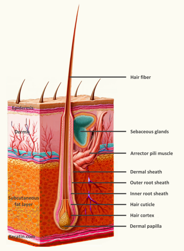
Follicular structure: The hair follicle can be recognized as a separate entity within the skin with formation and maintenance based on interaction between dermal mesenchyme cells and epidermal cells. Development of hair follicles will be described in detail on another page on this website, but we know that during development, cells in the dermis of the skin start to cluster together and then communicate with the epithelial cells in the outermost layer of the skin. Together they form the hair follicle structure and grow down into the skin tissue before they start to make a hair fiber.
In general, the outer “sock” of mesenchymal cells holds the follicle together, and sends out signals to regulate the epithelial cells inside the sock. The epithelial cells inside the follicle form several layers and in the center there are keratinocyte cells that make the actual hair fiber. So we can split the hair follicle appendage into different parts; the mesenchymal outer layer of cells which includes the dermal sheath and dermal papilla, the internal sheaths (layers) of epithelial cells, and the epithelial cells that make the hair fiber itself in the center of the follicle.
Dermal papilla: It is the dermal papilla (often abbreviated to DP) which directs and dictates the embryonic generation of a hair follicle and it also retains this instructive ability throughout the life of the hair follicle. The DP presents as a healthy “pear” shape at the bottom of the sock of normal hair follicles. As the name suggests, derived from the dermis mesenchyme the DP consists of a highly active group of cells shown to be capable of inducing follicle development from the epidermis and production of hair fiber.
The DP consists of a small group of fibroblast cells derived from the mesoderm during embryogenesis. The cells are held close to the base of the epidermal derived cells that produce the hair fiber and root sheaths but there is a thin layer, called the basement membrane (or basement lamina, or glassy membrane) that separates the DP cells from the hair fiber/sheath cells. In other words, the basement membrane provides a physical dividing line between cells descendant from embryonic ectoderm (epidermis) and embryonic mesoderm (dermis). This physical barrier has a role to play in our immunological protection and is continuous throughout our skin, separating the epithelial cells in the outer skin layers from the dermal cells..
Anchoring the DP cells in place is a dermal sheath capsule (the sock) that contains the DP cells in a cup (sometimes called the dermal sheath cup). The DP pear shape sits at the bottom of the dermal sheath sock and is connected to it by a stalk containing a few cells. The dermal sheath extends up the sides of the hair follicle to the skin epidermis. The whole follicle structure sits on a pad of fibrous tissue called the Arao-Perkins body. Nerve fibers and blood vessels penetrate through small gaps in the base of the sock and invade into the DP area.
Under the influence of the DP, epidermal cell differentiation during the anagen hair growth phase produces a keratinized hair fiber and associated products. The source epidermal cells, called matrix cells, that lie in the immediate vicinity of the dermal papilla are a living, actively proliferating group of cells which differentiate and become keratinized to form the hair cortex (Co) and surrounding hair cuticle (Hc) of the hair shaft at the center of which is situated the medulla (M). Cells around the hair shaft comprise the inner root sheath (IRS) which can be divided into three layers the cuticle (Cu), Huxley layer (Hu) and Henle layer (He) based on structure, patterns of keratinization and incorporation of a product called trichohyalin. The IRS breaks down at the level of the sebaceous gland to leave only the hair cortex and surrounding cuticle to protrude above the epidermis.
Root sheaths: The Outer Root Sheath (ORS) is distinct from other epidermal components of the hair follicle being continuous with the epidermis. The “bulge” region in the ORS is the site at which the arrector pili muscle is attached. The arrector pili muscle is anchored to the basement membrane under the epidermis at the other end. This is the muscle that makes hair stand erect and produces goose bumps in your skin when you are cold. The contraction of the muscle pulls on both the hair to make it erect and pulls on the skin making a bumpy surface.
The ORS bulge region is believed to be the storage area for hair follicle stem cells. Hair follicles go through a cycle of growth (anagen) and rest (telogen). With each renewed attempt to produce hair fiber, the hair follicle must obtain a source of cells to form the matrix cell population that make hair fibers. The source of these cells is believed to be the bulge region. Stem cells in the bulge region proliferate for a brief time period at the onset of a new anagen growth phase and produce daughter cells called transit amplifying cells. These daughter cells then multiply and migrate downwards to take up position in the lowest parts of the follicle, sitting above and around the dermal papilla, at which point they are usually called matrix cells.
In a mature anagen hair follicle, the ORS surrounds the hair fiber and inner root sheath until deep into the dermis. Just above the bulb region containing the dermal papilla, the ORS tapers and ends so the ORS does not quite entirely cover the hair fiber and inner root sheath. The ORS itself consists of several layers of cells that can be identified with unique ultrastructural properties, but I won’t go into the details here.
The inner root sheath (IRS) is produced by the epithelial matrix cells sitting over the dermal papilla. Those matrix cells in the center, sitting towards the top of the dermal papilla, proliferate and produce the hair fiber and cuticle. The matrix cells sitting around the lower sides and bottom of the dermal papilla proliferate and produce the IRS. As with cells making up the hair fiber, the cells destined to be IRS gradually become differentiated and keratinized as they are pushed away and upwards from the bulb region. As keratinization occurs, the cells form the IRS surrounding and protecting the development of the hair fiber. The IRS can be subdivided into several layers. Adjacent to the hair fiber we see a single cell thick IRS cuticle layer that closely interdigitates with the hair fiber cuticle layer. The next IRS layer is called the Huxley layer that may consist of up to four cell layers. Outside of this there a single cell layer called the IRS Henle layer. The Henle layer runs adjacent to the ORS layer. The IRS cells fully keratinize, die off, and the IRS disintegrates at the level of the sebaceous gland.
Back near the upper region of the hair follicle, also extending out from the ORS is the sebaceous gland. It consists of a few cells focused on production of oils (lipids). These cells are large with their cytoplasm filled with vacuoles containing lipid. The cells are often divided into several lobes of the sebaceous gland connected together by a sebaceous duct. The duct has a single opening into the tube where the hair fiber sits.
The products of the sebaceous gland are believed to help break down the IRS. The IRS does not extend out of the hair follicle. Only the hair fiber itself protrudes above the skin surface. The IRS disintegrates at the level of the sebaceous duct opening. It is thought that the sebaceous gland oils help to break down the IRS material and allow it to be extruded onto the scalp surface. This is one of the reasons why scalp hair gets so dirty around the roots if the hair is not washed regularly.
The debris from the IRS break down mixes with the sebaceous gland oils and the result is sebum. Sebum is an oily solid that is expelled from the hair follicle and normally scraped or washed away in our general skin care habits. Sometimes overproduction of sebum can build up in the hair follicle and harden into plugs of material called comedones (blackheads). Sebum is a nutrient rich material and an ideal habitat for bacteria. Colonies of bacteria, particularly one called Propionibacterium acnes may proliferate in hair follicles using sebum as a nutrient supply. If this occurs, the immune system responds to the antigens in the modified sebum and the bacteria resulting in inflammation and acne (whiteheads).
Hair fiber: The hair fiber is the core part of any hair follicle. Epidermal derived matrix cells close to the top of the DP remain undifferentiated cells, called matrix cells that focus on multiplying and proliferating to produce more cells. Those cells made towards the middle of the hair follicle are destined to become part of the hair fiber and are called cortex (cortical) cells. As the cells multiply the constant stream of production pushes the cells upwards towards the skin surface. As they move up the hair follicle they begin to differentiate into particular cell types.
The cortex cells change from a round shape into a flattened appearance. They are squeezed together into layers (lamella). These cortex cells become keratinized and harden. As they do so it becomes impossible for the cells to function properly and the cells die. The keratinized cells are then pushed away from the hair bulb region and upwards as new cells come in behind. The cortex cells are now part of the dead keratinized fiber.
Some large terminal hair follicles also have a central strand of cells that are loosely organized and not packed together. This tube in the very center of the hair fiber is called the medulla. These are seen most often in thick, non-pigmented grey hair fibers. Occasionally you can also find medullas in pigmented hair fiber, but more usually pigmented hairs do not have a central medulla.
Around the outside of hair fiber we see a cuticle. The cuticle is made up of more keratinized cells, but they arrange themselves in a slightly different way to cortex cells. As the cuticle cells are produced, they lay over the cortex cells and flatten into an overlapping roof tile fashion. Cuticle cells become progressively flatter as they get older. As with cortex cells, when they keratinize the cells can no longer function properly and die. As they die and flatten the keratinization process fixes the cuticle cells into position to form the hard, chemical resistant outer layer of the hair fiber.
Danforth CH. Hair : with special reference to hypertrichosis. Chicago: American Medical Association; 1925. 152 p.
1.
Lillie FR, Wang H. Physiology of Development of the Feather V. Experimental Morphogenesis. Physiological Zoology. 1941;14(2):103–35.
1.
Myers RJ, Hamilton JB. Regeneration and rate of growth of hairs in man. Ann N Y Acad Sci. 1951 Mar;53(3):562–8.
1.
Garn SM. Types and distribution of the hair in man. Ann N Y Acad Sci. 1951 Mar;53(3):498–507.
1.
Kligman AM. The human hair cycle. J Invest Dermatol. 1959 Dec;33:307–16.
1.
Szabo G. The regional anatomy of the human integument with special reference to the distribution of hair follicles, sweat glands and melanocytes. Phil Trans R Soc Lond B. 1967 Sep 22;252(779):447–85.
1.
Urmacher C. Histology of normal skin. Am J Surg Pathol. 1990 Jul;14(7):671–86.
Whiting DA. The Structure of the Human Hair Follicle: Light Microscopy of Vertical and Horizontal Sections of Scalp Biopsies. Canfield; 2004. 32 p.
1.
Vogt A, McElwee KJ, Blume-Peytavi U. Biology of the Hair Follicle. In: Whitting DA, Blume-Peytavi U, Tosti A, Trüeb RM, editors. Hair Growth and Disorders. Berlin, Heidelberg: Springer; 2008. p. 1–22.
Throughout human history, long hair has been a coveted characteristic, perceived as an emblem of beauty, strength, and social status. Most people cannot grow their…
Eyelashes, while often seen as a mere aesthetic feature, play a significant role in the biological scheme of mammals. These hair follicles are a distinct…
The complex tapestry of human evolution is significantly interwoven with physical attributes, where hair biology plays a pivotal role. Hair is a remarkable biological feature…
Manage Cookie Consent
We use technologies like cookies to store and/or access device information. We do this to improve browsing experience and to show (non-) personalized ads. Consenting to these technologies will allow us to process data such as browsing behavior or unique IDs on this site. Not consenting or withdrawing consent, may adversely affect certain features and functions.
Functional Always active
The technical storage or access is strictly necessary for the legitimate purpose of enabling the use of a specific service explicitly requested by the subscriber or user, or for the sole purpose of carrying out the transmission of a communication over an electronic communications network.
Preferences
The technical storage or access is necessary for the legitimate purpose of storing preferences that are not requested by the subscriber or user.
Statistics
The technical storage or access that is used exclusively for statistical purposes.The technical storage or access that is used exclusively for anonymous statistical purposes. Without a subpoena, voluntary compliance on the part of your Internet Service Provider, or additional records from a third party, information stored or retrieved for this purpose alone cannot usually be used to identify you.
Marketing
The technical storage or access is required to create user profiles to send advertising, or to track the user on a website or across several websites for similar marketing purposes.