Dermatology, the branch of medicine that focuses on the skin, hair, and nails, is a critical field for diagnosing and treating a broad array of conditions. One such subset of conditions relates to hair disorders, which can manifest in various forms such as hair thinning, balding, or unhealthy hair quality. This article aims to provide a detailed walkthrough of the visual examination process a dermatologist employs to assess a hair disorder.
Contrary to popular expectations of lengthy and intricate medical examinations, the evaluation of hair and hair loss conditions is often concise, typically spanning just a few minutes. The key reason behind this brevity lies in the visual nature of hair disorders. The dermatologist can glean ample information from a quick yet careful examination of the scalp’s condition.
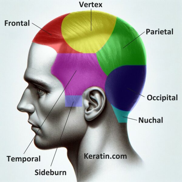
The visual examination often commences with an assessment of the overall hair loss pattern. This involves discerning whether the hair loss is localized to specific areas, like a focal patchy type of alopecia, or dispersed across the scalp in a diffuse pattern. The dermatologist further scrutinizes the extent of the hair loss. For instance, is it limited to the top of the scalp, temples, or the occipital area at the back of the head? The symmetry of the hair loss, whether it is symmetrical or asymmetrical, also provides valuable insights into the possible underlying conditions.
Following the hair loss pattern analysis, the dermatologist proceeds to examine the form of the hair loss. This includes an evaluation of inflammation, any signs of scar tissue, and the presence of crusting or scaling on the skin. Lumps and bumps on the skin could potentially indicate other underlying health issues and are therefore noted as well.
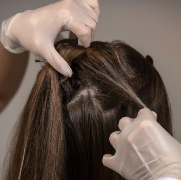
Next, the dermatologist observes the hairs’ condition. An assessment of the hairs’ overall quality, such as its shine, thickness, and texture, helps in identifying potential disorders. For instance, hair that appears lackluster, thin, or unusually textured, whether straight, curly, or kinked, might be symptomatic of a specific hair condition.
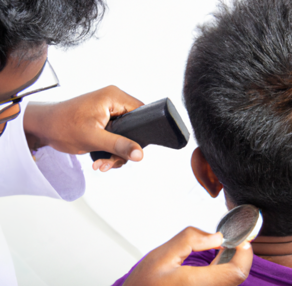
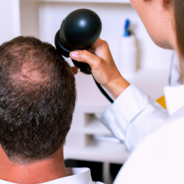
If a fungal infection is suspected, the dermatologist may resort to using a specialized light source known as a Wood’s lamp. Under this light, fungal material fluoresces, making it distinctly visible and enabling a clear identification of fungal infections. It can also detect skin pigment disorders such as vitiligo and other skin irregularities. The examination is usually conducted in a darkened room where the Wood’s lamp, a handheld device that uses black light, is held over an area of the skin. The presence of certain bacteria or fungi or changes in the pigmentation of the skin will cause the affected area to change color under the light. This tool adds an additional layer of precision in the examination process, however, the Wood’s lamp does not test for all fungal and bacterial infections, so further tests might be needed to confirm a diagnosis.
Apart from examining the hair and scalp, a comprehensive dermatological examination also encompasses related body structures. The dermatologist may investigate the quality of the patient’s nails, teeth, and eyes. These structures share a biological connection with the hair follicles and can occasionally display abnormalities in some hair conditions.
For example, brittle nails could indicate a deficiency in essential nutrients, which could also affect hair health. Similarly, certain disorders like alopecia areata can present changes in the nails, such as pitting or white spots. Eye and teeth examination may reveal symptoms of conditions like ectodermal dysplasia, which can affect hair growth.
To conclude, a dermatological visual examination for hair disorders, while brief, is a comprehensive and efficient process. It encompasses an in-depth assessment of the hair loss pattern, the form of hair loss, and the hair’s overall condition. Additional examinations may involve the use of specialized tools like a Wood’s lamp for fungal infection detection and an evaluation of related body structures like nails, teeth, and eyes. Despite the process’s brevity, the information it reveals is crucial in diagnosing and treating various hair conditions. Dermatologists are trained to understand these visual cues and derive insightful information that aids in providing effective treatment for patients dealing with hair disorders.
Neste RPRD Dominique Van. Hair and Scalp Disorders: Common Presenting Signs, Differential Diagnosis. 2nd ed. London: CRC Press; 2004. 294 p.
1.
Mubki T, Rudnicka L, Olszewska M, Shapiro J. Evaluation and diagnosis of the hair loss patient: part II. Trichoscopic and laboratory evaluations. J Am Acad Dermatol. 2014 Sep;71(3):431.e1-431.e11.
1.
Mubki T, Rudnicka L, Olszewska M, Shapiro J. Evaluation and diagnosis of the hair loss patient: part I. History and clinical examination. J Am Acad Dermatol. 2014 Sep;71(3):415.e1-415.e15.
The human scalp is a complex and vital area of the body, especially when considering hair and skin health. For healthcare professionals, particularly those specializing…
The hair pull test, hair tug test, and hair mount test are key diagnostic procedures that dermatologists use during consultations with patients experiencing hair disorders.…
When it comes to hair loss, many of us experience a sense of apprehension. It’s not merely about aesthetics; losing your hair can be a…
Manage Cookie Consent
We use technologies like cookies to store and/or access device information. We do this to improve browsing experience and to show (non-) personalized ads. Consenting to these technologies will allow us to process data such as browsing behavior or unique IDs on this site. Not consenting or withdrawing consent, may adversely affect certain features and functions.
Functional Always active
The technical storage or access is strictly necessary for the legitimate purpose of enabling the use of a specific service explicitly requested by the subscriber or user, or for the sole purpose of carrying out the transmission of a communication over an electronic communications network.
Preferences
The technical storage or access is necessary for the legitimate purpose of storing preferences that are not requested by the subscriber or user.
Statistics
The technical storage or access that is used exclusively for statistical purposes.The technical storage or access that is used exclusively for anonymous statistical purposes. Without a subpoena, voluntary compliance on the part of your Internet Service Provider, or additional records from a third party, information stored or retrieved for this purpose alone cannot usually be used to identify you.
Marketing
The technical storage or access is required to create user profiles to send advertising, or to track the user on a website or across several websites for similar marketing purposes.