The human body is a marvel of interconnected systems, where seemingly unrelated parts share intriguing connections. One such fascinating link exists between hair and eyeballs. At first glance, these two structures might appear distinct and unrelated, but a closer look reveals several shared origins and functions, pointing to a deeper biological relationship. This article delves into the compelling intersections of hair and eyeballs from their embryonic development to their molecular pathways.
Embryonic Origins: The Ectodermal Connection: The journey of both hair and eyes starts early in embryonic development. Both structures have their roots in the ectoderm layer, the outermost of the three germ layers in an embryo. As the embryo develops, the ectoderm differentiates into various structures, including the epidermis from which hair follicles arise. Concurrently, the same ectoderm layer gives birth to the primary structures of the eye, such as the lens and cornea. This shared embryonic origin is the foundation of the biological connection between hair and eyes.
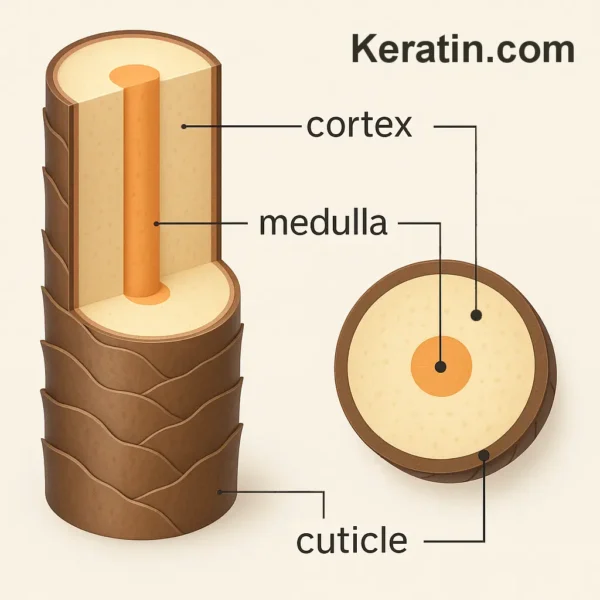
The Keratin Confluence: Keratin is associated with the strength and structure of hair, but its presence isn’t limited to the locks on our heads. These keratin proteins, though primarily known as the backbone of hair, finds their place in the eyes as well. The cornea, the clear front surface of the eye, contains its own unique types of keratin. While different from hair keratin in composition, the commonality of this protein family in both structures underscores their biological kinship.
The Genetic Ties that Bind: Certain genetic disorders reveal a deeper connection between hair and eyes. Ectodermal dysplasia, for instance, can manifest as sparse hair along with eye abnormalities. This highlights how mutations in specific genes can concurrently affect both structures, indicating a shared genetic basis or interconnected developmental pathways.
Shared Pathways – Beyond Development: Even beyond the embryonic stage, hair and eyes continue to share molecular pathways crucial to their maintenance and function. Prominent examples include the Wnt signaling pathway and TGF alpha signaling. These complex pathways play a pivotal role in many physiological processes. Pertinently, they are crucial for both hair follicle development and the formation of certain structures within the eye. Aberrations in these pathways can lead to disorders in either or both structures, underscoring their interwoven biology.
Shared Pathways – Immune Privilege: The eyes and hair follicles also share a unique biological characteristic known as “immune privilege.” This protective feature allows both organs to harbor foreign tissues or tumors that would typically be rejected in other parts of the body. Both ocular tissues and hair follicles produce an array of anti-inflammatory and immunosuppressive molecules. Transforming growth factor-beta (TGF-β) stands out as a common cytokine expressed by both. Additionally, the eye secretes molecules like CD95L (FasL), macrophage migration inhibitory factor, and alpha-melanocyte-stimulating hormone. In parallel, the hair follicle releases these same cytokines. These molecules collectively contribute to the immune-tolerant environment in both structures. The cells of the hair follicle exhibit restricted expression of class Ia MHC molecules. This limited display is strikingly similar to the cells lining the anterior chamber of the eye, offering them protection against CD8+ cytotoxic T lymphocyte attacks.
Medicinal Crossovers: The medical field has inadvertently stumbled upon the link between hair and eyes in the form of certain medications. Bimatoprost, for example, was initially an eye drop formulated for glaucoma treatment. However, a noted side effect was the enhanced growth of eyelashes. This discovery led to the drug’s repurposing as a cosmetic enhancer for eyelashes, emphasizing the delicate balance of molecules that influence both hair growth and eye function.
Diseases and Intersections: Certain diseases, especially those of an autoimmune nature, can cast a spotlight on the hair-eye connection. Alopecia areata, an autoimmune condition characterized by hair loss, can sometimes coexist with inflammatory eye conditions. The immune system’s simultaneous targeting of hair follicles and ocular tissues in some patients underscores a shared vulnerability, hinting at a deeper interconnected biology.
Hairy eyeballs: The term “hairy eyeballs” might sound quite peculiar and, in a literal sense, there is no medical condition where healthy eyeballs actually grow hair. The cells in the eyes are different from the cells that produce hair follicles, and thus it’s not biologically feasible for hair to grow directly on or from healthy eyeballs. However, it should be noted that there are abnormal benign growths that can occur in the eye which might look hairy including Pterygium, Pinguecula, and Dermoid tumors. Dermoid tumors are rare and human dermoid tumors with hair growing from them are extremely rare with only a few case reports published. However, it is not so unusual for dermoid tumors in animals, particularly dogs, cattle, and rabbits, to have very obvious hair growth. These growths can be surgically removed when they occur.
Floaters: As a side-note, “floaters” inside the eye, particularly in the vitreous humor (the clear gel that fills the back part of the eye), might look like shadows of “hair” to some people. Small flakes and fibers of tissue can break off and float in the eye jelly, casting shadows on the retina, these shadows are perceived as floaters that move around and as you move your eyeball around. You can’t quite see them clearly as they are inside the eye, but they can sometimes look like little hairs. Floaters are generally harmless and very common, especially as we get older. I mention this as I have, on occasion, been contacted by people who think they have hair growing inside their eyes, but usually it turns out to be floaters.
Conclusion: While hair and eyeballs serve very different primary functions in our physiology, their shared developmental origins, molecular pathways, and susceptibilities to certain diseases highlight a profound interconnectedness. This understanding not only offers a fascinating insight into human biology but also holds promise for innovative treatments. By studying the overlaps and intersections between hair and eyes, researchers can potentially unearth novel therapeutic strategies for disorders that affect either or both of these vital structures. Recognizing these links also reminds us of a broader truth: the human body’s intricate web of connections often holds the key to understanding, treating, and ultimately celebrating its complexity.
Bibliography
11711645 {11711645:R6MZ47DA},{11711645:SBXTAB8Q},{11711645:S2EHJ4C4},{11711645:TPXBNT3I},{11711645:IF53PFM6},{11711645:ZXDMTIAT},{11711645:IEGG2GAR},{11711645:M6EDB5XU},{11711645:IHR72BQC},{11711645:P9HHCIK6},{11711645:Z4E4ZHRW},{11711645:B6ZV4EMC},{11711645:RVQ2DGKW} 1 vancouver 50 date asc 528 https://www.keratin.com/wp-content/plugins/zotpress/ %7B%22status%22%3A%22success%22%2C%22updateneeded%22%3Afalse%2C%22instance%22%3Afalse%2C%22meta%22%3A%7B%22request_last%22%3A0%2C%22request_next%22%3A0%2C%22used_cache%22%3Atrue%7D%2C%22data%22%3A%5B%7B%22key%22%3A%22P9HHCIK6%22%2C%22library%22%3A%7B%22id%22%3A11711645%7D%2C%22meta%22%3A%7B%22creatorSummary%22%3A%22Dobson%22%2C%22parsedDate%22%3A%221879-10%22%2C%22numChildren%22%3A0%7D%2C%22bib%22%3A%22%26lt%3Bdiv%20class%3D%26quot%3Bcsl-bib-body%26quot%3B%20style%3D%26quot%3Bline-height%3A%201.35%3B%20%26quot%3B%26gt%3B%5Cn%20%20%26lt%3Bdiv%20class%3D%26quot%3Bcsl-entry%26quot%3B%20style%3D%26quot%3Bclear%3A%20left%3B%20%26quot%3B%26gt%3B%5Cn%20%20%20%20%26lt%3Bdiv%20class%3D%26quot%3Bcsl-left-margin%26quot%3B%20style%3D%26quot%3Bfloat%3A%20left%3B%20padding-right%3A%200.5em%3B%20text-align%3A%20right%3B%20width%3A%201em%3B%26quot%3B%26gt%3B1.%26lt%3B%5C%2Fdiv%26gt%3B%26lt%3Bdiv%20class%3D%26quot%3Bcsl-right-inline%26quot%3B%20style%3D%26quot%3Bmargin%3A%200%20.4em%200%201.5em%3B%26quot%3B%26gt%3BDobson%20GE.%20Case%20of%20Development%20of%20Hair%20on%20the%20Eyeball%20of%20a%20Dog.%20J%20Anat%20Physiol.%201879%20Oct%3B14%28Pt%201%29%3A143.%26lt%3B%5C%2Fdiv%26gt%3B%5Cn%20%20%20%26lt%3B%5C%2Fdiv%26gt%3B%5Cn%26lt%3B%5C%2Fdiv%26gt%3B%22%2C%22data%22%3A%7B%22itemType%22%3A%22journalArticle%22%2C%22title%22%3A%22Case%20of%20Development%20of%20Hair%20on%20the%20Eyeball%20of%20a%20Dog%22%2C%22creators%22%3A%5B%7B%22creatorType%22%3A%22author%22%2C%22firstName%22%3A%22G.%20E.%22%2C%22lastName%22%3A%22Dobson%22%7D%5D%2C%22abstractNote%22%3A%22%22%2C%22date%22%3A%221879-10%22%2C%22section%22%3A%22%22%2C%22partNumber%22%3A%22%22%2C%22partTitle%22%3A%22%22%2C%22DOI%22%3A%22%22%2C%22citationKey%22%3A%22%22%2C%22url%22%3A%22%22%2C%22PMID%22%3A%22%22%2C%22PMCID%22%3A%22%22%2C%22ISSN%22%3A%22%22%2C%22language%22%3A%22eng%22%2C%22collections%22%3A%5B%22WF88W4VA%22%5D%2C%22dateModified%22%3A%222023-09-06T16%3A16%3A06Z%22%7D%7D%2C%7B%22key%22%3A%22B6ZV4EMC%22%2C%22library%22%3A%7B%22id%22%3A11711645%7D%2C%22meta%22%3A%7B%22creatorSummary%22%3A%22Chan%22%2C%22parsedDate%22%3A%221932-06-01%22%2C%22numChildren%22%3A0%7D%2C%22bib%22%3A%22%26lt%3Bdiv%20class%3D%26quot%3Bcsl-bib-body%26quot%3B%20style%3D%26quot%3Bline-height%3A%201.35%3B%20%26quot%3B%26gt%3B%5Cn%20%20%26lt%3Bdiv%20class%3D%26quot%3Bcsl-entry%26quot%3B%20style%3D%26quot%3Bclear%3A%20left%3B%20%26quot%3B%26gt%3B%5Cn%20%20%20%20%26lt%3Bdiv%20class%3D%26quot%3Bcsl-left-margin%26quot%3B%20style%3D%26quot%3Bfloat%3A%20left%3B%20padding-right%3A%200.5em%3B%20text-align%3A%20right%3B%20width%3A%201em%3B%26quot%3B%26gt%3B1.%26lt%3B%5C%2Fdiv%26gt%3B%26lt%3Bdiv%20class%3D%26quot%3Bcsl-right-inline%26quot%3B%20style%3D%26quot%3Bmargin%3A%200%20.4em%200%201.5em%3B%26quot%3B%26gt%3BChan%20E.%20A%20Corneo-Scleral%20Dermoid%20in%20A%20Guinea%20Pig.%20American%20Journal%20of%20Ophthalmology.%201932%20June%201%3B15%286%29%3A525%26%23x2013%3B6.%26lt%3B%5C%2Fdiv%26gt%3B%5Cn%20%20%20%26lt%3B%5C%2Fdiv%26gt%3B%5Cn%26lt%3B%5C%2Fdiv%26gt%3B%22%2C%22data%22%3A%7B%22itemType%22%3A%22journalArticle%22%2C%22title%22%3A%22A%20Corneo-Scleral%20Dermoid%20in%20A%20Guinea%20Pig%22%2C%22creators%22%3A%5B%7B%22creatorType%22%3A%22author%22%2C%22firstName%22%3A%22Eugene%22%2C%22lastName%22%3A%22Chan%22%7D%5D%2C%22abstractNote%22%3A%22A%20dermoid%20which%20was%20found%20on%20the%20eye%20of%20a%20guinea%20pig%20was%20studied%20histologically.%20Several%20similar%20tumors%20on%20the%20eyes%20of%20other%20mammals%20have%20been%20reported%20in%20the%20literature.%20From%20the%20Wilmer%20Ophthalmological%20institute%20of%20the%20Johns%20Hopkins%20University%20and%20Hospital%2C%20Baltimore.%22%2C%22date%22%3A%221932-06-01%22%2C%22section%22%3A%22%22%2C%22partNumber%22%3A%22%22%2C%22partTitle%22%3A%22%22%2C%22DOI%22%3A%2210.1016%5C%2FS0002-9394%2832%2990742-9%22%2C%22citationKey%22%3A%22%22%2C%22url%22%3A%22%22%2C%22PMID%22%3A%22%22%2C%22PMCID%22%3A%22%22%2C%22ISSN%22%3A%220002-9394%22%2C%22language%22%3A%22%22%2C%22collections%22%3A%5B%22WF88W4VA%22%5D%2C%22dateModified%22%3A%222023-09-06T16%3A19%3A54Z%22%7D%7D%2C%7B%22key%22%3A%22ZXDMTIAT%22%2C%22library%22%3A%7B%22id%22%3A11711645%7D%2C%22meta%22%3A%7B%22creatorSummary%22%3A%22Wray%20et%20al.%22%2C%22parsedDate%22%3A%221976-02%22%2C%22numChildren%22%3A0%7D%2C%22bib%22%3A%22%26lt%3Bdiv%20class%3D%26quot%3Bcsl-bib-body%26quot%3B%20style%3D%26quot%3Bline-height%3A%201.35%3B%20%26quot%3B%26gt%3B%5Cn%20%20%26lt%3Bdiv%20class%3D%26quot%3Bcsl-entry%26quot%3B%20style%3D%26quot%3Bclear%3A%20left%3B%20%26quot%3B%26gt%3B%5Cn%20%20%20%20%26lt%3Bdiv%20class%3D%26quot%3Bcsl-left-margin%26quot%3B%20style%3D%26quot%3Bfloat%3A%20left%3B%20padding-right%3A%200.5em%3B%20text-align%3A%20right%3B%20width%3A%201em%3B%26quot%3B%26gt%3B1.%26lt%3B%5C%2Fdiv%26gt%3B%26lt%3Bdiv%20class%3D%26quot%3Bcsl-right-inline%26quot%3B%20style%3D%26quot%3Bmargin%3A%200%20.4em%200%201.5em%3B%26quot%3B%26gt%3BWray%20SH%2C%20Kuwabara%20T%2C%20Sanderson%20P.%20Menkes%26%23x2019%3B%20kinky%20hair%20disease%3A%20a%20light%20and%20electron%20microscopic%20study%20of%20the%20eye.%20Invest%20Ophthalmol.%201976%20Feb%3B15%282%29%3A128%26%23x2013%3B38.%26lt%3B%5C%2Fdiv%26gt%3B%5Cn%20%20%20%26lt%3B%5C%2Fdiv%26gt%3B%5Cn%26lt%3B%5C%2Fdiv%26gt%3B%22%2C%22data%22%3A%7B%22itemType%22%3A%22journalArticle%22%2C%22title%22%3A%22Menkes%27%20kinky%20hair%20disease%3A%20a%20light%20and%20electron%20microscopic%20study%20of%20the%20eye%22%2C%22creators%22%3A%5B%7B%22creatorType%22%3A%22author%22%2C%22firstName%22%3A%22S.%20H.%22%2C%22lastName%22%3A%22Wray%22%7D%2C%7B%22creatorType%22%3A%22author%22%2C%22firstName%22%3A%22T.%22%2C%22lastName%22%3A%22Kuwabara%22%7D%2C%7B%22creatorType%22%3A%22author%22%2C%22firstName%22%3A%22P.%22%2C%22lastName%22%3A%22Sanderson%22%7D%5D%2C%22abstractNote%22%3A%22Light%20and%20electron%20microscopic%20studies%20of%20the%20ocular%20tissue%20of%20a%20case%20of%20Menkes%26%23039%3B%20kinky%20hair%20disease%20are%20described.%20The%20copper%20deficiency%20responsible%20for%20this%20systemic%20and%20neurologic%20disease%20appears%20to%20cause%20a%20progressive%20degeneration%20of%20retinal%20ganglion%20cells%2C%20loss%20of%20nerve%20fibers%2C%20and%20optic%20atrophy.%20The%20pigment%20epithelium%20is%20also%20abnormal%20with%20only%20small%20and%20irregular%20melanin%20granules%20present%20among%20electron-dense%20inclusion%20bodies.%20Abnormal%20elastica%20is%20present%20in%20Bruch%26%23039%3Bs%20membrane.%22%2C%22date%22%3A%221976-02%22%2C%22section%22%3A%22%22%2C%22partNumber%22%3A%22%22%2C%22partTitle%22%3A%22%22%2C%22DOI%22%3A%22%22%2C%22citationKey%22%3A%22%22%2C%22url%22%3A%22%22%2C%22PMID%22%3A%22%22%2C%22PMCID%22%3A%22%22%2C%22ISSN%22%3A%220020-9988%22%2C%22language%22%3A%22eng%22%2C%22collections%22%3A%5B%22WF88W4VA%22%5D%2C%22dateModified%22%3A%222023-09-07T14%3A28%3A30Z%22%7D%7D%2C%7B%22key%22%3A%22TPXBNT3I%22%2C%22library%22%3A%7B%22id%22%3A11711645%7D%2C%22meta%22%3A%7B%22creatorSummary%22%3A%22Luetteke%20et%20al.%22%2C%22parsedDate%22%3A%221993-04-23%22%2C%22numChildren%22%3A0%7D%2C%22bib%22%3A%22%26lt%3Bdiv%20class%3D%26quot%3Bcsl-bib-body%26quot%3B%20style%3D%26quot%3Bline-height%3A%201.35%3B%20%26quot%3B%26gt%3B%5Cn%20%20%26lt%3Bdiv%20class%3D%26quot%3Bcsl-entry%26quot%3B%20style%3D%26quot%3Bclear%3A%20left%3B%20%26quot%3B%26gt%3B%5Cn%20%20%20%20%26lt%3Bdiv%20class%3D%26quot%3Bcsl-left-margin%26quot%3B%20style%3D%26quot%3Bfloat%3A%20left%3B%20padding-right%3A%200.5em%3B%20text-align%3A%20right%3B%20width%3A%201em%3B%26quot%3B%26gt%3B1.%26lt%3B%5C%2Fdiv%26gt%3B%26lt%3Bdiv%20class%3D%26quot%3Bcsl-right-inline%26quot%3B%20style%3D%26quot%3Bmargin%3A%200%20.4em%200%201.5em%3B%26quot%3B%26gt%3BLuetteke%20NC%2C%20Qiu%20TH%2C%20Peiffer%20RL%2C%20Oliver%20P%2C%20Smithies%20O%2C%20Lee%20DC.%20TGF%20alpha%20deficiency%20results%20in%20hair%20follicle%20and%20eye%20abnormalities%20in%20targeted%20and%20waved-1%20mice.%20Cell.%201993%20Apr%2023%3B73%282%29%3A263%26%23x2013%3B78.%26lt%3B%5C%2Fdiv%26gt%3B%5Cn%20%20%20%26lt%3B%5C%2Fdiv%26gt%3B%5Cn%26lt%3B%5C%2Fdiv%26gt%3B%22%2C%22data%22%3A%7B%22itemType%22%3A%22journalArticle%22%2C%22title%22%3A%22TGF%20alpha%20deficiency%20results%20in%20hair%20follicle%20and%20eye%20abnormalities%20in%20targeted%20and%20waved-1%20mice%22%2C%22creators%22%3A%5B%7B%22creatorType%22%3A%22author%22%2C%22firstName%22%3A%22N.%20C.%22%2C%22lastName%22%3A%22Luetteke%22%7D%2C%7B%22creatorType%22%3A%22author%22%2C%22firstName%22%3A%22T.%20H.%22%2C%22lastName%22%3A%22Qiu%22%7D%2C%7B%22creatorType%22%3A%22author%22%2C%22firstName%22%3A%22R.%20L.%22%2C%22lastName%22%3A%22Peiffer%22%7D%2C%7B%22creatorType%22%3A%22author%22%2C%22firstName%22%3A%22P.%22%2C%22lastName%22%3A%22Oliver%22%7D%2C%7B%22creatorType%22%3A%22author%22%2C%22firstName%22%3A%22O.%22%2C%22lastName%22%3A%22Smithies%22%7D%2C%7B%22creatorType%22%3A%22author%22%2C%22firstName%22%3A%22D.%20C.%22%2C%22lastName%22%3A%22Lee%22%7D%5D%2C%22abstractNote%22%3A%22To%20explore%20the%20physiological%20roles%20of%20transforming%20growth%20factor%20alpha%20%28TGF%20alpha%29%2C%20we%20disrupted%20the%20mouse%20gene%20by%20homologous%20recombination%20in%20embryonic%20stem%20cells.%20Homozygous%20mutant%20mice%20were%20viable%20and%20fertile%2C%20but%20displayed%20pronounced%20waviness%20of%20the%20whiskers%20and%20fur%2C%20accompanied%20by%20abnormal%20curvature%2C%20disorientation%2C%20and%20misalignment%20of%20the%20hair%20follicles.%20Homozygous%20and%2C%20to%20a%20lesser%20extent%2C%20heterozygous%20mice%20displayed%20eye%20abnormalities%20of%20variable%20incidence%20and%20severity%2C%20including%20open%20eyelids%20at%20birth%2C%20reduced%20eyeball%20size%2C%20and%20superficial%20opacity.%20Histological%20examination%20revealed%20eyelid%20and%20anterior%20segment%20dysgenesis%2C%20corneal%20inflammation%20and%20scarring%2C%20and%20lens%20and%20retinal%20defects.%20Although%20TGF%20alpha%20deficiency%20affected%20skin%20and%20eyes%2C%20wound%20healing%20in%20these%20tissues%20was%20not%20impaired.%20Similar%20hair%20and%20eye%20defects%20have%20been%20previously%20associated%20with%20the%20recessive%20mutation%20waved-1%20%28wa-1%29%2C%20and%20Northern%20analysis%20revealed%20reduced%20expression%20of%20TGF%20alpha%20in%20wa-1%20mice.%20Crosses%20between%20wa-1%20homozygotes%20and%20TGF%20alpha-targeted%20mice%20confirmed%20that%20wa-1%20and%20TGF%20alpha%20are%20allelic.%22%2C%22date%22%3A%221993-04-23%22%2C%22section%22%3A%22%22%2C%22partNumber%22%3A%22%22%2C%22partTitle%22%3A%22%22%2C%22DOI%22%3A%2210.1016%5C%2F0092-8674%2893%2990228-i%22%2C%22citationKey%22%3A%22%22%2C%22url%22%3A%22%22%2C%22PMID%22%3A%22%22%2C%22PMCID%22%3A%22%22%2C%22ISSN%22%3A%220092-8674%22%2C%22language%22%3A%22eng%22%2C%22collections%22%3A%5B%22WF88W4VA%22%5D%2C%22dateModified%22%3A%222023-09-06T16%3A52%3A44Z%22%7D%7D%2C%7B%22key%22%3A%22M6EDB5XU%22%2C%22library%22%3A%7B%22id%22%3A11711645%7D%2C%22meta%22%3A%7B%22creatorSummary%22%3A%22Mohammad%20and%20Kroosh%22%2C%22parsedDate%22%3A%222002-12%22%2C%22numChildren%22%3A0%7D%2C%22bib%22%3A%22%26lt%3Bdiv%20class%3D%26quot%3Bcsl-bib-body%26quot%3B%20style%3D%26quot%3Bline-height%3A%201.35%3B%20%26quot%3B%26gt%3B%5Cn%20%20%26lt%3Bdiv%20class%3D%26quot%3Bcsl-entry%26quot%3B%20style%3D%26quot%3Bclear%3A%20left%3B%20%26quot%3B%26gt%3B%5Cn%20%20%20%20%26lt%3Bdiv%20class%3D%26quot%3Bcsl-left-margin%26quot%3B%20style%3D%26quot%3Bfloat%3A%20left%3B%20padding-right%3A%200.5em%3B%20text-align%3A%20right%3B%20width%3A%201em%3B%26quot%3B%26gt%3B1.%26lt%3B%5C%2Fdiv%26gt%3B%26lt%3Bdiv%20class%3D%26quot%3Bcsl-right-inline%26quot%3B%20style%3D%26quot%3Bmargin%3A%200%20.4em%200%201.5em%3B%26quot%3B%26gt%3BMohammad%20AEA%2C%20Kroosh%20SS.%20Huge%20corneal%20dermoid%20in%20a%20well-formed%20eye%3A%20a%20case%20report%20and%20review%20of%20the%20literature.%20Orbit.%202002%20Dec%3B21%284%29%3A295%26%23x2013%3B9.%26lt%3B%5C%2Fdiv%26gt%3B%5Cn%20%20%20%26lt%3B%5C%2Fdiv%26gt%3B%5Cn%26lt%3B%5C%2Fdiv%26gt%3B%22%2C%22data%22%3A%7B%22itemType%22%3A%22journalArticle%22%2C%22title%22%3A%22Huge%20corneal%20dermoid%20in%20a%20well-formed%20eye%3A%20a%20case%20report%20and%20review%20of%20the%20literature%22%2C%22creators%22%3A%5B%7B%22creatorType%22%3A%22author%22%2C%22firstName%22%3A%22Abd-Elnasser%20A.%22%2C%22lastName%22%3A%22Mohammad%22%7D%2C%7B%22creatorType%22%3A%22author%22%2C%22firstName%22%3A%22Sana%20S.%22%2C%22lastName%22%3A%22Kroosh%22%7D%5D%2C%22abstractNote%22%3A%22A%2025-day-old%20boy%20presented%20with%20a%20left%20corneal%20mass%20and%20left%20nasal%20obstruction.%20The%20mass%20involved%20the%20entire%20cornea%20with%20a%20skin-like%20surface%20and%20protruded%20outside%20the%20palpebral%20fissure.%20CT%20of%20the%20orbits%20disclosed%20a%20large%20cyst%20coating%20the%20entire%20left%20cornea%2C%20in%20an%20eye%20with%20a%20well-formed%20anterior%20chamber%20and%20a%20clearly%20evident%20lens.%20CT%20also%20revealed%20left%20nasal%20meningo-encephalocele.%20The%20eye%20with%20the%20mass%20was%20excised.%20The%20histopathologic%20report%20confirmed%20the%20diagnosis%20of%20corneal%20dermoid%20in%20an%20otherwise%20normally%20developed%20eye.%20This%20report%20of%20a%20huge%20dermoid%20involving%20the%20entire%20corneal%20diameter%20and%20extending%20into%20the%20sclera%20without%20ocular%20alteration%20posterior%20to%20Descemet%26%23039%3Bs%20membrane%20is%20the%20first%20such%20report%20in%20the%20literature.%20The%20literature%20on%20corneal%20dermoids%20is%20also%20reviewed.%22%2C%22date%22%3A%222002-12%22%2C%22section%22%3A%22%22%2C%22partNumber%22%3A%22%22%2C%22partTitle%22%3A%22%22%2C%22DOI%22%3A%2210.1076%5C%2Forbi.21.4.295.8561%22%2C%22citationKey%22%3A%22%22%2C%22url%22%3A%22%22%2C%22PMID%22%3A%22%22%2C%22PMCID%22%3A%22%22%2C%22ISSN%22%3A%220167-6830%22%2C%22language%22%3A%22eng%22%2C%22collections%22%3A%5B%22WF88W4VA%22%5D%2C%22dateModified%22%3A%222023-09-06T16%3A37%3A51Z%22%7D%7D%2C%7B%22key%22%3A%22IEGG2GAR%22%2C%22library%22%3A%7B%22id%22%3A11711645%7D%2C%22meta%22%3A%7B%22creatorSummary%22%3A%22Niederkorn%22%2C%22parsedDate%22%3A%222003-10%22%2C%22numChildren%22%3A0%7D%2C%22bib%22%3A%22%26lt%3Bdiv%20class%3D%26quot%3Bcsl-bib-body%26quot%3B%20style%3D%26quot%3Bline-height%3A%201.35%3B%20%26quot%3B%26gt%3B%5Cn%20%20%26lt%3Bdiv%20class%3D%26quot%3Bcsl-entry%26quot%3B%20style%3D%26quot%3Bclear%3A%20left%3B%20%26quot%3B%26gt%3B%5Cn%20%20%20%20%26lt%3Bdiv%20class%3D%26quot%3Bcsl-left-margin%26quot%3B%20style%3D%26quot%3Bfloat%3A%20left%3B%20padding-right%3A%200.5em%3B%20text-align%3A%20right%3B%20width%3A%201em%3B%26quot%3B%26gt%3B1.%26lt%3B%5C%2Fdiv%26gt%3B%26lt%3Bdiv%20class%3D%26quot%3Bcsl-right-inline%26quot%3B%20style%3D%26quot%3Bmargin%3A%200%20.4em%200%201.5em%3B%26quot%3B%26gt%3BNiederkorn%20JY.%20Mechanisms%20of%20immune%20privilege%20in%20the%20eye%20and%20hair%20follicle.%20J%20Investig%20Dermatol%20Symp%20Proc.%202003%20Oct%3B8%282%29%3A168%26%23x2013%3B72.%26lt%3B%5C%2Fdiv%26gt%3B%5Cn%20%20%20%26lt%3B%5C%2Fdiv%26gt%3B%5Cn%26lt%3B%5C%2Fdiv%26gt%3B%22%2C%22data%22%3A%7B%22itemType%22%3A%22journalArticle%22%2C%22title%22%3A%22Mechanisms%20of%20immune%20privilege%20in%20the%20eye%20and%20hair%20follicle%22%2C%22creators%22%3A%5B%7B%22creatorType%22%3A%22author%22%2C%22firstName%22%3A%22Jerry%20Y.%22%2C%22lastName%22%3A%22Niederkorn%22%7D%5D%2C%22abstractNote%22%3A%22It%20has%20been%20recognized%20for%20over%20a%20century%20that%20the%20eye%20is%20endowed%20with%20remarkable%20properties%20that%20permit%20the%20long-term%20survival%20of%20foreign%20tumor%20and%20tissue%20grafts%20that%20are%20normally%20rejected%20at%20extraocular%20sites.%20This%20ocular%20immune%20privilege%20was%20originally%20attributed%20to%20a%20putative%20sequestration%20of%20antigens%20in%20the%20eye%20as%20a%20result%20of%20the%20conspicuous%20absence%20of%20intraocular%20lymphatic%20drainage%20channels.%20In%20the%20last%2030%20years%2C%20a%20sizeable%20body%20of%20information%20indicates%20that%20ocular%20immune%20privilege%20is%20a%20product%20of%20multiple%20anatomical%2C%20physiological%2C%20and%20immunoregulatory%20processes.%20Ocular%20tissues%20and%20fluids%20express%20a%20wide%20variety%20of%20anti-inflammatory%20and%20immunosuppressive%20molecules%2C%20including%20CD95L%20%28FasL%29%2C%20transforming%20growth%20factor-beta%2C%20macrophage%20migration%20inhibitory%20factor%2C%20alpha-melanocyte-stimulating%20hormone%2C%20calcitonin%20gene-related%20peptide%2C%20somatostatin%2C%20and%20complement%20regulatory%20proteins.%20Moreover%2C%20antigens%20entering%20the%20anterior%20chamber%20of%20the%20eye%20evoke%20a%20unique%20form%20of%20immune%20deviation%20that%20culminates%20in%20the%20antigen-specific%20suppression%20of%20TH1%20immune%20responses.%20Finally%2C%20the%20intraocular%20milieu%20contains%20both%20cell%20membrane%20and%20soluble%20factors%20that%20inhibit%20both%20the%20adaptive%20and%20innate%20immune%20systems.%20The%20hair%20follicle%20is%20also%20recognized%20for%20its%20immune%20privilege.%20Like%20the%20anterior%20chamber%20of%20the%20eye%2C%20it%20produces%20anti-inflammatory%20and%20immunosuppressive%20cytokines%2C%20such%20as%20transforming%20growth%20factor-beta%20and%20adrenocorticotrophic%20hormone.%20The%20cells%20of%20the%20hair%20follicle%20display%20limited%20expression%20of%20class%20Ia%20MHC%20molecules%20and%2C%20like%20cells%20that%20line%20the%20anterior%20chamber%20of%20the%20eye%2C%20are%20protected%20against%20CD8%2B%20cytotoxic%20T%20lymphocyte%20attack.%20Gaining%20a%20better%20understanding%20of%20the%20immune%20privilege%20of%20the%20hair%20follicle%20may%20provide%20insights%20into%20the%20regulation%20and%20pathogenesis%20of%20immune-mediated%20diseases%20of%20the%20skin.%22%2C%22date%22%3A%222003-10%22%2C%22section%22%3A%22%22%2C%22partNumber%22%3A%22%22%2C%22partTitle%22%3A%22%22%2C%22DOI%22%3A%2210.1046%5C%2Fj.1087-0024.2003.00803.x%22%2C%22citationKey%22%3A%22%22%2C%22url%22%3A%22%22%2C%22PMID%22%3A%22%22%2C%22PMCID%22%3A%22%22%2C%22ISSN%22%3A%221087-0024%22%2C%22language%22%3A%22eng%22%2C%22collections%22%3A%5B%22WF88W4VA%22%5D%2C%22dateModified%22%3A%222023-09-07T14%3A29%3A48Z%22%7D%7D%2C%7B%22key%22%3A%22R6MZ47DA%22%2C%22library%22%3A%7B%22id%22%3A11711645%7D%2C%22meta%22%3A%7B%22creatorSummary%22%3A%22Ito%20et%20al.%22%2C%22parsedDate%22%3A%222007-05-17%22%2C%22numChildren%22%3A0%7D%2C%22bib%22%3A%22%26lt%3Bdiv%20class%3D%26quot%3Bcsl-bib-body%26quot%3B%20style%3D%26quot%3Bline-height%3A%201.35%3B%20%26quot%3B%26gt%3B%5Cn%20%20%26lt%3Bdiv%20class%3D%26quot%3Bcsl-entry%26quot%3B%20style%3D%26quot%3Bclear%3A%20left%3B%20%26quot%3B%26gt%3B%5Cn%20%20%20%20%26lt%3Bdiv%20class%3D%26quot%3Bcsl-left-margin%26quot%3B%20style%3D%26quot%3Bfloat%3A%20left%3B%20padding-right%3A%200.5em%3B%20text-align%3A%20right%3B%20width%3A%201em%3B%26quot%3B%26gt%3B1.%26lt%3B%5C%2Fdiv%26gt%3B%26lt%3Bdiv%20class%3D%26quot%3Bcsl-right-inline%26quot%3B%20style%3D%26quot%3Bmargin%3A%200%20.4em%200%201.5em%3B%26quot%3B%26gt%3BIto%20M%2C%20Yang%20Z%2C%20Andl%20T%2C%20Cui%20C%2C%20Kim%20N%2C%20Millar%20SE%2C%20et%20al.%20Wnt-dependent%20de%20novo%20hair%20follicle%20regeneration%20in%20adult%20mouse%20skin%20after%20wounding.%20Nature.%202007%20May%2017%3B447%287142%29%3A316%26%23x2013%3B20.%26lt%3B%5C%2Fdiv%26gt%3B%5Cn%20%20%20%26lt%3B%5C%2Fdiv%26gt%3B%5Cn%26lt%3B%5C%2Fdiv%26gt%3B%22%2C%22data%22%3A%7B%22itemType%22%3A%22journalArticle%22%2C%22title%22%3A%22Wnt-dependent%20de%20novo%20hair%20follicle%20regeneration%20in%20adult%20mouse%20skin%20after%20wounding%22%2C%22creators%22%3A%5B%7B%22creatorType%22%3A%22author%22%2C%22firstName%22%3A%22Mayumi%22%2C%22lastName%22%3A%22Ito%22%7D%2C%7B%22creatorType%22%3A%22author%22%2C%22firstName%22%3A%22Zaixin%22%2C%22lastName%22%3A%22Yang%22%7D%2C%7B%22creatorType%22%3A%22author%22%2C%22firstName%22%3A%22Thomas%22%2C%22lastName%22%3A%22Andl%22%7D%2C%7B%22creatorType%22%3A%22author%22%2C%22firstName%22%3A%22Chunhua%22%2C%22lastName%22%3A%22Cui%22%7D%2C%7B%22creatorType%22%3A%22author%22%2C%22firstName%22%3A%22Noori%22%2C%22lastName%22%3A%22Kim%22%7D%2C%7B%22creatorType%22%3A%22author%22%2C%22firstName%22%3A%22Sarah%20E.%22%2C%22lastName%22%3A%22Millar%22%7D%2C%7B%22creatorType%22%3A%22author%22%2C%22firstName%22%3A%22George%22%2C%22lastName%22%3A%22Cotsarelis%22%7D%5D%2C%22abstractNote%22%3A%22The%20mammalian%20hair%20follicle%20is%20a%20complex%20%26%23039%3Bmini-organ%26%23039%3B%20thought%20to%20form%20only%20during%20development%3B%20loss%20of%20an%20adult%20follicle%20is%20considered%20permanent.%20However%2C%20the%20possibility%20that%20hair%20follicles%20develop%20de%20novo%20following%20wounding%20was%20raised%20in%20studies%20on%20rabbits%2C%20mice%20and%20even%20humans%20fifty%20years%20ago.%20Subsequently%2C%20these%20observations%20were%20generally%20discounted%20because%20definitive%20evidence%20for%20follicular%20neogenesis%20was%20not%20presented.%20Here%20we%20show%20that%2C%20after%20wounding%2C%20hair%20follicles%20form%20de%20novo%20in%20genetically%20normal%20adult%20mice.%20The%20regenerated%20hair%20follicles%20establish%20a%20stem%20cell%20population%2C%20express%20known%20molecular%20markers%20of%20follicle%20differentiation%2C%20produce%20a%20hair%20shaft%20and%20progress%20through%20all%20stages%20of%20the%20hair%20follicle%20cycle.%20Lineage%20analysis%20demonstrated%20that%20the%20nascent%20follicles%20arise%20from%20epithelial%20cells%20outside%20of%20the%20hair%20follicle%20stem%20cell%20niche%2C%20suggesting%20that%20epidermal%20cells%20in%20the%20wound%20assume%20a%20hair%20follicle%20stem%20cell%20phenotype.%20Inhibition%20of%20Wnt%20signalling%20after%20re-epithelialization%20completely%20abrogates%20this%20wounding-induced%20folliculogenesis%2C%20whereas%20overexpression%20of%20Wnt%20ligand%20in%20the%20epidermis%20increases%20the%20number%20of%20regenerated%20hair%20follicles.%20These%20remarkable%20regenerative%20capabilities%20of%20the%20adult%20support%20the%20notion%20that%20wounding%20induces%20an%20embryonic%20phenotype%20in%20skin%2C%20and%20that%20this%20provides%20a%20window%20for%20manipulation%20of%20hair%20follicle%20neogenesis%20by%20Wnt%20proteins.%20These%20findings%20suggest%20treatments%20for%20wounds%2C%20hair%20loss%20and%20other%20degenerative%20skin%20disorders.%22%2C%22date%22%3A%222007-05-17%22%2C%22section%22%3A%22%22%2C%22partNumber%22%3A%22%22%2C%22partTitle%22%3A%22%22%2C%22DOI%22%3A%2210.1038%5C%2Fnature05766%22%2C%22citationKey%22%3A%22%22%2C%22url%22%3A%22%22%2C%22PMID%22%3A%22%22%2C%22PMCID%22%3A%22%22%2C%22ISSN%22%3A%221476-4687%22%2C%22language%22%3A%22eng%22%2C%22collections%22%3A%5B%22WF88W4VA%22%5D%2C%22dateModified%22%3A%222023-09-07T14%3A29%3A23Z%22%7D%7D%2C%7B%22key%22%3A%22SBXTAB8Q%22%2C%22library%22%3A%7B%22id%22%3A11711645%7D%2C%22meta%22%3A%7B%22creatorSummary%22%3A%22Fuhrmann%22%2C%22parsedDate%22%3A%222008%22%2C%22numChildren%22%3A0%7D%2C%22bib%22%3A%22%26lt%3Bdiv%20class%3D%26quot%3Bcsl-bib-body%26quot%3B%20style%3D%26quot%3Bline-height%3A%201.35%3B%20%26quot%3B%26gt%3B%5Cn%20%20%26lt%3Bdiv%20class%3D%26quot%3Bcsl-entry%26quot%3B%20style%3D%26quot%3Bclear%3A%20left%3B%20%26quot%3B%26gt%3B%5Cn%20%20%20%20%26lt%3Bdiv%20class%3D%26quot%3Bcsl-left-margin%26quot%3B%20style%3D%26quot%3Bfloat%3A%20left%3B%20padding-right%3A%200.5em%3B%20text-align%3A%20right%3B%20width%3A%201em%3B%26quot%3B%26gt%3B1.%26lt%3B%5C%2Fdiv%26gt%3B%26lt%3Bdiv%20class%3D%26quot%3Bcsl-right-inline%26quot%3B%20style%3D%26quot%3Bmargin%3A%200%20.4em%200%201.5em%3B%26quot%3B%26gt%3BFuhrmann%20S.%20Wnt%20signaling%20in%20eye%20organogenesis.%20Organogenesis.%202008%3B4%282%29%3A60%26%23x2013%3B7.%26lt%3B%5C%2Fdiv%26gt%3B%5Cn%20%20%20%26lt%3B%5C%2Fdiv%26gt%3B%5Cn%26lt%3B%5C%2Fdiv%26gt%3B%22%2C%22data%22%3A%7B%22itemType%22%3A%22journalArticle%22%2C%22title%22%3A%22Wnt%20signaling%20in%20eye%20organogenesis%22%2C%22creators%22%3A%5B%7B%22creatorType%22%3A%22author%22%2C%22firstName%22%3A%22Sabine%22%2C%22lastName%22%3A%22Fuhrmann%22%7D%5D%2C%22abstractNote%22%3A%22The%20vertebrate%20eye%20consists%20of%20multiple%20tissues%20with%20distinct%20embryonic%20origins.%20To%20ensure%20formation%20of%20the%20eye%20as%20a%20functional%20organ%2C%20development%20of%20ocular%20tissues%20must%20be%20precisely%20coordinated.%20Besides%20intrinsic%20regulators%2C%20several%20extracellular%20pathways%20have%20been%20shown%20to%20participate%20in%20controlling%20critical%20steps%20during%20eye%20development.%20Many%20components%20of%20Wnt%5C%2FFrizzled%20signaling%20pathways%20are%20expressed%20in%20developing%20ocular%20tissues%2C%20and%20substantial%20progress%20has%20been%20made%20in%20the%20past%20few%20years%20in%20understanding%20their%20function%20during%20vertebrate%20eye%20development.%20Here%2C%20I%20summarize%20recent%20work%20using%20functional%20experiments%20to%20elucidate%20the%20roles%20of%20Wnt%5C%2FFrizzled%20pathways%20during%20development%20of%20ocular%20tissues%20in%20different%20vertebrates.%22%2C%22date%22%3A%2204%5C%2F2008%22%2C%22section%22%3A%22%22%2C%22partNumber%22%3A%22%22%2C%22partTitle%22%3A%22%22%2C%22DOI%22%3A%2210.4161%5C%2Forg.4.2.5850%22%2C%22citationKey%22%3A%22%22%2C%22url%22%3A%22%22%2C%22PMID%22%3A%22%22%2C%22PMCID%22%3A%22%22%2C%22ISSN%22%3A%221547-6278%2C%201555-8592%22%2C%22language%22%3A%22en%22%2C%22collections%22%3A%5B%22WF88W4VA%22%5D%2C%22dateModified%22%3A%222023-09-07T14%3A27%3A26Z%22%7D%7D%2C%7B%22key%22%3A%22Z4E4ZHRW%22%2C%22library%22%3A%7B%22id%22%3A11711645%7D%2C%22meta%22%3A%7B%22creatorSummary%22%3A%22Barr%5Cu00f3n-Hern%5Cu00e1ndez%20and%20Tosti%22%2C%22parsedDate%22%3A%222017-04%22%2C%22numChildren%22%3A0%7D%2C%22bib%22%3A%22%26lt%3Bdiv%20class%3D%26quot%3Bcsl-bib-body%26quot%3B%20style%3D%26quot%3Bline-height%3A%201.35%3B%20%26quot%3B%26gt%3B%5Cn%20%20%26lt%3Bdiv%20class%3D%26quot%3Bcsl-entry%26quot%3B%20style%3D%26quot%3Bclear%3A%20left%3B%20%26quot%3B%26gt%3B%5Cn%20%20%20%20%26lt%3Bdiv%20class%3D%26quot%3Bcsl-left-margin%26quot%3B%20style%3D%26quot%3Bfloat%3A%20left%3B%20padding-right%3A%200.5em%3B%20text-align%3A%20right%3B%20width%3A%201em%3B%26quot%3B%26gt%3B1.%26lt%3B%5C%2Fdiv%26gt%3B%26lt%3Bdiv%20class%3D%26quot%3Bcsl-right-inline%26quot%3B%20style%3D%26quot%3Bmargin%3A%200%20.4em%200%201.5em%3B%26quot%3B%26gt%3BBarr%26%23xF3%3Bn-Hern%26%23xE1%3Bndez%20YL%2C%20Tosti%20A.%20Bimatoprost%20for%20the%20treatment%20of%20eyelash%2C%20eyebrow%20and%20scalp%20alopecia.%20Expert%20Opin%20Investig%20Drugs.%202017%20Apr%3B26%284%29%3A515%26%23x2013%3B22.%26lt%3B%5C%2Fdiv%26gt%3B%5Cn%20%20%20%26lt%3B%5C%2Fdiv%26gt%3B%5Cn%26lt%3B%5C%2Fdiv%26gt%3B%22%2C%22data%22%3A%7B%22itemType%22%3A%22journalArticle%22%2C%22title%22%3A%22Bimatoprost%20for%20the%20treatment%20of%20eyelash%2C%20eyebrow%20and%20scalp%20alopecia%22%2C%22creators%22%3A%5B%7B%22creatorType%22%3A%22author%22%2C%22firstName%22%3A%22Yevher%20Lorena%22%2C%22lastName%22%3A%22Barr%5Cu00f3n-Hern%5Cu00e1ndez%22%7D%2C%7B%22creatorType%22%3A%22author%22%2C%22firstName%22%3A%22Antonella%22%2C%22lastName%22%3A%22Tosti%22%7D%5D%2C%22abstractNote%22%3A%22Alopecia%20is%20a%20common%20condition%20observed%20among%20people%20of%20all%20ages.%20It%20is%20a%20disorder%20that%20can%20involve%20only%20the%20scalp%20as%20observed%20in%20androgenetic%20alopecia%20or%20scalp%20and%20body%20as%20in%20alopecia%20areata%20or%20patients%20under%20chemotherapy%20treatment.%20There%20are%20several%20treatment%20options%20with%20different%20safety%20and%20efficacy%20outcomes.%20Bimatoprost%2C%20a%20synthetic%20prostamide%20F2%5Cu03b1%20analog%20originally%20approved%20for%20the%20treatment%20of%20ocular%20hypertension%20and%20open-angle%20glaucoma%2C%20is%20now%20FDA%20approved%20as%20a%200.03%25%2C%20solution%20to%20be%20applied%20once%20daily%20to%20increase%20eyelashes%20growth.%20Areas%20covered%3A%20In%20this%20review%2C%20the%20authors%20evaluate%20the%20role%20of%20bimatoprost%20in%20idiopathic%20hypotrichosis%20of%20the%20eyelashes%2C%20in%20hypotrichosis%20of%20the%20eyelashes%20associated%20to%20chemotherapy%2C%20in%20alopecia%20areata%20of%20the%20eyelashes%20and%20eyebrows%20and%20in%20androgenetic%20alopecia.%20In%20addition%2C%20pharmacokinetics%2C%20pharmacodynamics%2C%20safety%20and%20tolerability%20of%20bimatoprost%20are%20discussed.%20Expert%20opinion%3A%20Bimatoprost%20will%20likely%20be%20the%20third%20FDA%20approved%20weapon%20in%20the%20fight%20against%20hair%20loss.%20Prostaglandin%20analogs%20are%20the%20only%20possible%20treatment%20for%20hypotrichosis%20and%20alopecia%20of%20the%20eyelashes%20regardless%20of%20its%20etiology.%20Eyebrow%20hypotrichosis%20due%20to%20alopecia%20areata%20or%20frontal%20fibrosis%20alopecia%20can%20also%20possibly%20benefit%20of%20these%20medications.%22%2C%22date%22%3A%222017-04%22%2C%22section%22%3A%22%22%2C%22partNumber%22%3A%22%22%2C%22partTitle%22%3A%22%22%2C%22DOI%22%3A%2210.1080%5C%2F13543784.2017.1303480%22%2C%22citationKey%22%3A%22%22%2C%22url%22%3A%22%22%2C%22PMID%22%3A%22%22%2C%22PMCID%22%3A%22%22%2C%22ISSN%22%3A%221744-7658%22%2C%22language%22%3A%22eng%22%2C%22collections%22%3A%5B%22WF88W4VA%22%5D%2C%22dateModified%22%3A%222023-09-07T14%3A44%3A52Z%22%7D%7D%2C%7B%22key%22%3A%22RVQ2DGKW%22%2C%22library%22%3A%7B%22id%22%3A11711645%7D%2C%22meta%22%3A%7B%22creatorSummary%22%3A%22Badanes%20and%20Ledbetter%22%2C%22parsedDate%22%3A%222019%22%2C%22numChildren%22%3A0%7D%2C%22bib%22%3A%22%26lt%3Bdiv%20class%3D%26quot%3Bcsl-bib-body%26quot%3B%20style%3D%26quot%3Bline-height%3A%201.35%3B%20%26quot%3B%26gt%3B%5Cn%20%20%26lt%3Bdiv%20class%3D%26quot%3Bcsl-entry%26quot%3B%20style%3D%26quot%3Bclear%3A%20left%3B%20%26quot%3B%26gt%3B%5Cn%20%20%20%20%26lt%3Bdiv%20class%3D%26quot%3Bcsl-left-margin%26quot%3B%20style%3D%26quot%3Bfloat%3A%20left%3B%20padding-right%3A%200.5em%3B%20text-align%3A%20right%3B%20width%3A%201em%3B%26quot%3B%26gt%3B1.%26lt%3B%5C%2Fdiv%26gt%3B%26lt%3Bdiv%20class%3D%26quot%3Bcsl-right-inline%26quot%3B%20style%3D%26quot%3Bmargin%3A%200%20.4em%200%201.5em%3B%26quot%3B%26gt%3BBadanes%20Z%2C%20Ledbetter%20EC.%20Ocular%20dermoids%20in%20dogs%3A%20A%20retrospective%20study.%20Vet%20Ophthalmol.%202019%3B22%286%29%3A760%26%23x2013%3B6.%26lt%3B%5C%2Fdiv%26gt%3B%5Cn%20%20%20%26lt%3B%5C%2Fdiv%26gt%3B%5Cn%26lt%3B%5C%2Fdiv%26gt%3B%22%2C%22data%22%3A%7B%22itemType%22%3A%22journalArticle%22%2C%22title%22%3A%22Ocular%20dermoids%20in%20dogs%3A%20A%20retrospective%20study%22%2C%22creators%22%3A%5B%7B%22creatorType%22%3A%22author%22%2C%22firstName%22%3A%22Zachary%22%2C%22lastName%22%3A%22Badanes%22%7D%2C%7B%22creatorType%22%3A%22author%22%2C%22firstName%22%3A%22Eric%20C.%22%2C%22lastName%22%3A%22Ledbetter%22%7D%5D%2C%22abstractNote%22%3A%22%22%2C%22date%22%3A%2211%5C%2F2019%22%2C%22section%22%3A%22%22%2C%22partNumber%22%3A%22%22%2C%22partTitle%22%3A%22%22%2C%22DOI%22%3A%2210.1111%5C%2Fvop.12647%22%2C%22citationKey%22%3A%22%22%2C%22url%22%3A%22%22%2C%22PMID%22%3A%22%22%2C%22PMCID%22%3A%22%22%2C%22ISSN%22%3A%221463-5216%2C%201463-5224%22%2C%22language%22%3A%22en%22%2C%22collections%22%3A%5B%22WF88W4VA%22%5D%2C%22dateModified%22%3A%222023-09-07T15%3A01%3A37Z%22%7D%7D%2C%7B%22key%22%3A%22IHR72BQC%22%2C%22library%22%3A%7B%22id%22%3A11711645%7D%2C%22meta%22%3A%7B%22creatorSummary%22%3A%22Bertolini%20et%20al.%22%2C%22parsedDate%22%3A%222020-08%22%2C%22numChildren%22%3A0%7D%2C%22bib%22%3A%22%26lt%3Bdiv%20class%3D%26quot%3Bcsl-bib-body%26quot%3B%20style%3D%26quot%3Bline-height%3A%201.35%3B%20%26quot%3B%26gt%3B%5Cn%20%20%26lt%3Bdiv%20class%3D%26quot%3Bcsl-entry%26quot%3B%20style%3D%26quot%3Bclear%3A%20left%3B%20%26quot%3B%26gt%3B%5Cn%20%20%20%20%26lt%3Bdiv%20class%3D%26quot%3Bcsl-left-margin%26quot%3B%20style%3D%26quot%3Bfloat%3A%20left%3B%20padding-right%3A%200.5em%3B%20text-align%3A%20right%3B%20width%3A%201em%3B%26quot%3B%26gt%3B1.%26lt%3B%5C%2Fdiv%26gt%3B%26lt%3Bdiv%20class%3D%26quot%3Bcsl-right-inline%26quot%3B%20style%3D%26quot%3Bmargin%3A%200%20.4em%200%201.5em%3B%26quot%3B%26gt%3BBertolini%20M%2C%20McElwee%20K%2C%20Gilhar%20A%2C%20Bulfone-Paus%20S%2C%20Paus%20R.%20Hair%20follicle%20immune%20privilege%20and%20its%20collapse%20in%20alopecia%20areata.%20Exp%20Dermatol.%202020%20Aug%3B29%288%29%3A703%26%23x2013%3B25.%26lt%3B%5C%2Fdiv%26gt%3B%5Cn%20%20%20%26lt%3B%5C%2Fdiv%26gt%3B%5Cn%26lt%3B%5C%2Fdiv%26gt%3B%22%2C%22data%22%3A%7B%22itemType%22%3A%22journalArticle%22%2C%22title%22%3A%22Hair%20follicle%20immune%20privilege%20and%20its%20collapse%20in%20alopecia%20areata%22%2C%22creators%22%3A%5B%7B%22creatorType%22%3A%22author%22%2C%22firstName%22%3A%22Marta%22%2C%22lastName%22%3A%22Bertolini%22%7D%2C%7B%22creatorType%22%3A%22author%22%2C%22firstName%22%3A%22Kevin%22%2C%22lastName%22%3A%22McElwee%22%7D%2C%7B%22creatorType%22%3A%22author%22%2C%22firstName%22%3A%22Amos%22%2C%22lastName%22%3A%22Gilhar%22%7D%2C%7B%22creatorType%22%3A%22author%22%2C%22firstName%22%3A%22Silvia%22%2C%22lastName%22%3A%22Bulfone-Paus%22%7D%2C%7B%22creatorType%22%3A%22author%22%2C%22firstName%22%3A%22Ralf%22%2C%22lastName%22%3A%22Paus%22%7D%5D%2C%22abstractNote%22%3A%22Anagen%20stage%20hair%20follicles%20%28HFs%29%20exhibit%20%26quot%3Bimmune%20privilege%20%28IP%29%26quot%3B%20from%20the%20level%20of%20the%20bulge%20downwards%20to%20the%20bulb.%20Both%20passive%20and%20active%20IP%20mechanisms%20protect%20HFs%20from%20physiologically%20undesired%20immune%20responses%20and%20limit%20immune%20surveillance.%20IP%20is%20relative%2C%20not%20absolute%2C%20and%20is%20primarily%20based%20on%20absent%2C%20or%20greatly%20reduced%2C%20intra-follicular%20antigen%20presentation%20via%20MHC%20class%20I%20and%20II%20molecules%2C%20along%20with%20prominent%20expression%20of%20%26quot%3Bno%20danger%26quot%3B%20signals%20like%20CD200%20and%20the%20creation%20of%20an%20immunoinhibitory%20signalling%20milieu%20generated%20by%20the%20secretory%20activities%20of%20HFs.%20Perifollicular%20mast%20cells%2C%20Tregs%20and%20other%20immunocytes%20may%20also%20contribute%20to%20HF%20IP%20maintenance%20in%20healthy%20human%20skin.%20Collapse%20of%20anagen%20hair%20bulb%20IP%20is%20an%20essential%20prerequisite%20for%20the%20development%20of%20alopecia%20areata%20%28AA%29.%20In%20AA%2C%20lesional%20HFs%20are%20rapidly%20infiltrated%20by%20NKG2D%5Cu00a0%2B%5Cu00a0T%20cells%20and%20natural%20killer%20%28NK%29%20cells%2C%20while%20perifollicular%20mast%20cells%20acquire%20a%20profoundly%20pro-inflammatory%20phenotype%20and%20interact%20with%20autoreactive%20CD8%2B%20T%20cells.%20Using%20animal%20models%2C%20significant%20functional%20evidence%20has%20accumulated%20that%20demonstrates%20the%20dominance%20of%20the%20immune%20system%20in%20AA%20pathogenesis.%20Purified%20CD8%2BT-cell%5Cu00a0and%20NK%20cell%20populations%20alone%2C%20which%20secrete%20f%5Cu03b3%2C%20suffice%20to%20induce%20the%20AA%20phenotype%2C%20while%20CD4%2BT-cells%20aggravate%20it%2C%20and%20Tregs%20and%20iNKT%20cells%20may%20provide%20relative%20protection%20against%20AA%20development.%20While%20IP%20collapse%20may%20be%20induced%20by%20exogenous%20agents%2C%20inherent%20IP%20deficiencies%20might%20confer%20increased%20susceptibility%20to%20AA%20for%20some%20individuals.%20Thus%2C%20a%20key%20goal%20for%20effective%20AA%20management%20is%20the%20re-establishment%20of%20a%20functional%20HF%20IP%2C%20which%20will%20also%20provide%20superior%20protection%20from%20disease%20relapse.%22%2C%22date%22%3A%222020-08%22%2C%22section%22%3A%22%22%2C%22partNumber%22%3A%22%22%2C%22partTitle%22%3A%22%22%2C%22DOI%22%3A%2210.1111%5C%2Fexd.14155%22%2C%22citationKey%22%3A%22%22%2C%22url%22%3A%22%22%2C%22PMID%22%3A%22%22%2C%22PMCID%22%3A%22%22%2C%22ISSN%22%3A%221600-0625%22%2C%22language%22%3A%22eng%22%2C%22collections%22%3A%5B%22WF88W4VA%22%5D%2C%22dateModified%22%3A%222023-09-07T14%3A35%3A17Z%22%7D%7D%2C%7B%22key%22%3A%22IF53PFM6%22%2C%22library%22%3A%7B%22id%22%3A11711645%7D%2C%22meta%22%3A%7B%22creatorSummary%22%3A%22Landau%20Prat%20et%20al.%22%2C%22parsedDate%22%3A%222021-05-01%22%2C%22numChildren%22%3A0%7D%2C%22bib%22%3A%22%26lt%3Bdiv%20class%3D%26quot%3Bcsl-bib-body%26quot%3B%20style%3D%26quot%3Bline-height%3A%201.35%3B%20%26quot%3B%26gt%3B%5Cn%20%20%26lt%3Bdiv%20class%3D%26quot%3Bcsl-entry%26quot%3B%20style%3D%26quot%3Bclear%3A%20left%3B%20%26quot%3B%26gt%3B%5Cn%20%20%20%20%26lt%3Bdiv%20class%3D%26quot%3Bcsl-left-margin%26quot%3B%20style%3D%26quot%3Bfloat%3A%20left%3B%20padding-right%3A%200.5em%3B%20text-align%3A%20right%3B%20width%3A%201em%3B%26quot%3B%26gt%3B1.%26lt%3B%5C%2Fdiv%26gt%3B%26lt%3Bdiv%20class%3D%26quot%3Bcsl-right-inline%26quot%3B%20style%3D%26quot%3Bmargin%3A%200%20.4em%200%201.5em%3B%26quot%3B%26gt%3BLandau%20Prat%20D%2C%20Katowitz%20WR%2C%20Strong%20A%2C%20Katowitz%20JA.%20Ocular%20manifestations%20of%20ectodermal%20dysplasia.%20Orphanet%20Journal%20of%20Rare%20Diseases.%202021%20May%201%3B16%281%29%3A197.%26lt%3B%5C%2Fdiv%26gt%3B%5Cn%20%20%20%26lt%3B%5C%2Fdiv%26gt%3B%5Cn%26lt%3B%5C%2Fdiv%26gt%3B%22%2C%22data%22%3A%7B%22itemType%22%3A%22journalArticle%22%2C%22title%22%3A%22Ocular%20manifestations%20of%20ectodermal%20dysplasia%22%2C%22creators%22%3A%5B%7B%22creatorType%22%3A%22author%22%2C%22firstName%22%3A%22Daphna%22%2C%22lastName%22%3A%22Landau%20Prat%22%7D%2C%7B%22creatorType%22%3A%22author%22%2C%22firstName%22%3A%22William%20R.%22%2C%22lastName%22%3A%22Katowitz%22%7D%2C%7B%22creatorType%22%3A%22author%22%2C%22firstName%22%3A%22Alanna%22%2C%22lastName%22%3A%22Strong%22%7D%2C%7B%22creatorType%22%3A%22author%22%2C%22firstName%22%3A%22James%20A.%22%2C%22lastName%22%3A%22Katowitz%22%7D%5D%2C%22abstractNote%22%3A%22The%20ectodermal%20dysplasias%20%28EDs%29%20constitute%20a%20group%20of%20disorders%20characterized%20by%20abnormalities%20in%20two%20or%20more%20ectodermal%20derivatives%2C%20including%20skin%2C%20hair%2C%20teeth%2C%20and%20sweat%20glands.%20The%20purpose%20of%20the%20current%20study%20was%20to%20evaluate%20ocular%20manifestations%20in%20pediatric%20patients%20with%20ED.%22%2C%22date%22%3A%222021-05-01%22%2C%22section%22%3A%22%22%2C%22partNumber%22%3A%22%22%2C%22partTitle%22%3A%22%22%2C%22DOI%22%3A%2210.1186%5C%2Fs13023-021-01824-2%22%2C%22citationKey%22%3A%22%22%2C%22url%22%3A%22%22%2C%22PMID%22%3A%22%22%2C%22PMCID%22%3A%22%22%2C%22ISSN%22%3A%221750-1172%22%2C%22language%22%3A%22%22%2C%22collections%22%3A%5B%22WF88W4VA%22%5D%2C%22dateModified%22%3A%222023-09-07T14%3A42%3A26Z%22%7D%7D%2C%7B%22key%22%3A%22S2EHJ4C4%22%2C%22library%22%3A%7B%22id%22%3A11711645%7D%2C%22meta%22%3A%7B%22creatorSummary%22%3A%22Oltulu%20et%20al.%22%2C%22parsedDate%22%3A%222022-01%22%2C%22numChildren%22%3A0%7D%2C%22bib%22%3A%22%26lt%3Bdiv%20class%3D%26quot%3Bcsl-bib-body%26quot%3B%20style%3D%26quot%3Bline-height%3A%201.35%3B%20%26quot%3B%26gt%3B%5Cn%20%20%26lt%3Bdiv%20class%3D%26quot%3Bcsl-entry%26quot%3B%20style%3D%26quot%3Bclear%3A%20left%3B%20%26quot%3B%26gt%3B%5Cn%20%20%20%20%26lt%3Bdiv%20class%3D%26quot%3Bcsl-left-margin%26quot%3B%20style%3D%26quot%3Bfloat%3A%20left%3B%20padding-right%3A%200.5em%3B%20text-align%3A%20right%3B%20width%3A%201em%3B%26quot%3B%26gt%3B1.%26lt%3B%5C%2Fdiv%26gt%3B%26lt%3Bdiv%20class%3D%26quot%3Bcsl-right-inline%26quot%3B%20style%3D%26quot%3Bmargin%3A%200%20.4em%200%201.5em%3B%26quot%3B%26gt%3BOltulu%20P%2C%20Oltulu%20R%2C%20Turk%20HB%2C%20Turk%20N%2C%20Kilinc%20F%2C%20Belviranli%20S%2C%20et%20al.%20The%20ocular%20surface%20findings%20in%20alopecia%20areata%20patients%3A%20clinical%20parameters%20and%20impression%20cytology.%20Int%20Ophthalmol.%202022%20Jan%3B42%281%29%3A7%26%23x2013%3B12.%26lt%3B%5C%2Fdiv%26gt%3B%5Cn%20%20%20%26lt%3B%5C%2Fdiv%26gt%3B%5Cn%26lt%3B%5C%2Fdiv%26gt%3B%22%2C%22data%22%3A%7B%22itemType%22%3A%22journalArticle%22%2C%22title%22%3A%22The%20ocular%20surface%20findings%20in%20alopecia%20areata%20patients%3A%20clinical%20parameters%20and%20impression%20cytology%22%2C%22creators%22%3A%5B%7B%22creatorType%22%3A%22author%22%2C%22firstName%22%3A%22Pembe%22%2C%22lastName%22%3A%22Oltulu%22%7D%2C%7B%22creatorType%22%3A%22author%22%2C%22firstName%22%3A%22Refik%22%2C%22lastName%22%3A%22Oltulu%22%7D%2C%7B%22creatorType%22%3A%22author%22%2C%22firstName%22%3A%22Huseyin%20Bugra%22%2C%22lastName%22%3A%22Turk%22%7D%2C%7B%22creatorType%22%3A%22author%22%2C%22firstName%22%3A%22Nazli%22%2C%22lastName%22%3A%22Turk%22%7D%2C%7B%22creatorType%22%3A%22author%22%2C%22firstName%22%3A%22Fahriye%22%2C%22lastName%22%3A%22Kilinc%22%7D%2C%7B%22creatorType%22%3A%22author%22%2C%22firstName%22%3A%22Selman%22%2C%22lastName%22%3A%22Belviranli%22%7D%2C%7B%22creatorType%22%3A%22author%22%2C%22firstName%22%3A%22Enver%22%2C%22lastName%22%3A%22Mirza%22%7D%2C%7B%22creatorType%22%3A%22author%22%2C%22firstName%22%3A%22Arzu%22%2C%22lastName%22%3A%22Ataseven%22%7D%5D%2C%22abstractNote%22%3A%22PURPOSE%3A%20The%20objective%20of%20this%20study%20was%20to%20examine%20the%20effects%20of%20alopecia%20areata%20%28AA%29%20on%20the%20ocular%20surface%20and%20conjunctival%20cytology.%5CnMETHODS%3A%20A%20total%20of%2048%20subjects%20were%20included%20in%20the%20present%20study.%20Twenty-three%20subjects%20were%20assigned%20to%20group%201%20as%20the%20patient%20group%2C%20and%2025%20healthy%20individuals%20were%20included%20in%20group%202%20as%20the%20control%20group.%20The%20ocular%20surface%20examination%20was%20performed%2C%20and%20the%20right%20eyes%20of%20all%20participants%20were%20included%20in%20the%20analysis.%20Both%20groups%20underwent%20the%20following%20tests%20for%20evaluation%20of%20ocular%20surface%3A%20tear%20break-up%20time%20%28TBUT%29%2C%20Schirmer%20I%20test%2C%20Ocular%20Surface%20Disease%20Index%20%28OSDI%29%2C%20and%20conjunctival%20impression%20cytology%20%28CIC%29.%20Results%20obtained%20from%20the%20tests%20were%20then%20analyzed%20and%20compared%20between%20the%20groups.%5CnRESULTS%3A%20The%20mean%20TBUT%20value%20was%20significantly%20lower%20in%20Group%201%20compared%20to%20Group%202%20%284.96%5Cu2009%5Cu00b1%5Cu20093.4%20vs%2010.52%5Cu2009%5Cu00b1%5Cu20094.8%5Cu00a0s%29%20%28p%5Cu2009%26lt%3B%5Cu20090.001%29.%20There%20was%20no%20significant%20difference%20between%20Group%201%20and%20Group%202%20in%20terms%20of%20the%20mean%20Schirmer%20I%20test%20score%20%28p%5Cu2009%3D%5Cu20090.129%29.%20The%20mean%20OSDI%20score%20was%20higher%20in%20Group%201%20compared%20to%20Group%202%20%2815.48%5Cu2009%5Cu00b1%5Cu200910.4%20vs%209.61%5Cu2009%5Cu00b1%5Cu200913.4%29%2C%20but%20the%20difference%20between%20both%20groups%20was%20not%20statistically%20significant%20%28p%5Cu2009%3D%5Cu20090.1%29.%20The%20mean%20CIC%20score%20was%20statistically%20significantly%20higher%20in%20Group%201%20than%20in%20Group%202%20%281.65%5Cu2009%5Cu00b1%5Cu20090.7%20vs%200.52%5Cu2009%5Cu00b1%5Cu20090.5%29%20%28p%5Cu2009%26lt%3B%5Cu20090.001%29.%5CnCONCLUSION%3A%20The%20results%20of%20this%20study%20showed%20that%20AA%20was%20correlated%20with%20significant%20disturbances%20in%20conjunctival%20cytology%20and%20the%20tear%20function.%22%2C%22date%22%3A%222022-01%22%2C%22section%22%3A%22%22%2C%22partNumber%22%3A%22%22%2C%22partTitle%22%3A%22%22%2C%22DOI%22%3A%2210.1007%5C%2Fs10792-021-01991-y%22%2C%22citationKey%22%3A%22%22%2C%22url%22%3A%22%22%2C%22PMID%22%3A%22%22%2C%22PMCID%22%3A%22%22%2C%22ISSN%22%3A%221573-2630%22%2C%22language%22%3A%22eng%22%2C%22collections%22%3A%5B%22WF88W4VA%22%5D%2C%22dateModified%22%3A%222023-09-07T14%3A51%3A32Z%22%7D%7D%5D%7D 1.
Dobson GE. Case of Development of Hair on the Eyeball of a Dog. J Anat Physiol. 1879 Oct;14(Pt 1):143.
1.
Chan E. A Corneo-Scleral Dermoid in A Guinea Pig. American Journal of Ophthalmology. 1932 June 1;15(6):525–6.
1.
Wray SH, Kuwabara T, Sanderson P. Menkes’ kinky hair disease: a light and electron microscopic study of the eye. Invest Ophthalmol. 1976 Feb;15(2):128–38.
1.
Luetteke NC, Qiu TH, Peiffer RL, Oliver P, Smithies O, Lee DC. TGF alpha deficiency results in hair follicle and eye abnormalities in targeted and waved-1 mice. Cell. 1993 Apr 23;73(2):263–78.
1.
Mohammad AEA, Kroosh SS. Huge corneal dermoid in a well-formed eye: a case report and review of the literature. Orbit. 2002 Dec;21(4):295–9.
1.
Niederkorn JY. Mechanisms of immune privilege in the eye and hair follicle. J Investig Dermatol Symp Proc. 2003 Oct;8(2):168–72.
1.
Ito M, Yang Z, Andl T, Cui C, Kim N, Millar SE, et al. Wnt-dependent de novo hair follicle regeneration in adult mouse skin after wounding. Nature. 2007 May 17;447(7142):316–20.
1.
Fuhrmann S. Wnt signaling in eye organogenesis. Organogenesis. 2008;4(2):60–7.
1.
Barrón-Hernández YL, Tosti A. Bimatoprost for the treatment of eyelash, eyebrow and scalp alopecia. Expert Opin Investig Drugs. 2017 Apr;26(4):515–22.
1.
Badanes Z, Ledbetter EC. Ocular dermoids in dogs: A retrospective study. Vet Ophthalmol. 2019;22(6):760–6.
1.
Bertolini M, McElwee K, Gilhar A, Bulfone-Paus S, Paus R. Hair follicle immune privilege and its collapse in alopecia areata. Exp Dermatol. 2020 Aug;29(8):703–25.
1.
Landau Prat D, Katowitz WR, Strong A, Katowitz JA. Ocular manifestations of ectodermal dysplasia. Orphanet Journal of Rare Diseases. 2021 May 1;16(1):197.
1.
Oltulu P, Oltulu R, Turk HB, Turk N, Kilinc F, Belviranli S, et al. The ocular surface findings in alopecia areata patients: clinical parameters and impression cytology. Int Ophthalmol. 2022 Jan;42(1):7–12.