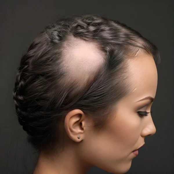
Alopecia areata (AA) is widely recognized as an autoimmune disorder. Autoimmunity arises when the immune system, which is designed to protect the body from infections and diseases, mistakenly attacks its own cells. There are many autoimmune diseases such as lupus, rheumatoid arthritis, and psoriasis, among many others. AA is also believed to be an autoimmune disease where the immune system targets the hair follicles, leading to hair loss.
Mechanisms of Immune Response: The immune system consists of various cell types like lymphocytes, monocytes, and macrophages, each playing a unique role in combating infections. To fight invading organisms the immune cells must be able to recognize bacteria (or whatever) as foreign and not part of the individual’s own body. They do this by recognizing structures called “antigens” (antigens are usually proteins) on the surface of the invader. Every individual organism, including ourselves, has a different set of antigens and the immune system as a whole can recognize all antigens with different immune cell clones/types recognizing different antigens.
Understanding Autoimmunity: Potentially the immune system can recognise any antigen on any bacteria, cell, or whatever, whether it is foreign or part of our own body. Those particular immune cells in our bodies that react with our own antigens must be prevented from coming into contact with the antigens or have their activity prohibited to stop them attacking and destroying our own organs. There are different mechanisms involved in this inhibitory process – most of which are complicated and not fully understood. In essence however, the self-reactive immune cells are “schooled” not to respond to self-antigens in the thymus organ and elsewhere in the body by regulatory cells. For the most part, prohibition of self-reactive cells or prevention of contact with self-antigens is done successfully, but inevitably within the complexities of the immune system things can go wrong. The development of an autoimmune disease can occur when potentially autoreactive cells escape our bodies’ usual mechanisms of rendering them ineffective. These autoreactive lymphocytes go to work with potentially catastrophic results on the organs where the antigens they are specific for are expressed.
Autoimmune Attack on Hair Follicles: The specific cause initiating the autoimmune response in AA remains unknown. However, it has been observed that lymphocytes and macrophages aggressively respond to antigens (proteins) in the hair follicle. Some limited studies suggest the proteins that are targeted are keratins in the hair fiber and root sheaths, and/or proteins related to melanogenesis – the process of hair pigment production. Theories suggest that this targeting of self-proteins by the immune system may involve a malfunction in the immune regulation or an abnormal presentation of hair follicle antigens to autoreactive lymphocytes.
Beyond Classical Autoimmune Theory: Unlike other autoimmune diseases where the target tissue is completely destroyed, AA shows only a disruption of hair follicle function. If the inflammation can be brought under control, the hair follicles can grow hair again. This has led to hypotheses that the immune response in AA might be more complex than in most other autoimmune diseases, possibly involving cytokines or a short-lived antigenic exposure initiating the disease. Notably, immune cells only target hair follicles when they are growing hair in an active anagen growth phase. Follicles that are in a resting state tend to be ignored by the immune cells in patients with AA. This suggests that the protein targets of the immune cells are only present when the hair follicle is busy making a hair fiber.
The Role of Hair Follicle Phases: A prevailing hypothesis suggests that, when hair follicles become inflamed, the hair follicles enter a resting phase (telogen), reducing antigen expression and hence immune attack. This may be one way that hair follicles avoid total destruction in alopecia areata. By going into a dormant (telogen) resting state, the follicles can “hide” from the pathogenic immune cells. This cyclic transition between active and resting phases might explain the non-permanent hair loss in AA and lack of scarring. In addition, hair follicles have natural regenerative abilities. Even with quite severe damage, as with some skin wounds, the follicles can recover and reform. Research has shown that the entire lower third of a hair follicle can be surgically removed – and yet the remnants of the follicle can remake the missing parts and start making new hair fiber.
Evaluating the Etiology of Alopecia Areata: While the exact cause of AA is still under investigation, the prevailing view is that it’s primarily an autoimmune disease. A few laboratory studies have shown that some immune cells taken from patients respond to hair follicle specific antigens including something called “trichohyalin”, as well as to another protein involved in pigment manufacture called “tyrosinase relate protein 2”. The claim that AA is an autoimmune disease is supported by the observation of immune cells in affected hair follicles and the effectiveness of immunosuppressive therapies in some patients. Additionally, AA often coexists with other autoimmune disorders. There seems to be a correlation between AA and autoimmune thyroid disorders, for example.
Alternative Pathways and Theories: Despite the strong autoimmune hypothesis, other potential pathways could contribute to AA. These include an external factor triggering both hair loss and immune response, abnormal presentation of antigens, or damage to the hair follicle exposing new antigens. These other possibilities have led to the suggestion that there may be a non-autoimmune initiation of AA – at least in a subset of patients. This could be brought about by high levels of a stress chemical called “substance P” or changes to the hair follicle microbiome, for example. However, alopecia areata begins, whether by classic autoimmune mechanisms or otherwise, the initial events lead to something called hair follicle “immune privilege collapse”.
The Concept of Immune Privilege in Hair Follicles: One of the pivotal aspects in understanding autoimmunity in alopecia areata (AA) is the concept of “immune privilege” of hair follicles. Immune privilege is a physiological state where certain body sites, like hair follicles, are less accessible to the immune system, thereby reducing the likelihood of inflammatory immune responses. This privilege is maintained through multiple mechanisms, including the expression of specific molecules that inhibit immune cell activation and the suppression of antigen presentation to immune cells. In the normal hair growth cycle, particularly during the anagen (growth) phase, hair follicles exhibit a strong immune privilege. Essentially, immune privilege means the cells of the immune system cannot see what is going on inside the hair follicle. This is probably a good idea as making hair fiber involves making a lot of unique proteins (which might look like pathogens), a lot of cell proliferation (which might look like cancer), and a lot of cell death as cells are keratinized into a hard fiber (which might look like tissue damage) – these things would probably be quite stimulatory to immune systems.
Hair Follicle Immune Privilege Collapse in Alopecia Areata: However, in AA, this immune privilege is disrupted or collapses, leading to the follicle becoming a target for autoimmune attacks. The immune cells can finally see what is going on inside the hair follicles and, it seems, they don’t like what they find. Recent research suggests that this breakdown of immune privilege might be a critical trigger for the onset of AA, wherein the follicle antigens are exposed to and attacked by the immune system. Understanding how and why this immune privilege is compromised in AA is crucial for developing targeted therapies to either restore or mimic this protective environment, offering a promising direction for future treatments.
Future Research Directions: A critical aspect of ongoing research is identifying specific antigens in hair follicles that trigger the autoimmune response in AA. Understanding these mechanisms can lead to targeted therapies and better management of the condition. Recent studies are exploring the complex interactions between the immune system and hair follicle biology. This includes investigating how immune cells and cytokines influence hair growth cycles and follicle health. This research to understand the disease mechanisms better should also led to new treatments to interfere with the mechanisms. For example, if we knew the specific hair follicle proteins that activate the immune response, it may be possible to develop a type of “reverse vaccine” to render immune cells non-responsive to those proteins.
Conclusion – Autoimmunity in Alopecia Areata is a Complex Puzzle: Autoimmunity plays a central role in the pathogenesis of alopecia areata. However, the condition presents a unique challenge due to its non-destructive nature and the cyclic pattern of hair loss. Ongoing research is crucial to unravel the intricate mechanisms involved, paving the way for more effective treatments and a deeper understanding of autoimmune diseases in general.
Alkhalifah A, Alsantali A, Wang E, McElwee KJ, Shapiro J. Alopecia areata update: part I. Clinical picture, histopathology, and pathogenesis. J Am Acad Dermatol. 2010 Feb;62(2):177–88, quiz 189–90.
1.
Kang H, Wu WY, Lo BKK, Yu M, Leung G, Shapiro J, et al. Hair follicles from alopecia areata patients exhibit alterations in immune privilege-associated gene expression in advance of hair loss. J Invest Dermatol. 2010 Nov;130(11):2677–80.
1.
Wang E, McElwee KJ. Etiopathogenesis of alopecia areata: Why do our patients get it? Dermatol Ther. 2011;24(3):337–47.
1.
McElwee KJ, Gilhar A, Tobin DJ, Ramot Y, Sundberg JP, Nakamura M, et al. What causes alopecia areata?: Section Editors: Ralf Paus, Manchester/Lübeck and Raymond Cho, San Francisco. Exp Dermatol. 2013;22(9):609–26.
1.
Zhang B, Zhao Y, Cai Z, Caulloo S, McElwee KJ, Li Y, et al. Early stage alopecia areata is associated with inflammation in the upper dermis and damage to the hair follicle infundibulum. Australas J Dermatol. 2013 Aug;54(3):184–91.
1.
Guo H, Cheng Y, Shapiro J, McElwee K. The role of lymphocytes in the development and treatment of alopecia areata. Expert Rev Clin Immunol. 2015;11(12):1335–51.
1.
Wang EHC, Yu M, Breitkopf T, Akhoundsadegh N, Wang X, Shi FT, et al. Identification of Autoantigen Epitopes in Alopecia Areata. J Invest Dermatol. 2016 Aug;136(8):1617–26.
1.
Alahmmed LM, Alibrahim EA, Alkhars AF, Almulhim MN, Ali SI, Kaliyadan F. Scanning electron microscopy study of hair shaft changes related to hardness of water. Indian J Dermatol Venereol Leprol. 2017;83(6):740.
1.
Broadley D, McElwee KJ. A “hair-raising” history of alopecia areata. Exp Dermatol. 2020 Mar;29(3):208–22.
1.
Bertolini M, McElwee K, Gilhar A, Bulfone-Paus S, Paus R. Hair follicle immune privilege and its collapse in alopecia areata. Exp Dermatol. 2020 Aug;29(8):703–25.
Alopecia areata (AA) is a chronic-relapsing non-scarring hair loss condition believed to stem from autoimmune origins. Certain forms of the disease, such as ophiasis, subtotalis,…
Alopecia areata (AA) is a complex autoimmune disorder, characterized by non-scarring hair loss. The etiology of AA is multifactorial, with genetic, immunological, and psychological factors,…
Introduction: Alopecia areata is an autoimmune condition characterized by non-scarring hair loss, affecting individuals of all ages, genders, and ethnicities. It is marked by the…
Manage Cookie Consent
We use technologies like cookies to store and/or access device information. We do this to improve browsing experience and to show (non-) personalized ads. Consenting to these technologies will allow us to process data such as browsing behavior or unique IDs on this site. Not consenting or withdrawing consent, may adversely affect certain features and functions.
Functional Always active
The technical storage or access is strictly necessary for the legitimate purpose of enabling the use of a specific service explicitly requested by the subscriber or user, or for the sole purpose of carrying out the transmission of a communication over an electronic communications network.
Preferences
The technical storage or access is necessary for the legitimate purpose of storing preferences that are not requested by the subscriber or user.
Statistics
The technical storage or access that is used exclusively for statistical purposes.The technical storage or access that is used exclusively for anonymous statistical purposes. Without a subpoena, voluntary compliance on the part of your Internet Service Provider, or additional records from a third party, information stored or retrieved for this purpose alone cannot usually be used to identify you.
Marketing
The technical storage or access is required to create user profiles to send advertising, or to track the user on a website or across several websites for similar marketing purposes.