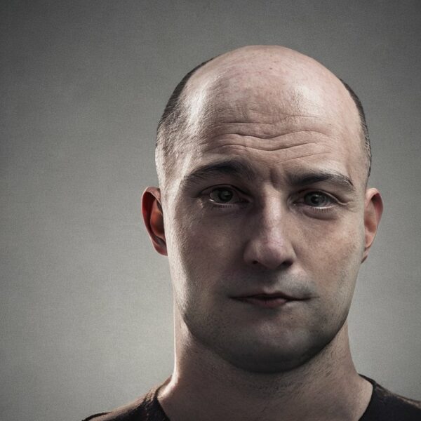
Androgenetic alopecia (AGA), commonly known as male or female pattern baldness, is a prevalent form of hair loss affecting millions worldwide. The condition is characterized by a distinct pattern of progressive hair thinning, primarily influenced by genetic predisposition and androgen hormones. This essay aims to elucidate the molecular mechanisms underlying the role of androgens in AGA.
Androgens are a group of hormones that play a crucial role in male sexual development and physiological processes. Testosterone, the primary androgen, can be converted to a more potent form, dihydrotestosterone (DHT), by the enzyme 5-alpha reductase. It is this DHT that is implicated in the pathogenesis of AGA.
In individuals genetically predisposed to AGA, hair follicles on top of the scalp are inherently sensitive to DHT. This sensitivity is primarily due to the presence of androgen receptors on the cells of the hair follicles. When DHT binds to these receptors, it triggers a cascade of cellular events that eventually lead to follicular miniaturization, a hallmark of AGA.
Follicular miniaturization refers to the progressive shrinking of the hair follicle, resulting in the production of thinner, shorter hairs. Over time, this process leads to a visible reduction in hair density. The exact molecular mechanisms driving this process are complex and not fully understood. However, several key pathways have been identified.
One such pathway involves the activation of dermal papilla cells (DPCs) in the hair follicle. DPCs are specialized cells that play a critical role in hair growth and cycling. Upon DHT binding, these cells undergo changes that negatively affect their ability to support hair growth. In some respects, the dermal papilla cells are pushed into old age, something called “cellular senescence”. As time goes on, and with continued negative signalling through their androgen receptors, the dermal papilla cells start to die off. Consequently, the cells are less able to send out positive hair growth signals to the hair follicle keratinocytes that make the hair fiber.
DHT has been shown to inhibit the production of growth factors necessary for hair growth, such as Insulin-like Growth Factor 1 (IGF-1). These growth factors are normally made by the dermal papilla cells and are then secreted and link up with growth factor receptors on hair follicle keratinocytes. Concurrently, DHT upregulates the expression of growth inhibitory factors, including Transforming Growth Factor-beta (TGF-β) and DKK1. This imbalance in growth factors contributes to the shortened growth phase of the hair cycle, and later leading to the production of miniaturized hair follicles and thinner hair fiber. Eventually the follicles are reduced in size and only survive as vellus follicles. In women, the vellus follicles tend to survive, but in men eventually even the vellus follicle structures can be completely destroyed.
DHT-induced activation of DPCs can also lead to increased expression of molecules involved in inflammation and fibrosis. Inflammation is quite variable from person to person, but where it is present, it has the potential to completely destroy the hair follicles. This inflammatory response contributes to the degradation of the extracellular matrix, a supportive network crucial for hair follicle integrity and function. Over time, this leads to the replacement of hair follicles with fibrous tissue.
In conclusion, androgens, specifically DHT, play a pivotal role in the pathogenesis of AGA. The binding of DHT to androgen receptors on hair follicle dermal papilla cells triggers a series of molecular events leading to follicular miniaturization and hair loss. While our understanding of these processes has significantly improved, further research is needed to fully elucidate the complex interplay of genetic and hormonal factors in AGA. Such insights will undoubtedly pave the way for the development of more effective treatments for this common condition.
Please note that this essay is a simplified explanation of a complex biological process. The actual mechanisms involved in androgenetic alopecia are much more intricate and are still the subject of ongoing research.
Bibliography
11711645 {11711645:QX57QSQA},{11711645:JJN9KXW2},{11711645:6UD83Z67},{11711645:M6AMQ42J},{11711645:TEKUUI3A},{11711645:5GWZQ46S} 1 vancouver 50 date asc 139 https://www.keratin.com/wp-content/plugins/zotpress/ %7B%22status%22%3A%22success%22%2C%22updateneeded%22%3Afalse%2C%22instance%22%3Afalse%2C%22meta%22%3A%7B%22request_last%22%3A0%2C%22request_next%22%3A0%2C%22used_cache%22%3Atrue%7D%2C%22data%22%3A%5B%7B%22key%22%3A%226UD83Z67%22%2C%22library%22%3A%7B%22id%22%3A11711645%7D%2C%22meta%22%3A%7B%22creatorSummary%22%3A%22Tr%5Cu00fceb%22%2C%22parsedDate%22%3A%222002%22%2C%22numChildren%22%3A0%7D%2C%22bib%22%3A%22%26lt%3Bdiv%20class%3D%26quot%3Bcsl-bib-body%26quot%3B%20style%3D%26quot%3Bline-height%3A%201.35%3B%20%26quot%3B%26gt%3B%5Cn%20%20%26lt%3Bdiv%20class%3D%26quot%3Bcsl-entry%26quot%3B%20style%3D%26quot%3Bclear%3A%20left%3B%20%26quot%3B%26gt%3B%5Cn%20%20%20%20%26lt%3Bdiv%20class%3D%26quot%3Bcsl-left-margin%26quot%3B%20style%3D%26quot%3Bfloat%3A%20left%3B%20padding-right%3A%200.5em%3B%20text-align%3A%20right%3B%20width%3A%201em%3B%26quot%3B%26gt%3B1.%26lt%3B%5C%2Fdiv%26gt%3B%26lt%3Bdiv%20class%3D%26quot%3Bcsl-right-inline%26quot%3B%20style%3D%26quot%3Bmargin%3A%200%20.4em%200%201.5em%3B%26quot%3B%26gt%3BTr%26%23xFC%3Beb%20RM.%20Molecular%20mechanisms%20of%20androgenetic%20alopecia.%20Exp%20Gerontol.%202002%3B37%288%26%23x2013%3B9%29%3A981%26%23x2013%3B90.%26lt%3B%5C%2Fdiv%26gt%3B%5Cn%20%20%20%26lt%3B%5C%2Fdiv%26gt%3B%5Cn%26lt%3B%5C%2Fdiv%26gt%3B%22%2C%22data%22%3A%7B%22itemType%22%3A%22journalArticle%22%2C%22title%22%3A%22Molecular%20mechanisms%20of%20androgenetic%20alopecia%22%2C%22creators%22%3A%5B%7B%22creatorType%22%3A%22author%22%2C%22firstName%22%3A%22Ralph%20M.%22%2C%22lastName%22%3A%22Tr%5Cu00fceb%22%7D%5D%2C%22abstractNote%22%3A%22Androgenetic%20alopecia%20%28AGA%29%20is%20hereditary%20and%20androgen-dependent%2C%20progressive%20thinning%20of%20the%20scalp%20hair%20that%20follows%20a%20defined%20pattern.%20While%20the%20genetic%20involvement%20is%20pronounced%20but%20poorly%20understood%2C%20major%20advances%20have%20been%20achieved%20in%20understanding%20principal%20elements%20of%20the%20androgen%20metabolism%20involved%3A%20androgen-dependent%20processes%20are%20predominantly%20due%20to%20the%20binding%20of%20dihydrotestosterone%20%28DHT%29%20to%20the%20androgen%20receptor%20%28AR%29.%20DHT-dependent%20cell%20functions%20depend%20on%20the%20availability%20of%20weak%20androgens%2C%20their%20conversion%20to%20more%20potent%20androgens%20via%20the%20action%20of%205%20alpha-reductase%2C%20low%20enzymatic%20activity%20of%20androgen%20inactivating%20enzymes%2C%20and%20functionally%20active%20AR%20present%20in%20high%20numbers.%20The%20predisposed%20scalp%20exhibits%20high%20levels%20of%20DHT%2C%20and%20increased%20expression%20of%20the%20AR.%20Conversion%20of%20testosterone%20to%20DHT%20within%20the%20dermal%20papilla%20plays%20a%20central%20role%2C%20while%20androgen-regulated%20factors%20deriving%20from%20dermal%20papilla%20cells%20are%20believed%20to%20influence%20growth%20of%20other%20components%20of%20the%20hair%20follicle.%20Current%20available%20treatment%20modalities%20with%20proven%20efficacy%20are%20oral%20finasteride%2C%20a%20competitive%20inhibitor%20of%20type%202%205%20alpha-reductase%2C%20and%20topical%20minoxidil%2C%20an%20adenosine-triphosphate-sensitive%20potassium%20channel%20opener%20which%20has%20been%20reported%20to%20stimulate%20the%20production%20of%20vascular%20endothelial%20growth%20factor%20in%20cultured%20dermal%20papilla%20cells.%20Since%20the%20clinical%20success%20rate%20of%20treatment%20of%20AGA%20with%20modulators%20of%20androgen%20metabolism%20or%20hair%20growth%20promoters%20is%20limited%2C%20sustained%20microscopic%20follicular%20inflammation%20with%20connective%20tissue%20remodeling%2C%20eventually%20resulting%20in%20permanent%20hair%20loss%2C%20is%20considered%20a%20possible%20cofactor%20in%20the%20complex%20etiology%20of%20AGA.%22%2C%22date%22%3A%222002%22%2C%22language%22%3A%22eng%22%2C%22DOI%22%3A%2210.1016%5C%2Fs0531-5565%2802%2900093-1%22%2C%22ISSN%22%3A%220531-5565%22%2C%22url%22%3A%22%22%2C%22collections%22%3A%5B%22VXZPE5TH%22%5D%2C%22dateModified%22%3A%222024-06-28T16%3A35%3A10Z%22%7D%7D%2C%7B%22key%22%3A%22QX57QSQA%22%2C%22library%22%3A%7B%22id%22%3A11711645%7D%2C%22meta%22%3A%7B%22creatorSummary%22%3A%22Ellis%20et%20al.%22%2C%22parsedDate%22%3A%222002-11-19%22%2C%22numChildren%22%3A0%7D%2C%22bib%22%3A%22%26lt%3Bdiv%20class%3D%26quot%3Bcsl-bib-body%26quot%3B%20style%3D%26quot%3Bline-height%3A%201.35%3B%20%26quot%3B%26gt%3B%5Cn%20%20%26lt%3Bdiv%20class%3D%26quot%3Bcsl-entry%26quot%3B%20style%3D%26quot%3Bclear%3A%20left%3B%20%26quot%3B%26gt%3B%5Cn%20%20%20%20%26lt%3Bdiv%20class%3D%26quot%3Bcsl-left-margin%26quot%3B%20style%3D%26quot%3Bfloat%3A%20left%3B%20padding-right%3A%200.5em%3B%20text-align%3A%20right%3B%20width%3A%201em%3B%26quot%3B%26gt%3B1.%26lt%3B%5C%2Fdiv%26gt%3B%26lt%3Bdiv%20class%3D%26quot%3Bcsl-right-inline%26quot%3B%20style%3D%26quot%3Bmargin%3A%200%20.4em%200%201.5em%3B%26quot%3B%26gt%3BEllis%20JA%2C%20Sinclair%20R%2C%20Harrap%20SB.%20Androgenetic%20alopecia%3A%20pathogenesis%20and%20potential%20for%20therapy.%20Expert%20Rev%20Mol%20Med.%202002%20Nov%2019%3B4%2822%29%3A1%26%23x2013%3B11.%26lt%3B%5C%2Fdiv%26gt%3B%5Cn%20%20%20%26lt%3B%5C%2Fdiv%26gt%3B%5Cn%26lt%3B%5C%2Fdiv%26gt%3B%22%2C%22data%22%3A%7B%22itemType%22%3A%22journalArticle%22%2C%22title%22%3A%22Androgenetic%20alopecia%3A%20pathogenesis%20and%20potential%20for%20therapy%22%2C%22creators%22%3A%5B%7B%22creatorType%22%3A%22author%22%2C%22firstName%22%3A%22Justine%20A.%22%2C%22lastName%22%3A%22Ellis%22%7D%2C%7B%22creatorType%22%3A%22author%22%2C%22firstName%22%3A%22Rodney%22%2C%22lastName%22%3A%22Sinclair%22%7D%2C%7B%22creatorType%22%3A%22author%22%2C%22firstName%22%3A%22Stephen%20B.%22%2C%22lastName%22%3A%22Harrap%22%7D%5D%2C%22abstractNote%22%3A%22Androgenetic%20alopecia%20occurs%20in%20men%20and%20women%2C%20and%20is%20characterised%20by%20the%20loss%20of%20hair%20from%20the%20scalp%20in%20a%20defined%20pattern.%20Determining%20factors%20appear%20to%20be%20genetic%20predisposition%20coupled%20with%20the%20presence%20of%20sufficient%20circulating%20androgens.%20The%20prevalence%20of%20this%20condition%20is%20high%20%28up%20to%2050%25%20of%20white%20males%20are%20affected%20by%2050%20years%20of%20age%29%20and%2C%20although%20there%20are%20no%20serious%20direct%20health%20consequences%2C%20the%20loss%20of%20scalp%20hair%20can%20be%20distressing.%20Knowledge%20of%20the%20pathogenesis%20of%20androgenetic%20alopecia%20has%20increased%20markedly%20in%20recent%20years.%20Pre-programmed%20follicles%20on%20the%20scalp%20undergo%20a%20transformation%20from%20long%20growth%20%28anagen%29%20and%20short%20rest%20%28telogen%29%20cycles%2C%20to%20long%20rest%20and%20short%20growth%20cycles.%20This%20process%20is%20coupled%20with%20progressive%20miniaturisation%20of%20the%20follicle.%20These%20changes%20are%20androgen%20dependent%2C%20and%20require%20the%20inheritance%20of%20several%20genes.%20To%20date%2C%20only%20one%20of%20these%20genes%2C%20which%20encodes%20the%20androgen%20receptor%20%28AR%29%2C%20has%20been%20identified.%20Of%20the%20many%20treatments%20available%20for%20androgenetic%20alopecia%2C%20only%20two%20%28finasteride%20and%20minoxidil%29%20have%20been%20scientifically%20shown%20to%20be%20useful%20in%20the%20treatment%20of%20hair%20loss.%20However%2C%20these%20therapies%20are%20variable%20in%20their%20effectiveness.%20Discovery%20of%20the%20involvement%20of%20the%20AR%20gene%2C%20and%20the%20identification%20of%20other%20genes%20contributing%20to%20the%20condition%2C%20might%20lead%20to%20the%20development%20of%20new%20and%20more%20effective%20therapies%20that%20target%20the%20condition%20at%20a%20more%20fundamental%20level.%22%2C%22date%22%3A%222002-11-19%22%2C%22language%22%3A%22eng%22%2C%22DOI%22%3A%2210.1017%5C%2FS1462399402005112%22%2C%22ISSN%22%3A%221462-3994%22%2C%22url%22%3A%22%22%2C%22collections%22%3A%5B%22VXZPE5TH%22%5D%2C%22dateModified%22%3A%222023-05-20T19%3A38%3A44Z%22%7D%7D%2C%7B%22key%22%3A%22M6AMQ42J%22%2C%22library%22%3A%7B%22id%22%3A11711645%7D%2C%22meta%22%3A%7B%22creatorSummary%22%3A%22Rebora%22%2C%22parsedDate%22%3A%222004-05%22%2C%22numChildren%22%3A0%7D%2C%22bib%22%3A%22%26lt%3Bdiv%20class%3D%26quot%3Bcsl-bib-body%26quot%3B%20style%3D%26quot%3Bline-height%3A%201.35%3B%20%26quot%3B%26gt%3B%5Cn%20%20%26lt%3Bdiv%20class%3D%26quot%3Bcsl-entry%26quot%3B%20style%3D%26quot%3Bclear%3A%20left%3B%20%26quot%3B%26gt%3B%5Cn%20%20%20%20%26lt%3Bdiv%20class%3D%26quot%3Bcsl-left-margin%26quot%3B%20style%3D%26quot%3Bfloat%3A%20left%3B%20padding-right%3A%200.5em%3B%20text-align%3A%20right%3B%20width%3A%201em%3B%26quot%3B%26gt%3B1.%26lt%3B%5C%2Fdiv%26gt%3B%26lt%3Bdiv%20class%3D%26quot%3Bcsl-right-inline%26quot%3B%20style%3D%26quot%3Bmargin%3A%200%20.4em%200%201.5em%3B%26quot%3B%26gt%3BRebora%20A.%20Pathogenesis%20of%20androgenetic%20alopecia.%20J%20Am%20Acad%20Dermatol.%202004%20May%3B50%285%29%3A777%26%23x2013%3B9.%26lt%3B%5C%2Fdiv%26gt%3B%5Cn%20%20%20%26lt%3B%5C%2Fdiv%26gt%3B%5Cn%26lt%3B%5C%2Fdiv%26gt%3B%22%2C%22data%22%3A%7B%22itemType%22%3A%22journalArticle%22%2C%22title%22%3A%22Pathogenesis%20of%20androgenetic%20alopecia%22%2C%22creators%22%3A%5B%7B%22creatorType%22%3A%22author%22%2C%22firstName%22%3A%22Alfredo%22%2C%22lastName%22%3A%22Rebora%22%7D%5D%2C%22abstractNote%22%3A%22Although%20it%20is%20universally%20accepted%20that%20dihydrotestosterone%20causes%20baldness%2C%20how%20it%20does%20so%20is%20not%20well%20understood.%20I%20propose%20that%203%20mechanisms%20are%20at%20work%3A%20miniaturization%20by%20a%20dihydrotestosterone-induced%20acceleration%20of%20the%20mitotic%20rate%20of%20the%20matrix%20that%20leaves%20less%20and%20less%20time%20for%20differentiation%3B%20an%20increased%20telogen%20shedding%20as%20a%20result%20of%20the%20shortening%20of%20the%20hair%20cycles%20that%20increases%20the%20telogen%20number%20per%20unit%20of%20time%3B%20and%20the%20increased%20number%20and%20duration%20of%20the%20lag%20phase%20or%20kenogen.%20That%20this%20last%20mechanism%20is%20related%20to%20dihydrotestosterone%20is%20unclear.%22%2C%22date%22%3A%222004-05%22%2C%22language%22%3A%22eng%22%2C%22DOI%22%3A%2210.1016%5C%2Fj.jaad.2003.11.073%22%2C%22ISSN%22%3A%220190-9622%22%2C%22url%22%3A%22%22%2C%22collections%22%3A%5B%22VXZPE5TH%22%5D%2C%22dateModified%22%3A%222023-05-20T19%3A39%3A30Z%22%7D%7D%2C%7B%22key%22%3A%22TEKUUI3A%22%2C%22library%22%3A%7B%22id%22%3A11711645%7D%2C%22meta%22%3A%7B%22creatorSummary%22%3A%22Yoo%20et%20al.%22%2C%22parsedDate%22%3A%222006-06%22%2C%22numChildren%22%3A0%7D%2C%22bib%22%3A%22%26lt%3Bdiv%20class%3D%26quot%3Bcsl-bib-body%26quot%3B%20style%3D%26quot%3Bline-height%3A%201.35%3B%20%26quot%3B%26gt%3B%5Cn%20%20%26lt%3Bdiv%20class%3D%26quot%3Bcsl-entry%26quot%3B%20style%3D%26quot%3Bclear%3A%20left%3B%20%26quot%3B%26gt%3B%5Cn%20%20%20%20%26lt%3Bdiv%20class%3D%26quot%3Bcsl-left-margin%26quot%3B%20style%3D%26quot%3Bfloat%3A%20left%3B%20padding-right%3A%200.5em%3B%20text-align%3A%20right%3B%20width%3A%201em%3B%26quot%3B%26gt%3B1.%26lt%3B%5C%2Fdiv%26gt%3B%26lt%3Bdiv%20class%3D%26quot%3Bcsl-right-inline%26quot%3B%20style%3D%26quot%3Bmargin%3A%200%20.4em%200%201.5em%3B%26quot%3B%26gt%3BYoo%20HG%2C%20Kim%20JS%2C%20Lee%20SR%2C%20Pyo%20HK%2C%20Moon%20HI%2C%20Lee%20JH%2C%20et%20al.%20Perifollicular%20fibrosis%3A%20pathogenetic%20role%20in%20androgenetic%20alopecia.%20Biol%20Pharm%20Bull.%202006%20Jun%3B29%286%29%3A1246%26%23x2013%3B50.%26lt%3B%5C%2Fdiv%26gt%3B%5Cn%20%20%20%26lt%3B%5C%2Fdiv%26gt%3B%5Cn%26lt%3B%5C%2Fdiv%26gt%3B%22%2C%22data%22%3A%7B%22itemType%22%3A%22journalArticle%22%2C%22title%22%3A%22Perifollicular%20fibrosis%3A%20pathogenetic%20role%20in%20androgenetic%20alopecia%22%2C%22creators%22%3A%5B%7B%22creatorType%22%3A%22author%22%2C%22firstName%22%3A%22Hyeon%20Gyeong%22%2C%22lastName%22%3A%22Yoo%22%7D%2C%7B%22creatorType%22%3A%22author%22%2C%22firstName%22%3A%22Jin%20Sook%22%2C%22lastName%22%3A%22Kim%22%7D%2C%7B%22creatorType%22%3A%22author%22%2C%22firstName%22%3A%22Se%20Rah%22%2C%22lastName%22%3A%22Lee%22%7D%2C%7B%22creatorType%22%3A%22author%22%2C%22firstName%22%3A%22Hyun%20Keol%22%2C%22lastName%22%3A%22Pyo%22%7D%2C%7B%22creatorType%22%3A%22author%22%2C%22firstName%22%3A%22Hyung%20In%22%2C%22lastName%22%3A%22Moon%22%7D%2C%7B%22creatorType%22%3A%22author%22%2C%22firstName%22%3A%22Jong%20Hee%22%2C%22lastName%22%3A%22Lee%22%7D%2C%7B%22creatorType%22%3A%22author%22%2C%22firstName%22%3A%22Oh%20Sang%22%2C%22lastName%22%3A%22Kwon%22%7D%2C%7B%22creatorType%22%3A%22author%22%2C%22firstName%22%3A%22Jin%20Ho%22%2C%22lastName%22%3A%22Chung%22%7D%2C%7B%22creatorType%22%3A%22author%22%2C%22firstName%22%3A%22Kyu%20Han%22%2C%22lastName%22%3A%22Kim%22%7D%2C%7B%22creatorType%22%3A%22author%22%2C%22firstName%22%3A%22Hee%20Chul%22%2C%22lastName%22%3A%22Eun%22%7D%2C%7B%22creatorType%22%3A%22author%22%2C%22firstName%22%3A%22Kwang%20Hyun%22%2C%22lastName%22%3A%22Cho%22%7D%5D%2C%22abstractNote%22%3A%22Androgenetic%20alopecia%20%28AGA%29%20is%20a%20dihydrotestosterone%20%28DHT%29-mediated%20process%2C%20characterized%20by%20continuous%20miniaturization%20of%20androgen%20reactive%20hair%20follicles%20and%20accompanied%20by%20perifollicular%20fibrosis%20of%20follicular%20units%20in%20histological%20examination.%20Testosterone%20%28T%3A%2010%28-9%29-10%28-7%29%20M%29%20treatment%20increased%20the%20expression%20of%20type%20I%20procollagen%20at%20mRNA%20and%20protein%20level.%20Pretreatment%20of%20finasteride%20%2810%28-8%29%20M%29%20inhibited%20the%20T-induced%20type%20I%20procollagen%20expression%20at%20mRNA%20%2840.2%25%29%20and%20protein%20levels%20%2824.9%25%29.%20T%20treatment%20increased%20the%20expression%20of%20transforming%20growth%20factor-beta%201%20%28TGF-beta1%29%20at%20protein%20levels%20by%2081.9%25%20in%20the%20human%20scalp%20dermal%20fibroblasts%20%28DFs%29.%20Pretreatment%20of%20finasteride%20decreased%20the%20expression%20of%20TGF-beta1%20protein%20induced%20by%20an%20average%20of%20T%20%2830.4%25%29.%20The%20type%20I%20procollagen%20expression%20after%20pretreatment%20of%20neutralizing%20TGF-beta1%20antibody%20%2810%20microg%5C%2Fml%29%20was%20inhibited%20by%20an%20average%20of%2054.3%25.%20Our%20findings%20suggest%20that%20T-induced%20TGF-beta1%20and%20type%20I%20procollagen%20expression%20may%20contribute%20to%20the%20development%20of%20perifollicular%20fibrosis%20in%20the%20AGA%2C%20and%20the%20inhibitory%20effects%20on%20T-induced%20procollagen%20and%20TGF-beta1%20expression%20may%20explain%20another%20possible%20mechanism%20how%20finasteride%20works%20in%20AGA.%22%2C%22date%22%3A%222006-06%22%2C%22language%22%3A%22eng%22%2C%22DOI%22%3A%2210.1248%5C%2Fbpb.29.1246%22%2C%22ISSN%22%3A%221347-5215%22%2C%22url%22%3A%22%22%2C%22collections%22%3A%5B%22VXZPE5TH%22%5D%2C%22dateModified%22%3A%222023-05-20T19%3A41%3A47Z%22%7D%7D%2C%7B%22key%22%3A%22JJN9KXW2%22%2C%22library%22%3A%7B%22id%22%3A11711645%7D%2C%22meta%22%3A%7B%22creatorSummary%22%3A%22Inui%20and%20Itami%22%2C%22parsedDate%22%3A%222011-01%22%2C%22numChildren%22%3A0%7D%2C%22bib%22%3A%22%26lt%3Bdiv%20class%3D%26quot%3Bcsl-bib-body%26quot%3B%20style%3D%26quot%3Bline-height%3A%201.35%3B%20%26quot%3B%26gt%3B%5Cn%20%20%26lt%3Bdiv%20class%3D%26quot%3Bcsl-entry%26quot%3B%20style%3D%26quot%3Bclear%3A%20left%3B%20%26quot%3B%26gt%3B%5Cn%20%20%20%20%26lt%3Bdiv%20class%3D%26quot%3Bcsl-left-margin%26quot%3B%20style%3D%26quot%3Bfloat%3A%20left%3B%20padding-right%3A%200.5em%3B%20text-align%3A%20right%3B%20width%3A%201em%3B%26quot%3B%26gt%3B1.%26lt%3B%5C%2Fdiv%26gt%3B%26lt%3Bdiv%20class%3D%26quot%3Bcsl-right-inline%26quot%3B%20style%3D%26quot%3Bmargin%3A%200%20.4em%200%201.5em%3B%26quot%3B%26gt%3BInui%20S%2C%20Itami%20S.%20Molecular%20basis%20of%20androgenetic%20alopecia%3A%20From%20androgen%20to%20paracrine%20mediators%20through%20dermal%20papilla.%20J%20Dermatol%20Sci.%202011%20Jan%3B61%281%29%3A1%26%23x2013%3B6.%26lt%3B%5C%2Fdiv%26gt%3B%5Cn%20%20%20%26lt%3B%5C%2Fdiv%26gt%3B%5Cn%26lt%3B%5C%2Fdiv%26gt%3B%22%2C%22data%22%3A%7B%22itemType%22%3A%22journalArticle%22%2C%22title%22%3A%22Molecular%20basis%20of%20androgenetic%20alopecia%3A%20From%20androgen%20to%20paracrine%20mediators%20through%20dermal%20papilla%22%2C%22creators%22%3A%5B%7B%22creatorType%22%3A%22author%22%2C%22firstName%22%3A%22Shigeki%22%2C%22lastName%22%3A%22Inui%22%7D%2C%7B%22creatorType%22%3A%22author%22%2C%22firstName%22%3A%22Satoshi%22%2C%22lastName%22%3A%22Itami%22%7D%5D%2C%22abstractNote%22%3A%22Androgenetic%20alopecia%20%28AGA%29%20is%20characterized%20by%20vellus%20transformation%20of%20scalp%20hairs%2C%20corresponding%20to%20hair%20follicle%20miniaturization%20during%20repeated%20hair%20cycles%20with%20shortened%20anagen%20phase.%20This%20phenomenon%20is%20mediated%20mainly%20by%20androgen.%20Then%2C%20the%20multi-step%20molecular%20pathway%20of%20androgen%20can%20be%20involved%20in%20the%20pathogenesis%20of%20AGA.%20The%20expression%20of%20type%20II%205%5Cu03b1-reductase%20is%20higher%20in%20dermal%20papilla%20cells%20from%20AGA%20and%20beard%20than%20those%20from%20other%20sites.%20On%20the%20other%20hand%2C%20type%20I%205%5Cu03b1-reductase%20expression%20is%20relatively%20low.%20Next%2C%20hormone%20binding%20assays%20and%20RT-PCR%20demonstrated%20that%20androgen%20receptor%20%28AR%29%20expression%20is%20significantly%20higher%20in%20bald%20dermal%20papilla%20cells%20than%20non-bald%20cells.%20Additionally%2C%20AR%20coactivator%20Hic-5%5C%2FARA55%20is%20highly%20expressed%20in%20dermal%20papilla%20cells%20of%20hair%20follicles%20from%20androgen-sensitive%20sites%20such%20as%20AGA%20and%20beard.%20Collectively%2C%20the%20enhanced%20expression%20of%20type%20II%205%5Cu03b1-reductase%2C%20AR%20and%20Hic-5%5C%2FARA55%20can%20upregulate%20sensitivity%20to%20androgen%20of%20dermal%20papilla%20cells%20in%20AGA.%20Furthermore%2C%20in%20the%20coculture%20of%20AR-overexpressing%20human%20dermal%20papilla%20cells%20from%20AGA%20and%20normal%20human%20keratinocytes%2C%20R1881%20suppresses%20keratinocyte%20growth%20through%20androgen-inducible%20TGF-%5Cu03b21%2C%20indicating%20that%20TGF-%5Cu03b21%20is%20one%20of%20the%20key%20players%20in%20pathogenesis%20of%20AGA.%20TGF-%5Cu03b22%20and%20DKK-1%20has%20been%20reported%20to%20be%20androgen-induced%20suppressor%20of%20growth%20of%20follicular%20epithelial%20cells.%20We%20expect%20that%20more%20pathogenic%20mediators%20will%20be%20identified%20in%20the%20future%2C%20enabling%20easier%20understanding%20of%20AGA%20pathogenesis%20and%20providing%20new%20therapeutic%20targets%20from%20aspect%20of%20andrology.%22%2C%22date%22%3A%222011-01%22%2C%22language%22%3A%22eng%22%2C%22DOI%22%3A%2210.1016%5C%2Fj.jdermsci.2010.10.015%22%2C%22ISSN%22%3A%221873-569X%22%2C%22url%22%3A%22%22%2C%22collections%22%3A%5B%22VXZPE5TH%22%5D%2C%22dateModified%22%3A%222023-05-20T19%3A42%3A54Z%22%7D%7D%2C%7B%22key%22%3A%225GWZQ46S%22%2C%22library%22%3A%7B%22id%22%3A11711645%7D%2C%22meta%22%3A%7B%22creatorSummary%22%3A%22Magro%20et%20al.%22%2C%22parsedDate%22%3A%222011-12%22%2C%22numChildren%22%3A0%7D%2C%22bib%22%3A%22%26lt%3Bdiv%20class%3D%26quot%3Bcsl-bib-body%26quot%3B%20style%3D%26quot%3Bline-height%3A%201.35%3B%20%26quot%3B%26gt%3B%5Cn%20%20%26lt%3Bdiv%20class%3D%26quot%3Bcsl-entry%26quot%3B%20style%3D%26quot%3Bclear%3A%20left%3B%20%26quot%3B%26gt%3B%5Cn%20%20%20%20%26lt%3Bdiv%20class%3D%26quot%3Bcsl-left-margin%26quot%3B%20style%3D%26quot%3Bfloat%3A%20left%3B%20padding-right%3A%200.5em%3B%20text-align%3A%20right%3B%20width%3A%201em%3B%26quot%3B%26gt%3B1.%26lt%3B%5C%2Fdiv%26gt%3B%26lt%3Bdiv%20class%3D%26quot%3Bcsl-right-inline%26quot%3B%20style%3D%26quot%3Bmargin%3A%200%20.4em%200%201.5em%3B%26quot%3B%26gt%3BMagro%20CM%2C%20Rossi%20A%2C%20Poe%20J%2C%20Manhas-Bhutani%20S%2C%20Sadick%20N.%20The%20role%20of%20inflammation%20and%20immunity%20in%20the%20pathogenesis%20of%20androgenetic%20alopecia.%20J%20Drugs%20Dermatol.%202011%20Dec%3B10%2812%29%3A1404%26%23x2013%3B11.%26lt%3B%5C%2Fdiv%26gt%3B%5Cn%20%20%20%26lt%3B%5C%2Fdiv%26gt%3B%5Cn%26lt%3B%5C%2Fdiv%26gt%3B%22%2C%22data%22%3A%7B%22itemType%22%3A%22journalArticle%22%2C%22title%22%3A%22The%20role%20of%20inflammation%20and%20immunity%20in%20the%20pathogenesis%20of%20androgenetic%20alopecia%22%2C%22creators%22%3A%5B%7B%22creatorType%22%3A%22author%22%2C%22firstName%22%3A%22Cynthia%20M.%22%2C%22lastName%22%3A%22Magro%22%7D%2C%7B%22creatorType%22%3A%22author%22%2C%22firstName%22%3A%22Anthony%22%2C%22lastName%22%3A%22Rossi%22%7D%2C%7B%22creatorType%22%3A%22author%22%2C%22firstName%22%3A%22Jonathan%22%2C%22lastName%22%3A%22Poe%22%7D%2C%7B%22creatorType%22%3A%22author%22%2C%22firstName%22%3A%22Suveena%22%2C%22lastName%22%3A%22Manhas-Bhutani%22%7D%2C%7B%22creatorType%22%3A%22author%22%2C%22firstName%22%3A%22Neil%22%2C%22lastName%22%3A%22Sadick%22%7D%5D%2C%22abstractNote%22%3A%22BACKGROUND%3A%20Female%20pattern%20hair%20loss%20affects%20many%20women%3B%20its%20pathogenetic%20basis%20has%20been%20held%20to%20be%20similar%20to%20men%20with%20common%20baldness.%5CnOBJECTIVE%3A%20The%20objective%20of%20this%20study%20was%20to%20determine%20the%20role%20of%20immunity%20and%20inflammation%20in%20androgenetic%20alopecia%20in%20women%20and%20modulate%20therapy%20according%20to%20inflammatory%20and%20immunoreactant%20profiles.%5CnMATERIALS%20AND%20METHODS%3A%2052%20women%20with%20androgenetic%20alopecia%20%28AA%29%20underwent%20scalp%20biopsies%20for%20routine%20light%20microscopic%20assessment%20and%20direct%20immunofluroescent%20studies.%20In%2018%20patients%2C%20serologic%20assessment%20for%20antibodies%20to%20androgen%20receptor%2C%20estrogen%20receptor%20and%20cytokeratin%2015%20was%20conducted.%5CnRESULTS%3A%20A%20lymphocytic%20folliculitis%20targeting%20the%20bulge%20epithelium%20was%20observed%20in%20many%20cases.%20Thirty-three%20of%2052%20female%20patients%20had%20significant%20deposits%20of%20IgM%20within%20the%20epidermal%20basement%20membrane%20zone%20typically%20accompanied%20by%20components%20of%20complement%20activation.%20The%20severity%20of%20changes%20light%20microscopically%20were%20more%20apparent%20in%20the%20positive%20immunoreactant%20group.%20Biopsies%20from%20men%20with%20androgenetic%20alopecia%20showed%20a%20similar%20pattern%20of%20inflammation%20and%20immunoreactant%20deposition.%20Serologic%20assessment%20for%20antibodies%20to%20androgen%20receptor%2C%20estrogen%20receptor%20or%20cytokeratin%2015%20were%20negative.%20Combined%20modality%20therapy%20with%20minocycline%20and%20topical%20steroids%20along%20with%20red%20light%20produced%20consistent%20good%20results%20in%20the%20positive%20immunoreactant%20group%20compared%20to%20the%20negative%20immunoreactant%20group.%5CnCONCLUSION%3A%20A%20lymphocytic%20microfolliculitis%20targeting%20the%20bulge%20epithelium%20along%20with%20deposits%20of%20epithelial%20basement%20membrane%20zone%20immunoreactants%20are%20frequent%20findings%20in%20androgenetic%20alopecia%20and%20could%20point%20toward%20an%20immunologically%20driven%20trigger.%20Cases%20showing%20a%20positive%20immunoreactant%20profile%20respond%20well%20to%20combined%20modality%20therapy%20compared%20to%20those%20with%20a%20negative%20result.%22%2C%22date%22%3A%222011-12%22%2C%22language%22%3A%22eng%22%2C%22DOI%22%3A%22%22%2C%22ISSN%22%3A%221545-9616%22%2C%22url%22%3A%22%22%2C%22collections%22%3A%5B%22VXZPE5TH%22%5D%2C%22dateModified%22%3A%222023-05-20T19%3A40%3A27Z%22%7D%7D%5D%7D 1.
Trüeb RM. Molecular mechanisms of androgenetic alopecia. Exp Gerontol. 2002;37(8–9):981–90.
1.
Ellis JA, Sinclair R, Harrap SB. Androgenetic alopecia: pathogenesis and potential for therapy. Expert Rev Mol Med. 2002 Nov 19;4(22):1–11.
1.
Rebora A. Pathogenesis of androgenetic alopecia. J Am Acad Dermatol. 2004 May;50(5):777–9.
1.
Yoo HG, Kim JS, Lee SR, Pyo HK, Moon HI, Lee JH, et al. Perifollicular fibrosis: pathogenetic role in androgenetic alopecia. Biol Pharm Bull. 2006 Jun;29(6):1246–50.
1.
Inui S, Itami S. Molecular basis of androgenetic alopecia: From androgen to paracrine mediators through dermal papilla. J Dermatol Sci. 2011 Jan;61(1):1–6.
1.
Magro CM, Rossi A, Poe J, Manhas-Bhutani S, Sadick N. The role of inflammation and immunity in the pathogenesis of androgenetic alopecia. J Drugs Dermatol. 2011 Dec;10(12):1404–11.