Introduction: Triangular alopecia, also known as congenital triangular alopecia, temporal triangular alopecia, or Brauer nevus, is a unique form of hair loss first described in 1905 by Raymond Sabouraud. triangular alopecia is characterized by a localized “triangular” pattern of hair loss, usually affecting the fronto-temporal regions. Despite being a noncicatricial (non-scarring) and benign form of alopecia, its distinctive pattern and prevalence warrant a deeper understanding. This article aims to provide a comprehensive overview of triangular alopecia, drawing upon various studies and clinical observations.
Epidemiology: Around 150 cases of triangular alopecia are presented in the medical literature up to the end of 2023. It predominantly affects the frontotemporal region, though cases involving the temporoparietal and occipital scalp have been reported in about 2.5% of people who have triangular alopecia. According to one article that looked at 6200 patients attending a dermatology clinic in Spain, triangular alopecia was only found in 7. That suggests a frequency of just 0.11% in the general population, with both males and females equally affected. While mostly reported in Caucasian patients, triangular alopecia has also been observed in Asian and African-American populations.
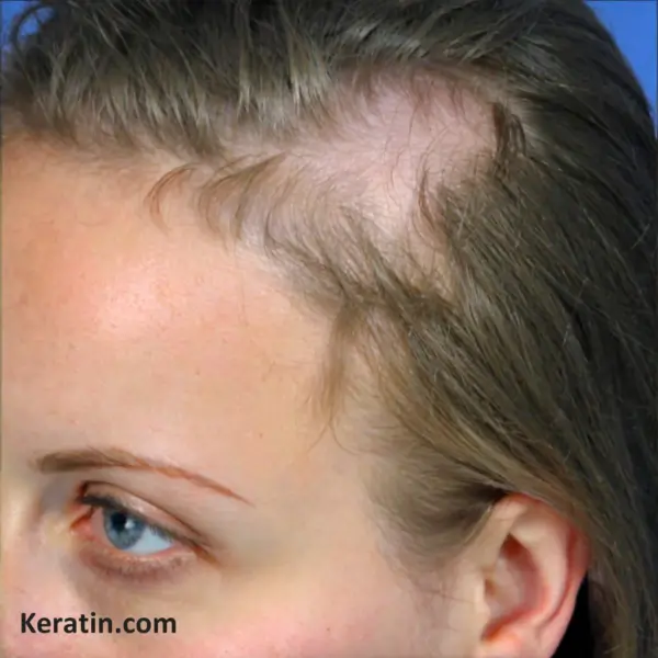
Clinical Features: triangular alopecia typically manifests as a triangular, oval, or lancet-shaped patch of alopecia. It can be present at birth or develop later in life and remains unchanged over time. Although often unilateral (just a single patch of alopecia), bilateral cases (two patches either side of the head) account for up to 20% of instances. The lesion may lack hair entirely, or it may contain some normal terminal or vellus hairs.
The condition usually becomes noticeable when the sparse hair contrasts with the surrounding terminal hair. This often means that parent don’t recognise it in their newborns right away – it can take 1-3 years or so before they can fully see the alopecia lesions as hair grows in around the patch.
Etiology: The condition was initially thought to be congenital, with some babies being born with the condition (and so described as having true congenital triangular alopecia), but recent studies suggest it is actually acquired in many individuals later in their lives.
In a study conducted by Yamazaki et al. in 2010, which involved a series of 53 patients from their clinic, it was found that a significant portion, approximately 59%, developed symptoms of the condition between the ages of two and nine. Additionally, 37% of the cases were evident at birth, and for a smaller fraction, about 3.8%, triangular alopecia first developed in adulthood.
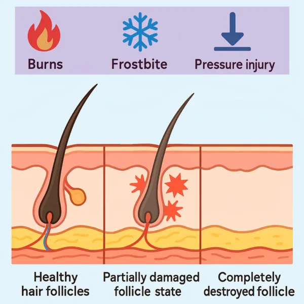
How exactly it develops remains unclear, with theories ranging from skin mosaicism to ectodermal defects during development. The miniaturization of hair follicles in the affected lesional area of skin indicates a localized process, but the exact stimulus for this hair follicle shrinking is unknown.
Histology: Histologically, triangular alopecia is characterized by the replacement of normal terminal hair follicles with sparse vellus hair follicles. The total number of hair follicles typically remains normal. The epidermis and dermis appear unremarkable, with no inflammatory infiltrate. This lack of inflammation is particularly important in making a diagnosis as inflammatory cells around hair follicles might suggest a diagnosis of alopecia areata or a scarring alopecia. Other structures like sweat glands and sebaceous glands look normal. The relatively benign histology findings suggest triangular alopecia could be a hamartomatous mosaic disease involving abnormal epithelial-mesenchymal interaction.
Diagnosis: Diagnosis of triangular alopecia is primarily clinical, based on its distinct appearance and location. The majority of the cases exhibit a triangular pattern (71.4%), followed by a quasi-circular alopecia patch (19%), and a small percentage of affected individuals present with a lance-shaped alopecia patch (9.5%).
Inui et al. proposed a classification system of 4 different features to properly identify triangular alopecia: (1) A patch of hair loss in the shape of a triangle or spear, primarily found in the frontotemporal area of the scalp; (2) the presence of normal hair follicles interspersed with fine, vellus-type hairs amidst an area of normal, thicker terminal hairs; (3) the absence of broken or ‘exclamation mark’ hairs, as well as the lack of black or yellow dots, while maintaining an intact follicular orifice; and (4) no significant regrowth of hair observed six months after the detection of vellus hairs through dermoscopic examination.
Associated Disorders: About 15% of triangular alopecia cases are associated with other disorders like phakomatosis pigmentovascularis, Down syndrome, and heart diseases. The link between triangular alopecia and these conditions remains an area of further research, but overall the apparent associations are limited to a very few individuals reported in the medical literature. Triangular alopecia can be associated with congenital heart diseases like atrial septal defect and ventricular septal defect, bone abnormalities, and café-au-lait patches. It has also been linked to neurological conditions such as epilepsy and mental retardation. Additionally, it can occur alongside genetic syndromes like Down’s syndrome and Turner syndrome. These associations underscore the importance of a comprehensive evaluation in patients presenting with triangular alopecia, as it can sometimes be indicative of broader systemic or developmental issues. However, as a reminder, 85% of people with triangular alopecia do not have any other associated conditions.
Investigations: Trichoscopic examination using a dermoscope (dermatoscope) is a valuable diagnostic tool, revealing features like short vellus hairs and absence of diagnostic features for other alopecia types. Videodermoscopy and hair pull tests also contribute to diagnosis. Interestingly, the nature of the hair in the affected area can be quite variable. While some people have a triangular alopecia patch that has no obvious hair at all, others can have a few isolated terminal hairs, a few intermediate hairs, and/or vellus hairs. Even the vellus hairs can be variable with normal short vellus hairs, and also sometimes long vellus hairs, being present. Dermoscopy shows no yellow or black spot signs or hair dystrophy that can be seen with alopecia areata. White spots can be seen and correspond to the sweat gland or sebaceous gland structures in the lesions.
Differential diagnoses: Unfortunately, the risk of misdiagnosis is high. One report suggests the misdiagnosis rate is as high as 65% in children. Most often triangular alopecia is misdiagnosed as alopecia areata and patient may even be given corticosteroid treatments, but these will have no effect on triangular alopecia. There are other potential differential diagnoses that triangular alopecia needs to be distinguished from:
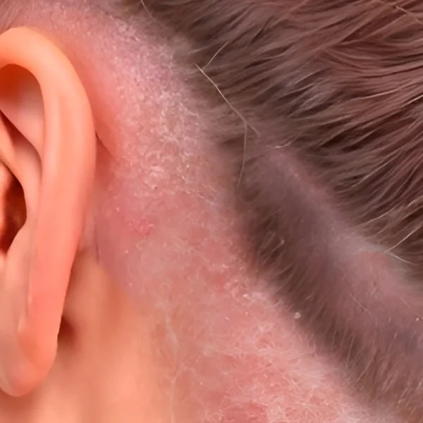
Congenital skin hypoplasia, for instance, is characterized by a translucent appearance of the skin and the absence of skin appendages. Sebaceous nevus presents with bright yellow dots that are not associated with hair follicles. Alopecia areata, a common differential diagnosis, exhibits several signs including yellow spot sign, black spot sign, broken hair, exclamation mark hair, and short vellus hair.
Trichotillomania, another condition to consider, may show signs like the black spot sign, broken hair, and splitting and curling of the hair stem stump. Telogen alopecia is identified by hair follicles with hairless trunks and the emergence of new hair. Tinea capitis, a fungal infection, can be differentiated by its presentation of broken hair, scale, spiral hair, and comma hair.
In addition to these, alopecia mucinosa, pressure alopecia, and traction alopecia are also conditions to consider in the differential diagnosis of triangular alopecia. Each of these conditions has distinct dermatoscopic features that aid in their identification and differentiation from triangular alopecia.
Genetics: The genetic basis of triangular alopecia is rather speculative, with paradominant inheritance and localized follicle miniaturization caused by an unknown gene signalling mechanism being the leading theories. With relatively few people having the condition, and few cases investigated, information on the genetics of the condition is very limited.
Treatment: triangular alopecia is often asymptomatic and permanent, with no specific treatment required. Parents should be informed about the nature of the condition to avoid unnecessary treatments. A few case reports have suggested various treatments including; topical minoxidil, sublingual minoxidil. In some cases, cosmetic interventions like hair restoration surgery may be considered.
Conclusion: triangular alopecia is likely an underreported and underdiagnosed condition due to its asymptomatic nature and diagnostic confusion with other nonscarring alopecias. Understanding its clinical presentation and potential associations is crucial for accurate diagnosis and management. Future research, particularly in molecular and genetic studies, is essential to unravel the etiology of triangular alopecia and to refine diagnostic and therapeutic approaches.
Bibliography
11711645 {11711645:PHRXFB8R},{11711645:MS2KTB63},{11711645:CKFQBEFD},{11711645:WQT59X7I},{11711645:DSWA3597},{11711645:8WE93T2X},{11711645:7W76D8T4},{11711645:GVH4WX57},{11711645:K545BGBR},{11711645:PNXST638},{11711645:H2XIEGZA},{11711645:JWRTN4MC},{11711645:H4U4EMIU},{11711645:2GEDM3FE},{11711645:MMMD24C5},{11711645:9CQZ24JU},{11711645:5EQFD65T},{11711645:SKFV6CPN} 1 vancouver 50 date asc 1055 https://www.keratin.com/wp-content/plugins/zotpress/ %7B%22status%22%3A%22success%22%2C%22updateneeded%22%3Afalse%2C%22instance%22%3Afalse%2C%22meta%22%3A%7B%22request_last%22%3A0%2C%22request_next%22%3A0%2C%22used_cache%22%3Atrue%7D%2C%22data%22%3A%5B%7B%22key%22%3A%22JWRTN4MC%22%2C%22library%22%3A%7B%22id%22%3A11711645%7D%2C%22meta%22%3A%7B%22creatorSummary%22%3A%22Sabouraud%22%2C%22parsedDate%22%3A%221905%22%2C%22numChildren%22%3A0%7D%2C%22bib%22%3A%22%26lt%3Bdiv%20class%3D%26quot%3Bcsl-bib-body%26quot%3B%20style%3D%26quot%3Bline-height%3A%201.35%3B%20%26quot%3B%26gt%3B%5Cn%20%20%26lt%3Bdiv%20class%3D%26quot%3Bcsl-entry%26quot%3B%20style%3D%26quot%3Bclear%3A%20left%3B%20%26quot%3B%26gt%3B%5Cn%20%20%20%20%26lt%3Bdiv%20class%3D%26quot%3Bcsl-left-margin%26quot%3B%20style%3D%26quot%3Bfloat%3A%20left%3B%20padding-right%3A%200.5em%3B%20text-align%3A%20right%3B%20width%3A%201em%3B%26quot%3B%26gt%3B1.%26lt%3B%5C%2Fdiv%26gt%3B%26lt%3Bdiv%20class%3D%26quot%3Bcsl-right-inline%26quot%3B%20style%3D%26quot%3Bmargin%3A%200%20.4em%200%201.5em%3B%26quot%3B%26gt%3BSabouraud%20R.%20Manuel%20%26%23xE9%3Bl%26%23xE9%3Bmentaire%20de%20dermatologie%20topographique%20r%26%23xE9%3Bgionale.%20Masson%20et%20cie%3B%201905.%20804%20p.%26lt%3B%5C%2Fdiv%26gt%3B%5Cn%20%20%20%26lt%3B%5C%2Fdiv%26gt%3B%5Cn%26lt%3B%5C%2Fdiv%26gt%3B%22%2C%22data%22%3A%7B%22itemType%22%3A%22book%22%2C%22title%22%3A%22Manuel%20%5Cu00e9l%5Cu00e9mentaire%20de%20dermatologie%20topographique%20r%5Cu00e9gionale%22%2C%22creators%22%3A%5B%7B%22creatorType%22%3A%22author%22%2C%22firstName%22%3A%22Raymond%22%2C%22lastName%22%3A%22Sabouraud%22%7D%5D%2C%22abstractNote%22%3A%22%22%2C%22date%22%3A%221905%22%2C%22originalDate%22%3A%22%22%2C%22originalPublisher%22%3A%22%22%2C%22originalPlace%22%3A%22%22%2C%22format%22%3A%22%22%2C%22ISBN%22%3A%22%22%2C%22DOI%22%3A%22%22%2C%22citationKey%22%3A%22%22%2C%22url%22%3A%22%22%2C%22ISSN%22%3A%22%22%2C%22language%22%3A%22fr%22%2C%22collections%22%3A%5B%223DPASB3D%22%5D%2C%22dateModified%22%3A%222024-01-09T17%3A12%3A37Z%22%7D%7D%2C%7B%22key%22%3A%22MS2KTB63%22%2C%22library%22%3A%7B%22id%22%3A11711645%7D%2C%22meta%22%3A%7B%22creatorSummary%22%3A%22Brauer%22%2C%22parsedDate%22%3A%221929%22%2C%22numChildren%22%3A0%7D%2C%22bib%22%3A%22%26lt%3Bdiv%20class%3D%26quot%3Bcsl-bib-body%26quot%3B%20style%3D%26quot%3Bline-height%3A%201.35%3B%20%26quot%3B%26gt%3B%5Cn%20%20%26lt%3Bdiv%20class%3D%26quot%3Bcsl-entry%26quot%3B%20style%3D%26quot%3Bclear%3A%20left%3B%20%26quot%3B%26gt%3B%5Cn%20%20%20%20%26lt%3Bdiv%20class%3D%26quot%3Bcsl-left-margin%26quot%3B%20style%3D%26quot%3Bfloat%3A%20left%3B%20padding-right%3A%200.5em%3B%20text-align%3A%20right%3B%20width%3A%201em%3B%26quot%3B%26gt%3B1.%26lt%3B%5C%2Fdiv%26gt%3B%26lt%3Bdiv%20class%3D%26quot%3Bcsl-right-inline%26quot%3B%20style%3D%26quot%3Bmargin%3A%200%20.4em%200%201.5em%3B%26quot%3B%26gt%3BBrauer%20A.%20Hereditaerer%20symmetrischer%20systematisierter%20Naevus%20aplasticus%20bei%2038%20Personen.%20Derm%20Wschr.%201929%3B89%3A1163%26%23x2013%3B8.%26lt%3B%5C%2Fdiv%26gt%3B%5Cn%20%20%20%26lt%3B%5C%2Fdiv%26gt%3B%5Cn%26lt%3B%5C%2Fdiv%26gt%3B%22%2C%22data%22%3A%7B%22itemType%22%3A%22journalArticle%22%2C%22title%22%3A%22Hereditaerer%20symmetrischer%20systematisierter%20Naevus%20aplasticus%20bei%2038%20Personen%22%2C%22creators%22%3A%5B%7B%22creatorType%22%3A%22author%22%2C%22firstName%22%3A%22A%22%2C%22lastName%22%3A%22Brauer%22%7D%5D%2C%22abstractNote%22%3A%22%22%2C%22date%22%3A%221929%22%2C%22section%22%3A%22%22%2C%22partNumber%22%3A%22%22%2C%22partTitle%22%3A%22%22%2C%22DOI%22%3A%22%22%2C%22citationKey%22%3A%22%22%2C%22url%22%3A%22%22%2C%22PMID%22%3A%22%22%2C%22PMCID%22%3A%22%22%2C%22ISSN%22%3A%22%22%2C%22language%22%3A%22%22%2C%22collections%22%3A%5B%223DPASB3D%22%5D%2C%22dateModified%22%3A%222024-01-09T20%3A58%3A25Z%22%7D%7D%2C%7B%22key%22%3A%222GEDM3FE%22%2C%22library%22%3A%7B%22id%22%3A11711645%7D%2C%22meta%22%3A%7B%22creatorSummary%22%3A%22Trakimas%20et%20al.%22%2C%22parsedDate%22%3A%221994-08%22%2C%22numChildren%22%3A0%7D%2C%22bib%22%3A%22%26lt%3Bdiv%20class%3D%26quot%3Bcsl-bib-body%26quot%3B%20style%3D%26quot%3Bline-height%3A%201.35%3B%20%26quot%3B%26gt%3B%5Cn%20%20%26lt%3Bdiv%20class%3D%26quot%3Bcsl-entry%26quot%3B%20style%3D%26quot%3Bclear%3A%20left%3B%20%26quot%3B%26gt%3B%5Cn%20%20%20%20%26lt%3Bdiv%20class%3D%26quot%3Bcsl-left-margin%26quot%3B%20style%3D%26quot%3Bfloat%3A%20left%3B%20padding-right%3A%200.5em%3B%20text-align%3A%20right%3B%20width%3A%201em%3B%26quot%3B%26gt%3B1.%26lt%3B%5C%2Fdiv%26gt%3B%26lt%3Bdiv%20class%3D%26quot%3Bcsl-right-inline%26quot%3B%20style%3D%26quot%3Bmargin%3A%200%20.4em%200%201.5em%3B%26quot%3B%26gt%3BTrakimas%20C%2C%20Sperling%20LC%2C%20Skelton%20HG%2C%20Smith%20KJ%2C%20Buker%20JL.%20Clinical%20and%20histologic%20findings%20in%20temporal%20triangular%20alopecia.%20J%20Am%20Acad%20Dermatol.%201994%20Aug%3B31%282%20Pt%201%29%3A205%26%23x2013%3B9.%26lt%3B%5C%2Fdiv%26gt%3B%5Cn%20%20%20%26lt%3B%5C%2Fdiv%26gt%3B%5Cn%26lt%3B%5C%2Fdiv%26gt%3B%22%2C%22data%22%3A%7B%22itemType%22%3A%22journalArticle%22%2C%22title%22%3A%22Clinical%20and%20histologic%20findings%20in%20temporal%20triangular%20alopecia%22%2C%22creators%22%3A%5B%7B%22creatorType%22%3A%22author%22%2C%22firstName%22%3A%22C.%22%2C%22lastName%22%3A%22Trakimas%22%7D%2C%7B%22creatorType%22%3A%22author%22%2C%22firstName%22%3A%22L.%20C.%22%2C%22lastName%22%3A%22Sperling%22%7D%2C%7B%22creatorType%22%3A%22author%22%2C%22firstName%22%3A%22H.%20G.%22%2C%22lastName%22%3A%22Skelton%22%7D%2C%7B%22creatorType%22%3A%22author%22%2C%22firstName%22%3A%22K.%20J.%22%2C%22lastName%22%3A%22Smith%22%7D%2C%7B%22creatorType%22%3A%22author%22%2C%22firstName%22%3A%22J.%20L.%22%2C%22lastName%22%3A%22Buker%22%7D%5D%2C%22abstractNote%22%3A%22BACKGROUND%3A%20Temporal%20triangular%20alopecia%20%28TTA%3B%20also%20called%20%26quot%3Bcongenital%20triangular%20alopecia%26quot%3B%29%20is%20a%20common%20disorder%20that%20is%20assumed%20to%20be%20congenital.%20Little%20is%20known%20about%20its%20histologic%20features.%5CnOBJECTIVE%3A%20Our%20purpose%20was%20to%20describe%20four%20new%20cases%2C%20review%20the%20literature%2C%20and%20present%20histologic%20features%20based%20on%20vertical%20and%20transverse%20sectioning.%5CnMETHODS%3A%20The%20history%2C%20clinical%20features%2C%20and%20histologic%20findings%20of%20four%20patients%20with%20TTA%20are%20described%20and%20the%20relevant%20literature%20reviewed.%5CnRESULTS%3A%20Lesions%20of%20TTA%20are%20seldom%20congenital%2C%20and%20most%20are%20best%20described%20as%20lancet-shaped.%20The%20%26quot%3Bbald%20spot%26quot%3B%20contains%20normal%20numbers%20of%20hairs%2C%20although%20virtually%20all%20are%20vellus%20or%20indeterminate%20follicles.%5CnCONCLUSION%3A%20Most%20cases%20of%20TTA%20appear%20to%20develop%20during%20the%20first%20few%20years%20of%20life%2C%20and%20the%20designation%20%26quot%3Bcongenital%26quot%3B%20is%20a%20misnomer.%20The%20appearance%20of%20alopecia%20can%20be%20best%20explained%20as%20a%20focal%20zone%20of%20hair%20miniaturization%20leading%20to%20vellus%20hair%20formation.%22%2C%22date%22%3A%221994-08%22%2C%22section%22%3A%22%22%2C%22partNumber%22%3A%22%22%2C%22partTitle%22%3A%22%22%2C%22DOI%22%3A%2210.1016%5C%2Fs0190-9622%2894%2970147-4%22%2C%22citationKey%22%3A%22%22%2C%22url%22%3A%22%22%2C%22PMID%22%3A%22%22%2C%22PMCID%22%3A%22%22%2C%22ISSN%22%3A%220190-9622%22%2C%22language%22%3A%22eng%22%2C%22collections%22%3A%5B%223DPASB3D%22%5D%2C%22dateModified%22%3A%222024-01-09T21%3A11%3A03Z%22%7D%7D%2C%7B%22key%22%3A%22DSWA3597%22%2C%22library%22%3A%7B%22id%22%3A11711645%7D%2C%22meta%22%3A%7B%22creatorSummary%22%3A%22Garc%5Cu00eda-Hern%5Cu00e1ndez%20et%20al.%22%2C%22parsedDate%22%3A%221995-12%22%2C%22numChildren%22%3A0%7D%2C%22bib%22%3A%22%26lt%3Bdiv%20class%3D%26quot%3Bcsl-bib-body%26quot%3B%20style%3D%26quot%3Bline-height%3A%201.35%3B%20%26quot%3B%26gt%3B%5Cn%20%20%26lt%3Bdiv%20class%3D%26quot%3Bcsl-entry%26quot%3B%20style%3D%26quot%3Bclear%3A%20left%3B%20%26quot%3B%26gt%3B%5Cn%20%20%20%20%26lt%3Bdiv%20class%3D%26quot%3Bcsl-left-margin%26quot%3B%20style%3D%26quot%3Bfloat%3A%20left%3B%20padding-right%3A%200.5em%3B%20text-align%3A%20right%3B%20width%3A%201em%3B%26quot%3B%26gt%3B1.%26lt%3B%5C%2Fdiv%26gt%3B%26lt%3Bdiv%20class%3D%26quot%3Bcsl-right-inline%26quot%3B%20style%3D%26quot%3Bmargin%3A%200%20.4em%200%201.5em%3B%26quot%3B%26gt%3BGarc%26%23xED%3Ba-Hern%26%23xE1%3Bndez%20MJ%2C%20Rodr%26%23xED%3Bguez-Pichardo%20A%2C%20Camacho%20F.%20Congenital%20triangular%20alopecia%20%28Brauer%20nevus%29.%20Pediatr%20Dermatol.%201995%20Dec%3B12%284%29%3A301%26%23x2013%3B3.%26lt%3B%5C%2Fdiv%26gt%3B%5Cn%20%20%20%26lt%3B%5C%2Fdiv%26gt%3B%5Cn%26lt%3B%5C%2Fdiv%26gt%3B%22%2C%22data%22%3A%7B%22itemType%22%3A%22journalArticle%22%2C%22title%22%3A%22Congenital%20triangular%20alopecia%20%28Brauer%20nevus%29%22%2C%22creators%22%3A%5B%7B%22creatorType%22%3A%22author%22%2C%22firstName%22%3A%22M.%20J.%22%2C%22lastName%22%3A%22Garc%5Cu00eda-Hern%5Cu00e1ndez%22%7D%2C%7B%22creatorType%22%3A%22author%22%2C%22firstName%22%3A%22A.%22%2C%22lastName%22%3A%22Rodr%5Cu00edguez-Pichardo%22%7D%2C%7B%22creatorType%22%3A%22author%22%2C%22firstName%22%3A%22F.%22%2C%22lastName%22%3A%22Camacho%22%7D%5D%2C%22abstractNote%22%3A%22Congenital%20triangular%20alopecia%20is%20manifested%20at%203%20to%205%20years%20of%20age%20by%20unilateral%20or%2C%20less%20frequently%2C%20bilateral%20patches%20of%20alopecia%20in%20the%20frontotemporal%20region.%20At%20this%20age%20the%20differential%20diagnosis%20is%20important%2C%20particularly%20as%20regards%20alopecia%20areata.%20Only%20about%2047%20cases%20have%20been%20reported%2C%20probably%20because%20the%20lesion%20is%20benign%20and%20nonprogressive.%20In%206200%20patients%20seen%20in%20index%20visits%2C%20we%20found%207%20with%20triangular%20alopecia%2C%20a%20frequency%20of%200.11%25.%20We%20believe%20that%20males%20do%20not%20require%20treatment%20because%20of%20the%20later%20development%20of%20androgenic%20alopecia%2C%20but%20in%20women%2C%20surgical%20treatment%20is%20successful.%22%2C%22date%22%3A%221995-12%22%2C%22section%22%3A%22%22%2C%22partNumber%22%3A%22%22%2C%22partTitle%22%3A%22%22%2C%22DOI%22%3A%2210.1111%5C%2Fj.1525-1470.1995.tb00187.x%22%2C%22citationKey%22%3A%22%22%2C%22url%22%3A%22%22%2C%22PMID%22%3A%22%22%2C%22PMCID%22%3A%22%22%2C%22ISSN%22%3A%220736-8046%22%2C%22language%22%3A%22eng%22%2C%22collections%22%3A%5B%223DPASB3D%22%5D%2C%22dateModified%22%3A%222024-01-09T20%3A36%3A52Z%22%7D%7D%2C%7B%22key%22%3A%22MMMD24C5%22%2C%22library%22%3A%7B%22id%22%3A11711645%7D%2C%22meta%22%3A%7B%22creatorSummary%22%3A%22Trakimas%20and%20Sperling%22%2C%22parsedDate%22%3A%221999-05%22%2C%22numChildren%22%3A0%7D%2C%22bib%22%3A%22%26lt%3Bdiv%20class%3D%26quot%3Bcsl-bib-body%26quot%3B%20style%3D%26quot%3Bline-height%3A%201.35%3B%20%26quot%3B%26gt%3B%5Cn%20%20%26lt%3Bdiv%20class%3D%26quot%3Bcsl-entry%26quot%3B%20style%3D%26quot%3Bclear%3A%20left%3B%20%26quot%3B%26gt%3B%5Cn%20%20%20%20%26lt%3Bdiv%20class%3D%26quot%3Bcsl-left-margin%26quot%3B%20style%3D%26quot%3Bfloat%3A%20left%3B%20padding-right%3A%200.5em%3B%20text-align%3A%20right%3B%20width%3A%201em%3B%26quot%3B%26gt%3B1.%26lt%3B%5C%2Fdiv%26gt%3B%26lt%3Bdiv%20class%3D%26quot%3Bcsl-right-inline%26quot%3B%20style%3D%26quot%3Bmargin%3A%200%20.4em%200%201.5em%3B%26quot%3B%26gt%3BTrakimas%20CA%2C%20Sperling%20LC.%20Temporal%20triangular%20alopecia%20acquired%20in%20adulthood.%20J%20Am%20Acad%20Dermatol.%201999%20May%3B40%285%20Pt%202%29%3A842%26%23x2013%3B4.%26lt%3B%5C%2Fdiv%26gt%3B%5Cn%20%20%20%26lt%3B%5C%2Fdiv%26gt%3B%5Cn%26lt%3B%5C%2Fdiv%26gt%3B%22%2C%22data%22%3A%7B%22itemType%22%3A%22journalArticle%22%2C%22title%22%3A%22Temporal%20triangular%20alopecia%20acquired%20in%20adulthood%22%2C%22creators%22%3A%5B%7B%22creatorType%22%3A%22author%22%2C%22firstName%22%3A%22C.%20A.%22%2C%22lastName%22%3A%22Trakimas%22%7D%2C%7B%22creatorType%22%3A%22author%22%2C%22firstName%22%3A%22L.%20C.%22%2C%22lastName%22%3A%22Sperling%22%7D%5D%2C%22abstractNote%22%3A%22Temporal%20triangular%20alopecia%20is%20a%20relatively%20common%2C%20nonscarring%20form%20of%20alopecia.%20Sometimes%20congenital%2C%20the%20vast%20majority%20of%20lesions%20appear%20during%20the%20first%206%20years%20of%20life%20and%20remain%20stable%20thereafter.%20We%20report%20a%20case%20of%20temporal%20triangular%20alopecia%20arising%20during%20adulthood.%22%2C%22date%22%3A%221999-05%22%2C%22section%22%3A%22%22%2C%22partNumber%22%3A%22%22%2C%22partTitle%22%3A%22%22%2C%22DOI%22%3A%2210.1053%5C%2Fjd.1999.v40.a97651%22%2C%22citationKey%22%3A%22%22%2C%22url%22%3A%22%22%2C%22PMID%22%3A%22%22%2C%22PMCID%22%3A%22%22%2C%22ISSN%22%3A%220190-9622%22%2C%22language%22%3A%22eng%22%2C%22collections%22%3A%5B%223DPASB3D%22%5D%2C%22dateModified%22%3A%222024-01-09T17%3A56%3A30Z%22%7D%7D%2C%7B%22key%22%3A%227W76D8T4%22%2C%22library%22%3A%7B%22id%22%3A11711645%7D%2C%22meta%22%3A%7B%22creatorSummary%22%3A%22Happle%22%2C%22parsedDate%22%3A%222003%22%2C%22numChildren%22%3A0%7D%2C%22bib%22%3A%22%26lt%3Bdiv%20class%3D%26quot%3Bcsl-bib-body%26quot%3B%20style%3D%26quot%3Bline-height%3A%201.35%3B%20%26quot%3B%26gt%3B%5Cn%20%20%26lt%3Bdiv%20class%3D%26quot%3Bcsl-entry%26quot%3B%20style%3D%26quot%3Bclear%3A%20left%3B%20%26quot%3B%26gt%3B%5Cn%20%20%20%20%26lt%3Bdiv%20class%3D%26quot%3Bcsl-left-margin%26quot%3B%20style%3D%26quot%3Bfloat%3A%20left%3B%20padding-right%3A%200.5em%3B%20text-align%3A%20right%3B%20width%3A%201em%3B%26quot%3B%26gt%3B1.%26lt%3B%5C%2Fdiv%26gt%3B%26lt%3Bdiv%20class%3D%26quot%3Bcsl-right-inline%26quot%3B%20style%3D%26quot%3Bmargin%3A%200%20.4em%200%201.5em%3B%26quot%3B%26gt%3BHapple%20R.%20Congenital%20triangular%20alopecia%20may%20be%20categorized%20as%20a%20paradominant%20trait.%20Eur%20J%20Dermatol.%202003%3B13%284%29%3A346%26%23x2013%3B7.%26lt%3B%5C%2Fdiv%26gt%3B%5Cn%20%20%20%26lt%3B%5C%2Fdiv%26gt%3B%5Cn%26lt%3B%5C%2Fdiv%26gt%3B%22%2C%22data%22%3A%7B%22itemType%22%3A%22journalArticle%22%2C%22title%22%3A%22Congenital%20triangular%20alopecia%20may%20be%20categorized%20as%20a%20paradominant%20trait%22%2C%22creators%22%3A%5B%7B%22creatorType%22%3A%22author%22%2C%22firstName%22%3A%22Rudolf%22%2C%22lastName%22%3A%22Happle%22%7D%5D%2C%22abstractNote%22%3A%22The%20genetic%20basis%20of%20congenital%20triangular%20alopecia%20%28CTA%29%20is%20so%20far%20not%20clear.%20The%20following%20arguments%20are%20presented%20in%20favor%20of%20the%20notion%20that%20this%20may%20be%20a%20paradominant%20trait.%20CTA%20usually%20occurs%20sporadically%20but%20may%20exceptionally%20affect%20several%20members%20of%20a%20family.%20The%20lesion%20is%20usually%20unilateral%20but%20bilateral%20involvement%20may%20likewise%20occur.%20CTA%20has%20been%20reported%20in%20association%20with%20phacomatosis%20pigmentovascularis%2C%20providing%20evidence%20that%20it%20may%20originate%20from%20loss%20of%20heterozygosity.%20Heterozygous%20individuals%20would%20be%20phenotypically%20normal.%20The%20trait%20would%20be%20expressed%20only%20when%20postzygotic%20loss%20of%20the%20corresponding%20wildtype%20allele%20occurred%20in%20a%20early%20developmental%20stage.%20Future%20molecular%20research%20may%20show%20whether%20the%20concept%20of%20paradominant%20inheritance%20holds%20true.%22%2C%22date%22%3A%222003%22%2C%22section%22%3A%22%22%2C%22partNumber%22%3A%22%22%2C%22partTitle%22%3A%22%22%2C%22DOI%22%3A%22%22%2C%22citationKey%22%3A%22%22%2C%22url%22%3A%22%22%2C%22PMID%22%3A%22%22%2C%22PMCID%22%3A%22%22%2C%22ISSN%22%3A%221167-1122%22%2C%22language%22%3A%22eng%22%2C%22collections%22%3A%5B%223DPASB3D%22%5D%2C%22dateModified%22%3A%222024-01-09T21%3A10%3A02Z%22%7D%7D%2C%7B%22key%22%3A%229CQZ24JU%22%2C%22library%22%3A%7B%22id%22%3A11711645%7D%2C%22meta%22%3A%7B%22creatorSummary%22%3A%22Wu%20et%20al.%22%2C%22parsedDate%22%3A%222009-08%22%2C%22numChildren%22%3A0%7D%2C%22bib%22%3A%22%26lt%3Bdiv%20class%3D%26quot%3Bcsl-bib-body%26quot%3B%20style%3D%26quot%3Bline-height%3A%201.35%3B%20%26quot%3B%26gt%3B%5Cn%20%20%26lt%3Bdiv%20class%3D%26quot%3Bcsl-entry%26quot%3B%20style%3D%26quot%3Bclear%3A%20left%3B%20%26quot%3B%26gt%3B%5Cn%20%20%20%20%26lt%3Bdiv%20class%3D%26quot%3Bcsl-left-margin%26quot%3B%20style%3D%26quot%3Bfloat%3A%20left%3B%20padding-right%3A%200.5em%3B%20text-align%3A%20right%3B%20width%3A%201em%3B%26quot%3B%26gt%3B1.%26lt%3B%5C%2Fdiv%26gt%3B%26lt%3Bdiv%20class%3D%26quot%3Bcsl-right-inline%26quot%3B%20style%3D%26quot%3Bmargin%3A%200%20.4em%200%201.5em%3B%26quot%3B%26gt%3BWu%20WY%2C%20Otberg%20N%2C%20Kang%20H%2C%20Zanet%20L%2C%20Shapiro%20J.%20Successful%20treatment%20of%20temporal%20triangular%20alopecia%20by%20hair%20restoration%20surgery%20using%20follicular%20unit%20transplantation.%20Dermatol%20Surg.%202009%20Aug%3B35%288%29%3A1307%26%23x2013%3B10.%26lt%3B%5C%2Fdiv%26gt%3B%5Cn%20%20%20%26lt%3B%5C%2Fdiv%26gt%3B%5Cn%26lt%3B%5C%2Fdiv%26gt%3B%22%2C%22data%22%3A%7B%22itemType%22%3A%22journalArticle%22%2C%22title%22%3A%22Successful%20treatment%20of%20temporal%20triangular%20alopecia%20by%20hair%20restoration%20surgery%20using%20follicular%20unit%20transplantation%22%2C%22creators%22%3A%5B%7B%22creatorType%22%3A%22author%22%2C%22firstName%22%3A%22Wen-Yu%22%2C%22lastName%22%3A%22Wu%22%7D%2C%7B%22creatorType%22%3A%22author%22%2C%22firstName%22%3A%22Nina%22%2C%22lastName%22%3A%22Otberg%22%7D%2C%7B%22creatorType%22%3A%22author%22%2C%22firstName%22%3A%22Hoon%22%2C%22lastName%22%3A%22Kang%22%7D%2C%7B%22creatorType%22%3A%22author%22%2C%22firstName%22%3A%22Lucianna%22%2C%22lastName%22%3A%22Zanet%22%7D%2C%7B%22creatorType%22%3A%22author%22%2C%22firstName%22%3A%22Jerry%22%2C%22lastName%22%3A%22Shapiro%22%7D%5D%2C%22abstractNote%22%3A%22%22%2C%22date%22%3A%222009-08%22%2C%22section%22%3A%22%22%2C%22partNumber%22%3A%22%22%2C%22partTitle%22%3A%22%22%2C%22DOI%22%3A%2210.1111%5C%2Fj.1524-4725.2009.01233.x%22%2C%22citationKey%22%3A%22%22%2C%22url%22%3A%22%22%2C%22PMID%22%3A%22%22%2C%22PMCID%22%3A%22%22%2C%22ISSN%22%3A%221524-4725%22%2C%22language%22%3A%22eng%22%2C%22collections%22%3A%5B%223DPASB3D%22%5D%2C%22dateModified%22%3A%222024-01-09T17%3A15%3A37Z%22%7D%7D%2C%7B%22key%22%3A%225EQFD65T%22%2C%22library%22%3A%7B%22id%22%3A11711645%7D%2C%22meta%22%3A%7B%22creatorSummary%22%3A%22Yamazaki%20et%20al.%22%2C%22parsedDate%22%3A%222010-04%22%2C%22numChildren%22%3A0%7D%2C%22bib%22%3A%22%26lt%3Bdiv%20class%3D%26quot%3Bcsl-bib-body%26quot%3B%20style%3D%26quot%3Bline-height%3A%201.35%3B%20%26quot%3B%26gt%3B%5Cn%20%20%26lt%3Bdiv%20class%3D%26quot%3Bcsl-entry%26quot%3B%20style%3D%26quot%3Bclear%3A%20left%3B%20%26quot%3B%26gt%3B%5Cn%20%20%20%20%26lt%3Bdiv%20class%3D%26quot%3Bcsl-left-margin%26quot%3B%20style%3D%26quot%3Bfloat%3A%20left%3B%20padding-right%3A%200.5em%3B%20text-align%3A%20right%3B%20width%3A%201em%3B%26quot%3B%26gt%3B1.%26lt%3B%5C%2Fdiv%26gt%3B%26lt%3Bdiv%20class%3D%26quot%3Bcsl-right-inline%26quot%3B%20style%3D%26quot%3Bmargin%3A%200%20.4em%200%201.5em%3B%26quot%3B%26gt%3BYamazaki%20M%2C%20Irisawa%20R%2C%20Tsuboi%20R.%20Temporal%20triangular%20alopecia%20and%20a%20review%20of%2052%20past%20cases.%20J%20Dermatol.%202010%20Apr%3B37%284%29%3A360%26%23x2013%3B2.%26lt%3B%5C%2Fdiv%26gt%3B%5Cn%20%20%20%26lt%3B%5C%2Fdiv%26gt%3B%5Cn%26lt%3B%5C%2Fdiv%26gt%3B%22%2C%22data%22%3A%7B%22itemType%22%3A%22journalArticle%22%2C%22title%22%3A%22Temporal%20triangular%20alopecia%20and%20a%20review%20of%2052%20past%20cases%22%2C%22creators%22%3A%5B%7B%22creatorType%22%3A%22author%22%2C%22firstName%22%3A%22Masashi%22%2C%22lastName%22%3A%22Yamazaki%22%7D%2C%7B%22creatorType%22%3A%22author%22%2C%22firstName%22%3A%22Ryokichi%22%2C%22lastName%22%3A%22Irisawa%22%7D%2C%7B%22creatorType%22%3A%22author%22%2C%22firstName%22%3A%22Ryoji%22%2C%22lastName%22%3A%22Tsuboi%22%7D%5D%2C%22abstractNote%22%3A%22Temporal%20triangular%20alopecia%20%28TTA%29%20is%20a%20circumscribed%2C%20non-cicatricial%20form%20of%20alopecia%20confined%20to%20the%20frontotemporal%20region.%20The%20patient%2C%20a%2015-year-old%20boy%2C%20was%20noticed%20at%20birth%20to%20have%20an%20alopecial%20area%2C%20sized%201.5%20cm%20x%202.5%20cm%2C%20in%20the%20right%20temporal%20region.%20Microscopic%20examination%20revealed%20miniaturized%20hair%20follicles%20accompanied%20by%20differentiated%20sebaceous%20glands.%20We%20have%20provided%20a%20synopsis%20of%20the%20past%2052%20cases.%20Of%20the%2053%20cases%20of%20TTA%20including%20our%20case%2C%20more%20than%20half%20%2855.8%25%29%20were%20detected%20in%20childhood%20between%20the%20ages%20of%202%20and%209%20years%2C%20while%2036.5%25%20were%20detected%20at%20birth%20and%20only%203.8%25%20%28only%20two%20cases%29%20in%20adulthood.%20There%20were%20three%20familial%20cases.%20Several%20congenital%20diseases%20were%20associated%20with%20the%20condition%2C%20for%20example%2C%20phakomatosis%20pigmentovascularis%2C%20Down%20syndrome%20and%20Dandy-Walker%20malformation.%20This%20information%20suggests%20that%20TTA%20can%20be%20recognized%20as%20a%20hamartomatous%20mosaic%20disease.%22%2C%22date%22%3A%222010-04%22%2C%22section%22%3A%22%22%2C%22partNumber%22%3A%22%22%2C%22partTitle%22%3A%22%22%2C%22DOI%22%3A%2210.1111%5C%2Fj.1346-8138.2010.00817.x%22%2C%22citationKey%22%3A%22%22%2C%22url%22%3A%22%22%2C%22PMID%22%3A%22%22%2C%22PMCID%22%3A%22%22%2C%22ISSN%22%3A%221346-8138%22%2C%22language%22%3A%22eng%22%2C%22collections%22%3A%5B%223DPASB3D%22%5D%2C%22dateModified%22%3A%222024-01-09T17%3A14%3A04Z%22%7D%7D%2C%7B%22key%22%3A%22GVH4WX57%22%2C%22library%22%3A%7B%22id%22%3A11711645%7D%2C%22meta%22%3A%7B%22creatorSummary%22%3A%22Inui%20et%20al.%22%2C%22parsedDate%22%3A%222012-06%22%2C%22numChildren%22%3A0%7D%2C%22bib%22%3A%22%26lt%3Bdiv%20class%3D%26quot%3Bcsl-bib-body%26quot%3B%20style%3D%26quot%3Bline-height%3A%201.35%3B%20%26quot%3B%26gt%3B%5Cn%20%20%26lt%3Bdiv%20class%3D%26quot%3Bcsl-entry%26quot%3B%20style%3D%26quot%3Bclear%3A%20left%3B%20%26quot%3B%26gt%3B%5Cn%20%20%20%20%26lt%3Bdiv%20class%3D%26quot%3Bcsl-left-margin%26quot%3B%20style%3D%26quot%3Bfloat%3A%20left%3B%20padding-right%3A%200.5em%3B%20text-align%3A%20right%3B%20width%3A%201em%3B%26quot%3B%26gt%3B1.%26lt%3B%5C%2Fdiv%26gt%3B%26lt%3Bdiv%20class%3D%26quot%3Bcsl-right-inline%26quot%3B%20style%3D%26quot%3Bmargin%3A%200%20.4em%200%201.5em%3B%26quot%3B%26gt%3BInui%20S%2C%20Nakajima%20T%2C%20Itami%20S.%20Temporal%20triangular%20alopecia%3A%20trichoscopic%20diagnosis.%20J%20Dermatol.%202012%20June%3B39%286%29%3A572%26%23x2013%3B4.%26lt%3B%5C%2Fdiv%26gt%3B%5Cn%20%20%20%26lt%3B%5C%2Fdiv%26gt%3B%5Cn%26lt%3B%5C%2Fdiv%26gt%3B%22%2C%22data%22%3A%7B%22itemType%22%3A%22journalArticle%22%2C%22title%22%3A%22Temporal%20triangular%20alopecia%3A%20trichoscopic%20diagnosis%22%2C%22creators%22%3A%5B%7B%22creatorType%22%3A%22author%22%2C%22firstName%22%3A%22Shigeki%22%2C%22lastName%22%3A%22Inui%22%7D%2C%7B%22creatorType%22%3A%22author%22%2C%22firstName%22%3A%22Takeshi%22%2C%22lastName%22%3A%22Nakajima%22%7D%2C%7B%22creatorType%22%3A%22author%22%2C%22firstName%22%3A%22Satoshi%22%2C%22lastName%22%3A%22Itami%22%7D%5D%2C%22abstractNote%22%3A%22%22%2C%22date%22%3A%222012-06%22%2C%22section%22%3A%22%22%2C%22partNumber%22%3A%22%22%2C%22partTitle%22%3A%22%22%2C%22DOI%22%3A%2210.1111%5C%2Fj.1346-8138.2011.01348.x%22%2C%22citationKey%22%3A%22%22%2C%22url%22%3A%22%22%2C%22PMID%22%3A%22%22%2C%22PMCID%22%3A%22%22%2C%22ISSN%22%3A%221346-8138%22%2C%22language%22%3A%22eng%22%2C%22collections%22%3A%5B%223DPASB3D%22%5D%2C%22dateModified%22%3A%222024-01-09T21%3A11%3A34Z%22%7D%7D%2C%7B%22key%22%3A%22CKFQBEFD%22%2C%22library%22%3A%7B%22id%22%3A11711645%7D%2C%22meta%22%3A%7B%22creatorSummary%22%3A%22Chung%20et%20al.%22%2C%22parsedDate%22%3A%222012-08%22%2C%22numChildren%22%3A0%7D%2C%22bib%22%3A%22%26lt%3Bdiv%20class%3D%26quot%3Bcsl-bib-body%26quot%3B%20style%3D%26quot%3Bline-height%3A%201.35%3B%20%26quot%3B%26gt%3B%5Cn%20%20%26lt%3Bdiv%20class%3D%26quot%3Bcsl-entry%26quot%3B%20style%3D%26quot%3Bclear%3A%20left%3B%20%26quot%3B%26gt%3B%5Cn%20%20%20%20%26lt%3Bdiv%20class%3D%26quot%3Bcsl-left-margin%26quot%3B%20style%3D%26quot%3Bfloat%3A%20left%3B%20padding-right%3A%200.5em%3B%20text-align%3A%20right%3B%20width%3A%201em%3B%26quot%3B%26gt%3B1.%26lt%3B%5C%2Fdiv%26gt%3B%26lt%3Bdiv%20class%3D%26quot%3Bcsl-right-inline%26quot%3B%20style%3D%26quot%3Bmargin%3A%200%20.4em%200%201.5em%3B%26quot%3B%26gt%3BChung%20J%2C%20Sim%20JH%2C%20Gye%20J%2C%20Namkoong%20S%2C%20Hong%20SP%2C%20Kim%20MH%2C%20et%20al.%20Successful%20hair%20transplantation%20for%20treatment%20of%20acquired%20temporal%20triangular%20alopecia.%20Dermatol%20Surg.%202012%20Aug%3B38%288%29%3A1404%26%23x2013%3B6.%26lt%3B%5C%2Fdiv%26gt%3B%5Cn%20%20%20%26lt%3B%5C%2Fdiv%26gt%3B%5Cn%26lt%3B%5C%2Fdiv%26gt%3B%22%2C%22data%22%3A%7B%22itemType%22%3A%22journalArticle%22%2C%22title%22%3A%22Successful%20hair%20transplantation%20for%20treatment%20of%20acquired%20temporal%20triangular%20alopecia%22%2C%22creators%22%3A%5B%7B%22creatorType%22%3A%22author%22%2C%22firstName%22%3A%22Jimin%22%2C%22lastName%22%3A%22Chung%22%7D%2C%7B%22creatorType%22%3A%22author%22%2C%22firstName%22%3A%22Ji%20Hyun%22%2C%22lastName%22%3A%22Sim%22%7D%2C%7B%22creatorType%22%3A%22author%22%2C%22firstName%22%3A%22Jiwon%22%2C%22lastName%22%3A%22Gye%22%7D%2C%7B%22creatorType%22%3A%22author%22%2C%22firstName%22%3A%22Sun%22%2C%22lastName%22%3A%22Namkoong%22%7D%2C%7B%22creatorType%22%3A%22author%22%2C%22firstName%22%3A%22Seung%20Phil%22%2C%22lastName%22%3A%22Hong%22%7D%2C%7B%22creatorType%22%3A%22author%22%2C%22firstName%22%3A%22Myung%20Hwa%22%2C%22lastName%22%3A%22Kim%22%7D%2C%7B%22creatorType%22%3A%22author%22%2C%22firstName%22%3A%22Byung%20Cheol%22%2C%22lastName%22%3A%22Park%22%7D%5D%2C%22abstractNote%22%3A%22%22%2C%22date%22%3A%222012-08%22%2C%22section%22%3A%22%22%2C%22partNumber%22%3A%22%22%2C%22partTitle%22%3A%22%22%2C%22DOI%22%3A%2210.1111%5C%2Fj.1524-4725.2012.02442.x%22%2C%22citationKey%22%3A%22%22%2C%22url%22%3A%22%22%2C%22PMID%22%3A%22%22%2C%22PMCID%22%3A%22%22%2C%22ISSN%22%3A%221524-4725%22%2C%22language%22%3A%22eng%22%2C%22collections%22%3A%5B%223DPASB3D%22%5D%2C%22dateModified%22%3A%222024-01-09T17%3A15%3A26Z%22%7D%7D%2C%7B%22key%22%3A%22PHRXFB8R%22%2C%22library%22%3A%7B%22id%22%3A11711645%7D%2C%22meta%22%3A%7B%22creatorSummary%22%3A%22Bang%20et%20al.%22%2C%22parsedDate%22%3A%222013-08%22%2C%22numChildren%22%3A0%7D%2C%22bib%22%3A%22%26lt%3Bdiv%20class%3D%26quot%3Bcsl-bib-body%26quot%3B%20style%3D%26quot%3Bline-height%3A%201.35%3B%20%26quot%3B%26gt%3B%5Cn%20%20%26lt%3Bdiv%20class%3D%26quot%3Bcsl-entry%26quot%3B%20style%3D%26quot%3Bclear%3A%20left%3B%20%26quot%3B%26gt%3B%5Cn%20%20%20%20%26lt%3Bdiv%20class%3D%26quot%3Bcsl-left-margin%26quot%3B%20style%3D%26quot%3Bfloat%3A%20left%3B%20padding-right%3A%200.5em%3B%20text-align%3A%20right%3B%20width%3A%201em%3B%26quot%3B%26gt%3B1.%26lt%3B%5C%2Fdiv%26gt%3B%26lt%3Bdiv%20class%3D%26quot%3Bcsl-right-inline%26quot%3B%20style%3D%26quot%3Bmargin%3A%200%20.4em%200%201.5em%3B%26quot%3B%26gt%3BBang%20CY%2C%20Byun%20JW%2C%20Kang%20MJ%2C%20Yang%20BH%2C%20Song%20HJ%2C%20Shin%20J%2C%20et%20al.%20Successful%20treatment%20of%20temporal%20triangular%20alopecia%20with%20topical%20minoxidil.%20Ann%20Dermatol.%202013%20Aug%3B25%283%29%3A387%26%23x2013%3B8.%26lt%3B%5C%2Fdiv%26gt%3B%5Cn%20%20%20%26lt%3B%5C%2Fdiv%26gt%3B%5Cn%26lt%3B%5C%2Fdiv%26gt%3B%22%2C%22data%22%3A%7B%22itemType%22%3A%22journalArticle%22%2C%22title%22%3A%22Successful%20treatment%20of%20temporal%20triangular%20alopecia%20with%20topical%20minoxidil%22%2C%22creators%22%3A%5B%7B%22creatorType%22%3A%22author%22%2C%22firstName%22%3A%22Chan-Yl%22%2C%22lastName%22%3A%22Bang%22%7D%2C%7B%22creatorType%22%3A%22author%22%2C%22firstName%22%3A%22Ji-Won%22%2C%22lastName%22%3A%22Byun%22%7D%2C%7B%22creatorType%22%3A%22author%22%2C%22firstName%22%3A%22Min-Ji%22%2C%22lastName%22%3A%22Kang%22%7D%2C%7B%22creatorType%22%3A%22author%22%2C%22firstName%22%3A%22Bo-Hee%22%2C%22lastName%22%3A%22Yang%22%7D%2C%7B%22creatorType%22%3A%22author%22%2C%22firstName%22%3A%22Hee-Jin%22%2C%22lastName%22%3A%22Song%22%7D%2C%7B%22creatorType%22%3A%22author%22%2C%22firstName%22%3A%22Jeonghyun%22%2C%22lastName%22%3A%22Shin%22%7D%2C%7B%22creatorType%22%3A%22author%22%2C%22firstName%22%3A%22Gwang%20Seong%22%2C%22lastName%22%3A%22Choi%22%7D%5D%2C%22abstractNote%22%3A%22%22%2C%22date%22%3A%222013-08%22%2C%22section%22%3A%22%22%2C%22partNumber%22%3A%22%22%2C%22partTitle%22%3A%22%22%2C%22DOI%22%3A%2210.5021%5C%2Fad.2013.25.3.387%22%2C%22citationKey%22%3A%22%22%2C%22url%22%3A%22%22%2C%22PMID%22%3A%22%22%2C%22PMCID%22%3A%22%22%2C%22ISSN%22%3A%221013-9087%22%2C%22language%22%3A%22eng%22%2C%22collections%22%3A%5B%223DPASB3D%22%5D%2C%22dateModified%22%3A%222024-01-09T20%3A51%3A08Z%22%7D%7D%2C%7B%22key%22%3A%22SKFV6CPN%22%2C%22library%22%3A%7B%22id%22%3A11711645%7D%2C%22meta%22%3A%7B%22creatorSummary%22%3A%22Yin%20Li%20and%20Yesudian%22%2C%22parsedDate%22%3A%222015%22%2C%22numChildren%22%3A0%7D%2C%22bib%22%3A%22%26lt%3Bdiv%20class%3D%26quot%3Bcsl-bib-body%26quot%3B%20style%3D%26quot%3Bline-height%3A%201.35%3B%20%26quot%3B%26gt%3B%5Cn%20%20%26lt%3Bdiv%20class%3D%26quot%3Bcsl-entry%26quot%3B%20style%3D%26quot%3Bclear%3A%20left%3B%20%26quot%3B%26gt%3B%5Cn%20%20%20%20%26lt%3Bdiv%20class%3D%26quot%3Bcsl-left-margin%26quot%3B%20style%3D%26quot%3Bfloat%3A%20left%3B%20padding-right%3A%200.5em%3B%20text-align%3A%20right%3B%20width%3A%201em%3B%26quot%3B%26gt%3B1.%26lt%3B%5C%2Fdiv%26gt%3B%26lt%3Bdiv%20class%3D%26quot%3Bcsl-right-inline%26quot%3B%20style%3D%26quot%3Bmargin%3A%200%20.4em%200%201.5em%3B%26quot%3B%26gt%3BYin%20Li%20VC%2C%20Yesudian%20PD.%20Congenital%20Triangular%20Alopecia.%20Int%20J%20Trichology.%202015%3B7%282%29%3A48%26%23x2013%3B53.%26lt%3B%5C%2Fdiv%26gt%3B%5Cn%20%20%20%26lt%3B%5C%2Fdiv%26gt%3B%5Cn%26lt%3B%5C%2Fdiv%26gt%3B%22%2C%22data%22%3A%7B%22itemType%22%3A%22journalArticle%22%2C%22title%22%3A%22Congenital%20Triangular%20Alopecia%22%2C%22creators%22%3A%5B%7B%22creatorType%22%3A%22author%22%2C%22firstName%22%3A%22Vincent%20Chum%22%2C%22lastName%22%3A%22Yin%20Li%22%7D%2C%7B%22creatorType%22%3A%22author%22%2C%22firstName%22%3A%22Paul%20Devakar%22%2C%22lastName%22%3A%22Yesudian%22%7D%5D%2C%22abstractNote%22%3A%22Congenital%20triangular%20alopecia%20%28CTA%29%20also%20known%20as%20temporal%20triangular%20alopecia%20is%20a%20benign%20noncicatricial%20pattern%20of%20hair%20loss.%20It%20typically%20affects%20the%20frontotemporal%20region%20and%20rarely%20involves%20the%20temporoparietal%20or%20occipital%20scalp.%20It%20is%20a%20nonprogressive%20disorder%20that%20presents%20as%20a%20triangular%2C%20oval%20or%20lancet-shaped%20patch%20of%20alopecia.%20CTA%20can%20manifest%20at%20birth%20or%20develop%20later%20in%20life.%20The%20exact%20etiology%20of%20this%20condition%20remains%20unknown.%20Rarely%2C%20it%20may%20be%20associated%20with%20other%20disorders%20such%20as%20Down%26%23039%3Bs%20syndrome%20and%20phakomatosis%20pigmentovascularis.%20The%20diagnosis%20is%20based%20on%20its%20distinct%20clinical%20appearance.%20Histologically%2C%20hair%20follicles%20are%20miniaturized%20and%20replaced%20by%20sparse%20vellus%20hair%20follicles.%20Tricoscopy%20using%20a%20polarized%20light%20handheld%20dermatoscope%20can%20be%20a%20useful%20diagnostic%20tool.%20CTA%20is%20often%20asymptomatic%20and%20remains%20unchanged%20throughout%20the%20life.%20No%20treatment%20is%20required.%20Surgical%20intervention%20with%20follicular%20unit%20hair%20transplantation%20can%20provide%20a%20satisfactory%20cosmetic%20result.%20In%20this%20paper%2C%20we%20have%20identified%20126%20cases%20of%20CTA%20in%20the%20published%20literature%20cited%20on%20PubMed%20between%201905%20and%202015.%20From%20the%20available%20evidence%2C%2079%25%20of%20patients%20with%20CTA%20presented%20with%20unilateral%20hair%20loss%2C%2018.5%25%20with%20bilateral%20involvement%20and%20rarely%2C%20with%20occipital%20alopecia%20%282.5%25%29.%20There%20was%20no%20gender%20predilection.%20These%20figures%20are%20entirely%20consistent%20with%20previously%20published%20data.%20Physicians%20should%20remember%20to%20consider%20CTA%20as%20a%20potential%20diagnosis%20in%20any%20patient%20presenting%20with%20a%20nonscarring%20alopecia%20in%20order%20to%20avoid%20unnecessary%20investigations%20and%20treatments.%22%2C%22date%22%3A%222015%22%2C%22section%22%3A%22%22%2C%22partNumber%22%3A%22%22%2C%22partTitle%22%3A%22%22%2C%22DOI%22%3A%2210.4103%5C%2F0974-7753.160089%22%2C%22citationKey%22%3A%22%22%2C%22url%22%3A%22%22%2C%22PMID%22%3A%22%22%2C%22PMCID%22%3A%22%22%2C%22ISSN%22%3A%220974-7753%22%2C%22language%22%3A%22eng%22%2C%22collections%22%3A%5B%223DPASB3D%22%5D%2C%22dateModified%22%3A%222024-01-09T17%3A11%3A13Z%22%7D%7D%2C%7B%22key%22%3A%22PNXST638%22%2C%22library%22%3A%7B%22id%22%3A11711645%7D%2C%22meta%22%3A%7B%22creatorSummary%22%3A%22Lacarrubba%20et%20al.%22%2C%22parsedDate%22%3A%222015%22%2C%22numChildren%22%3A0%7D%2C%22bib%22%3A%22%26lt%3Bdiv%20class%3D%26quot%3Bcsl-bib-body%26quot%3B%20style%3D%26quot%3Bline-height%3A%201.35%3B%20%26quot%3B%26gt%3B%5Cn%20%20%26lt%3Bdiv%20class%3D%26quot%3Bcsl-entry%26quot%3B%20style%3D%26quot%3Bclear%3A%20left%3B%20%26quot%3B%26gt%3B%5Cn%20%20%20%20%26lt%3Bdiv%20class%3D%26quot%3Bcsl-left-margin%26quot%3B%20style%3D%26quot%3Bfloat%3A%20left%3B%20padding-right%3A%200.5em%3B%20text-align%3A%20right%3B%20width%3A%201em%3B%26quot%3B%26gt%3B1.%26lt%3B%5C%2Fdiv%26gt%3B%26lt%3Bdiv%20class%3D%26quot%3Bcsl-right-inline%26quot%3B%20style%3D%26quot%3Bmargin%3A%200%20.4em%200%201.5em%3B%26quot%3B%26gt%3BLacarrubba%20F%2C%20Micali%20G%2C%20Tosti%20A.%20Scalp%20dermoscopy%20or%20trichoscopy.%20Curr%20Probl%20Dermatol.%202015%3B47%3A21%26%23x2013%3B32.%26lt%3B%5C%2Fdiv%26gt%3B%5Cn%20%20%20%26lt%3B%5C%2Fdiv%26gt%3B%5Cn%26lt%3B%5C%2Fdiv%26gt%3B%22%2C%22data%22%3A%7B%22itemType%22%3A%22journalArticle%22%2C%22title%22%3A%22Scalp%20dermoscopy%20or%20trichoscopy%22%2C%22creators%22%3A%5B%7B%22creatorType%22%3A%22author%22%2C%22firstName%22%3A%22Francesco%22%2C%22lastName%22%3A%22Lacarrubba%22%7D%2C%7B%22creatorType%22%3A%22author%22%2C%22firstName%22%3A%22Giuseppe%22%2C%22lastName%22%3A%22Micali%22%7D%2C%7B%22creatorType%22%3A%22author%22%2C%22firstName%22%3A%22Antonella%22%2C%22lastName%22%3A%22Tosti%22%7D%5D%2C%22abstractNote%22%3A%22Scalp%20dermoscopy%20or%20%60trichoscopy%26%23039%3B%20represents%20a%20valuable%2C%20noninvasive%20technique%20for%20the%20evaluation%20of%20patients%20with%20hair%20loss%20that%20allows%20for%20magnified%20visualization%20of%20the%20hair%20and%20scalp%20skin.%20It%20may%20be%20performed%20with%20a%20manual%20dermoscope%20%28%5Cu00d710%20magnification%29%20or%20a%20videodermoscope%20%28up%20to%20%5Cu00d71%2C000%20magnification%29.%20In%20particular%2C%20trichoscopy%20enhances%20the%20diagnosis%20of%20androgenetic%20alopecia%2C%20alopecia%20areata%2C%20telogen%20effluvium%2C%20trichotillomania%2C%20congenital%20triangular%20alopecia%2C%20scarring%20alopecia%2C%20tinea%20capitis%20and%20hair%20shaft%20disorders.%20This%20method%20is%20simple%2C%20quick%20and%20easy%20to%20perform%2C%20reduces%20the%20need%20for%20scalp%20biopsy%2C%20is%20well%20accepted%20by%20patients%2C%20and%20is%20useful%20for%20monitoring%20treatment%20and%20follow-up.%22%2C%22date%22%3A%222015%22%2C%22section%22%3A%22%22%2C%22partNumber%22%3A%22%22%2C%22partTitle%22%3A%22%22%2C%22DOI%22%3A%2210.1159%5C%2F000369402%22%2C%22citationKey%22%3A%22%22%2C%22url%22%3A%22%22%2C%22PMID%22%3A%22%22%2C%22PMCID%22%3A%22%22%2C%22ISSN%22%3A%221662-2944%22%2C%22language%22%3A%22eng%22%2C%22collections%22%3A%5B%223DPASB3D%22%5D%2C%22dateModified%22%3A%222024-01-09T21%3A09%3A25Z%22%7D%7D%2C%7B%22key%22%3A%22K545BGBR%22%2C%22library%22%3A%7B%22id%22%3A11711645%7D%2C%22meta%22%3A%7B%22creatorSummary%22%3A%22Kumar%20et%20al.%22%2C%22parsedDate%22%3A%222020%22%2C%22numChildren%22%3A0%7D%2C%22bib%22%3A%22%26lt%3Bdiv%20class%3D%26quot%3Bcsl-bib-body%26quot%3B%20style%3D%26quot%3Bline-height%3A%201.35%3B%20%26quot%3B%26gt%3B%5Cn%20%20%26lt%3Bdiv%20class%3D%26quot%3Bcsl-entry%26quot%3B%20style%3D%26quot%3Bclear%3A%20left%3B%20%26quot%3B%26gt%3B%5Cn%20%20%20%20%26lt%3Bdiv%20class%3D%26quot%3Bcsl-left-margin%26quot%3B%20style%3D%26quot%3Bfloat%3A%20left%3B%20padding-right%3A%200.5em%3B%20text-align%3A%20right%3B%20width%3A%201em%3B%26quot%3B%26gt%3B1.%26lt%3B%5C%2Fdiv%26gt%3B%26lt%3Bdiv%20class%3D%26quot%3Bcsl-right-inline%26quot%3B%20style%3D%26quot%3Bmargin%3A%200%20.4em%200%201.5em%3B%26quot%3B%26gt%3BKumar%20S%2C%20Kapoor%20P%2C%20Kaur%20N.%20Brauer%20Nevus%20of%20Eyebrow%3A%20A%20Rare%20Entity.%20Int%20J%20Trichology.%202020%3B12%283%29%3A124%26%23x2013%3B5.%26lt%3B%5C%2Fdiv%26gt%3B%5Cn%20%20%20%26lt%3B%5C%2Fdiv%26gt%3B%5Cn%26lt%3B%5C%2Fdiv%26gt%3B%22%2C%22data%22%3A%7B%22itemType%22%3A%22journalArticle%22%2C%22title%22%3A%22Brauer%20Nevus%20of%20Eyebrow%3A%20A%20Rare%20Entity%22%2C%22creators%22%3A%5B%7B%22creatorType%22%3A%22author%22%2C%22firstName%22%3A%22Sumir%22%2C%22lastName%22%3A%22Kumar%22%7D%2C%7B%22creatorType%22%3A%22author%22%2C%22firstName%22%3A%22Priya%22%2C%22lastName%22%3A%22Kapoor%22%7D%2C%7B%22creatorType%22%3A%22author%22%2C%22firstName%22%3A%22Narvinderjeet%22%2C%22lastName%22%3A%22Kaur%22%7D%5D%2C%22abstractNote%22%3A%22Brauer%20nevus%20presents%20as%20a%20local%20circumscribed%20area%20of%20noncicatricial%20alopecia.%20It%20usually%20involves%20the%20frontotemporal%20scalp.%20We%20report%20an%20unusual%20case%20of%20Brauer%20nevus%20involving%20the%20right%20eyebrow.%20Trichoscopy%20helps%20to%20confirm%20the%20diagnosis%20and%20differentiate%20this%20condition%20from%20other%20types%20of%20nonscarring%20alopecias.%20It%20also%20helps%20to%20avoid%20unnecessary%20diagnostic%20and%20therapeutic%20interventions.%22%2C%22date%22%3A%222020%22%2C%22section%22%3A%22%22%2C%22partNumber%22%3A%22%22%2C%22partTitle%22%3A%22%22%2C%22DOI%22%3A%2210.4103%5C%2Fijt.ijt_8_20%22%2C%22citationKey%22%3A%22%22%2C%22url%22%3A%22%22%2C%22PMID%22%3A%22%22%2C%22PMCID%22%3A%22%22%2C%22ISSN%22%3A%220974-7753%22%2C%22language%22%3A%22eng%22%2C%22collections%22%3A%5B%223DPASB3D%22%5D%2C%22dateModified%22%3A%222024-01-09T20%3A34%3A47Z%22%7D%7D%2C%7B%22key%22%3A%22H2XIEGZA%22%2C%22library%22%3A%7B%22id%22%3A11711645%7D%2C%22meta%22%3A%7B%22creatorSummary%22%3A%22Pathania%22%2C%22parsedDate%22%3A%222020-05%22%2C%22numChildren%22%3A0%7D%2C%22bib%22%3A%22%26lt%3Bdiv%20class%3D%26quot%3Bcsl-bib-body%26quot%3B%20style%3D%26quot%3Bline-height%3A%201.35%3B%20%26quot%3B%26gt%3B%5Cn%20%20%26lt%3Bdiv%20class%3D%26quot%3Bcsl-entry%26quot%3B%20style%3D%26quot%3Bclear%3A%20left%3B%20%26quot%3B%26gt%3B%5Cn%20%20%20%20%26lt%3Bdiv%20class%3D%26quot%3Bcsl-left-margin%26quot%3B%20style%3D%26quot%3Bfloat%3A%20left%3B%20padding-right%3A%200.5em%3B%20text-align%3A%20right%3B%20width%3A%201em%3B%26quot%3B%26gt%3B1.%26lt%3B%5C%2Fdiv%26gt%3B%26lt%3Bdiv%20class%3D%26quot%3Bcsl-right-inline%26quot%3B%20style%3D%26quot%3Bmargin%3A%200%20.4em%200%201.5em%3B%26quot%3B%26gt%3BPathania%20YS.%20Rapid%20response%20of%20topical%20minoxidil%20in%20congenital%20triangular%20alopecia.%20Dermatol%20Ther.%202020%20May%3B33%283%29%3Ae13371.%26lt%3B%5C%2Fdiv%26gt%3B%5Cn%20%20%20%26lt%3B%5C%2Fdiv%26gt%3B%5Cn%26lt%3B%5C%2Fdiv%26gt%3B%22%2C%22data%22%3A%7B%22itemType%22%3A%22journalArticle%22%2C%22title%22%3A%22Rapid%20response%20of%20topical%20minoxidil%20in%20congenital%20triangular%20alopecia%22%2C%22creators%22%3A%5B%7B%22creatorType%22%3A%22author%22%2C%22firstName%22%3A%22Yashdeep%20Singh%22%2C%22lastName%22%3A%22Pathania%22%7D%5D%2C%22abstractNote%22%3A%22%22%2C%22date%22%3A%222020-05%22%2C%22section%22%3A%22%22%2C%22partNumber%22%3A%22%22%2C%22partTitle%22%3A%22%22%2C%22DOI%22%3A%2210.1111%5C%2Fdth.13371%22%2C%22citationKey%22%3A%22%22%2C%22url%22%3A%22%22%2C%22PMID%22%3A%22%22%2C%22PMCID%22%3A%22%22%2C%22ISSN%22%3A%221529-8019%22%2C%22language%22%3A%22eng%22%2C%22collections%22%3A%5B%223DPASB3D%22%5D%2C%22dateModified%22%3A%222024-01-09T20%3A48%3A55Z%22%7D%7D%2C%7B%22key%22%3A%22H4U4EMIU%22%2C%22library%22%3A%7B%22id%22%3A11711645%7D%2C%22meta%22%3A%7B%22creatorSummary%22%3A%22Sharma%20and%20Ramam%22%2C%22parsedDate%22%3A%222023%22%2C%22numChildren%22%3A0%7D%2C%22bib%22%3A%22%26lt%3Bdiv%20class%3D%26quot%3Bcsl-bib-body%26quot%3B%20style%3D%26quot%3Bline-height%3A%201.35%3B%20%26quot%3B%26gt%3B%5Cn%20%20%26lt%3Bdiv%20class%3D%26quot%3Bcsl-entry%26quot%3B%20style%3D%26quot%3Bclear%3A%20left%3B%20%26quot%3B%26gt%3B%5Cn%20%20%20%20%26lt%3Bdiv%20class%3D%26quot%3Bcsl-left-margin%26quot%3B%20style%3D%26quot%3Bfloat%3A%20left%3B%20padding-right%3A%200.5em%3B%20text-align%3A%20right%3B%20width%3A%201em%3B%26quot%3B%26gt%3B1.%26lt%3B%5C%2Fdiv%26gt%3B%26lt%3Bdiv%20class%3D%26quot%3Bcsl-right-inline%26quot%3B%20style%3D%26quot%3Bmargin%3A%200%20.4em%200%201.5em%3B%26quot%3B%26gt%3BSharma%20A%2C%20Ramam%20M.%20Temporal%20Triangular%20Alopecia.%20Indian%20Dermatol%20Online%20J.%202023%3B14%283%29%3A452%26%23x2013%3B3.%26lt%3B%5C%2Fdiv%26gt%3B%5Cn%20%20%20%26lt%3B%5C%2Fdiv%26gt%3B%5Cn%26lt%3B%5C%2Fdiv%26gt%3B%22%2C%22data%22%3A%7B%22itemType%22%3A%22journalArticle%22%2C%22title%22%3A%22Temporal%20Triangular%20Alopecia%22%2C%22creators%22%3A%5B%7B%22creatorType%22%3A%22author%22%2C%22firstName%22%3A%22Ananya%22%2C%22lastName%22%3A%22Sharma%22%7D%2C%7B%22creatorType%22%3A%22author%22%2C%22firstName%22%3A%22M.%22%2C%22lastName%22%3A%22Ramam%22%7D%5D%2C%22abstractNote%22%3A%22%22%2C%22date%22%3A%222023%22%2C%22section%22%3A%22%22%2C%22partNumber%22%3A%22%22%2C%22partTitle%22%3A%22%22%2C%22DOI%22%3A%2210.4103%5C%2Fidoj.idoj_310_22%22%2C%22citationKey%22%3A%22%22%2C%22url%22%3A%22%22%2C%22PMID%22%3A%22%22%2C%22PMCID%22%3A%22%22%2C%22ISSN%22%3A%222229-5178%22%2C%22language%22%3A%22eng%22%2C%22collections%22%3A%5B%223DPASB3D%22%5D%2C%22dateModified%22%3A%222024-01-09T17%3A57%3A29Z%22%7D%7D%2C%7B%22key%22%3A%22WQT59X7I%22%2C%22library%22%3A%7B%22id%22%3A11711645%7D%2C%22meta%22%3A%7B%22creatorSummary%22%3A%22Clarysse%20and%20Sinclair%22%2C%22parsedDate%22%3A%222023-02%22%2C%22numChildren%22%3A0%7D%2C%22bib%22%3A%22%26lt%3Bdiv%20class%3D%26quot%3Bcsl-bib-body%26quot%3B%20style%3D%26quot%3Bline-height%3A%201.35%3B%20%26quot%3B%26gt%3B%5Cn%20%20%26lt%3Bdiv%20class%3D%26quot%3Bcsl-entry%26quot%3B%20style%3D%26quot%3Bclear%3A%20left%3B%20%26quot%3B%26gt%3B%5Cn%20%20%20%20%26lt%3Bdiv%20class%3D%26quot%3Bcsl-left-margin%26quot%3B%20style%3D%26quot%3Bfloat%3A%20left%3B%20padding-right%3A%200.5em%3B%20text-align%3A%20right%3B%20width%3A%201em%3B%26quot%3B%26gt%3B1.%26lt%3B%5C%2Fdiv%26gt%3B%26lt%3Bdiv%20class%3D%26quot%3Bcsl-right-inline%26quot%3B%20style%3D%26quot%3Bmargin%3A%200%20.4em%200%201.5em%3B%26quot%3B%26gt%3BClarysse%20KLJ%2C%20Sinclair%20R.%20Regrowth%20of%20hair%20in%20congenital%20triangular%20alopecia%20induced%20by%20sublingual%20minoxidil.%20Australas%20J%20Dermatol.%202023%20Feb%3B64%281%29%3A153%26%23x2013%3B4.%26lt%3B%5C%2Fdiv%26gt%3B%5Cn%20%20%20%26lt%3B%5C%2Fdiv%26gt%3B%5Cn%26lt%3B%5C%2Fdiv%26gt%3B%22%2C%22data%22%3A%7B%22itemType%22%3A%22journalArticle%22%2C%22title%22%3A%22Regrowth%20of%20hair%20in%20congenital%20triangular%20alopecia%20induced%20by%20sublingual%20minoxidil%22%2C%22creators%22%3A%5B%7B%22creatorType%22%3A%22author%22%2C%22firstName%22%3A%22Karlijn%20L.%20J.%22%2C%22lastName%22%3A%22Clarysse%22%7D%2C%7B%22creatorType%22%3A%22author%22%2C%22firstName%22%3A%22Rodney%22%2C%22lastName%22%3A%22Sinclair%22%7D%5D%2C%22abstractNote%22%3A%22%22%2C%22date%22%3A%222023-02%22%2C%22section%22%3A%22%22%2C%22partNumber%22%3A%22%22%2C%22partTitle%22%3A%22%22%2C%22DOI%22%3A%2210.1111%5C%2Fajd.13976%22%2C%22citationKey%22%3A%22%22%2C%22url%22%3A%22%22%2C%22PMID%22%3A%22%22%2C%22PMCID%22%3A%22%22%2C%22ISSN%22%3A%221440-0960%22%2C%22language%22%3A%22eng%22%2C%22collections%22%3A%5B%223DPASB3D%22%5D%2C%22dateModified%22%3A%222024-01-09T20%3A17%3A17Z%22%7D%7D%2C%7B%22key%22%3A%228WE93T2X%22%2C%22library%22%3A%7B%22id%22%3A11711645%7D%2C%22meta%22%3A%7B%22creatorSummary%22%3A%22Guan%20et%20al.%22%2C%22parsedDate%22%3A%222023-03%22%2C%22numChildren%22%3A0%7D%2C%22bib%22%3A%22%26lt%3Bdiv%20class%3D%26quot%3Bcsl-bib-body%26quot%3B%20style%3D%26quot%3Bline-height%3A%201.35%3B%20%26quot%3B%26gt%3B%5Cn%20%20%26lt%3Bdiv%20class%3D%26quot%3Bcsl-entry%26quot%3B%20style%3D%26quot%3Bclear%3A%20left%3B%20%26quot%3B%26gt%3B%5Cn%20%20%20%20%26lt%3Bdiv%20class%3D%26quot%3Bcsl-left-margin%26quot%3B%20style%3D%26quot%3Bfloat%3A%20left%3B%20padding-right%3A%200.5em%3B%20text-align%3A%20right%3B%20width%3A%201em%3B%26quot%3B%26gt%3B1.%26lt%3B%5C%2Fdiv%26gt%3B%26lt%3Bdiv%20class%3D%26quot%3Bcsl-right-inline%26quot%3B%20style%3D%26quot%3Bmargin%3A%200%20.4em%200%201.5em%3B%26quot%3B%26gt%3BGuan%20Z%2C%20Shi%20W%2C%20Ren%20M%2C%20Bi%20T%2C%20Su%20H.%20Clinical%20and%20dermatoscopic%20features%20of%20temporal%20triangular%20alopecia%20in%20infants.%20Skin%20Res%20Technol.%202023%20Mar%3B29%283%29%3Ae13294.%26lt%3B%5C%2Fdiv%26gt%3B%5Cn%20%20%20%26lt%3B%5C%2Fdiv%26gt%3B%5Cn%26lt%3B%5C%2Fdiv%26gt%3B%22%2C%22data%22%3A%7B%22itemType%22%3A%22journalArticle%22%2C%22title%22%3A%22Clinical%20and%20dermatoscopic%20features%20of%20temporal%20triangular%20alopecia%20in%20infants%22%2C%22creators%22%3A%5B%7B%22creatorType%22%3A%22author%22%2C%22firstName%22%3A%22Zhiwei%22%2C%22lastName%22%3A%22Guan%22%7D%2C%7B%22creatorType%22%3A%22author%22%2C%22firstName%22%3A%22Weijie%22%2C%22lastName%22%3A%22Shi%22%7D%2C%7B%22creatorType%22%3A%22author%22%2C%22firstName%22%3A%22Min%22%2C%22lastName%22%3A%22Ren%22%7D%2C%7B%22creatorType%22%3A%22author%22%2C%22firstName%22%3A%22Tiantian%22%2C%22lastName%22%3A%22Bi%22%7D%2C%7B%22creatorType%22%3A%22author%22%2C%22firstName%22%3A%22Haihui%22%2C%22lastName%22%3A%22Su%22%7D%5D%2C%22abstractNote%22%3A%22OBJECTIVE%3A%20To%20summarize%20the%20clinical%20and%20dermatoscopic%20features%20of%20temporal%20triangular%20alopecia%20in%20infants%20and%20explore%20the%20clinical%20significance%20of%20dermatoscopy%20in%20the%20diagnosis%20of%20triangular%20alopecia%20temporalis%20in%20infants.%5CnMETHODS%3A%20A%20retrospective%20analysis%20was%20performed%20on%2020%20children%20with%20temporal%20triangular%20alopecia%20diagnosed%20in%20the%20dermatology%20clinic%20of%20Tianjin%20Children%26%23039%3Bs%20Hospital%20from%20January%202015%20to%20December%202021.%20Dermatoscopy%20was%20performed%20on%20all%20children%2C%20and%20images%20were%20collected.%5CnRESULTS%3A%20The%20clinical%20features%20of%2020%20children%20were%2015%20males%20and%20five%20females%2C%20all%20of%20which%20were%20born%20immediately%20after%20birth%3B%20There%20were%20eight%20cases%20%2840%25%29%20in%20the%20left%20temporal%20region%2C%2010%20cases%20%2850%25%29%20in%20the%20right%20temporal%20region%2C%20one%20case%20%285%25%29%20in%20the%20head%20region%2C%20and%20one%20case%20%285%25%29%20in%20the%20occipital%20region%3B%2019%20cases%20were%20single%20%2895%25%29%2C%20one%20case%20was%20multiple%20%285%25%29%3B%20There%20were%2021%20skin%20lesions%2C%2015%20triangular%20lesions%20%2871.4%25%29%2C%20four%20quasi-circular%20lesions%20%2819%25%29%2C%20and%20two%20lance-shaped%20lesions%20%289.5%25%29.%20Trichoscopic%20features%3A%20The%20hair%20follicle%20opening%20in%20all%20skin%20lesions%20is%20normal%2C%20and%20the%20hair%20follicle%20opening%20can%20be%20seen%20with%20fluffy%20hair%20%28vellus%20hair%29.%20The%20vellus%20hair%20is%20evenly%20distributed%2C%20and%20the%20length%20is%20diverse%20%28both%20short%20and%20long%20vellus%20hair%20exist%20in%20the%20same%20hair%20loss%20area%29.%20There%20are%2014%20cases%20of%20white%20vellus%20hair%20%2870%25%29%2C%20five%20cases%20of%20white%20spots%20%2825%25%29%2C%20one%20case%20of%20honeycomb%20pigment%20pattern%20%285%25%29%2C%20and%20one%20case%20of%20vascular%20dilation%20pattern%20%285%25%29.%5CnCONCLUSION%3A%20Temporal%20triangular%20alopecia%20in%20infants%20has%20typical%20clinical%20and%20dermatoscopic%20characteristics%2C%20and%20the%20dermatoscopy%20can%20provide%20clinical%20basis%20for%20its%20diagnosis%20and%20differential%20diagnosis.%22%2C%22date%22%3A%222023-03%22%2C%22section%22%3A%22%22%2C%22partNumber%22%3A%22%22%2C%22partTitle%22%3A%22%22%2C%22DOI%22%3A%2210.1111%5C%2Fsrt.13294%22%2C%22citationKey%22%3A%22%22%2C%22url%22%3A%22%22%2C%22PMID%22%3A%22%22%2C%22PMCID%22%3A%22%22%2C%22ISSN%22%3A%221600-0846%22%2C%22language%22%3A%22eng%22%2C%22collections%22%3A%5B%223DPASB3D%22%5D%2C%22dateModified%22%3A%222024-01-09T20%3A17%3A34Z%22%7D%7D%5D%7D 1.
Sabouraud R. Manuel élémentaire de dermatologie topographique régionale. Masson et cie; 1905. 804 p.
1.
Brauer A. Hereditaerer symmetrischer systematisierter Naevus aplasticus bei 38 Personen. Derm Wschr. 1929;89:1163–8.
1.
Trakimas C, Sperling LC, Skelton HG, Smith KJ, Buker JL. Clinical and histologic findings in temporal triangular alopecia. J Am Acad Dermatol. 1994 Aug;31(2 Pt 1):205–9.
1.
García-Hernández MJ, Rodríguez-Pichardo A, Camacho F. Congenital triangular alopecia (Brauer nevus). Pediatr Dermatol. 1995 Dec;12(4):301–3.
1.
Trakimas CA, Sperling LC. Temporal triangular alopecia acquired in adulthood. J Am Acad Dermatol. 1999 May;40(5 Pt 2):842–4.
1.
Happle R. Congenital triangular alopecia may be categorized as a paradominant trait. Eur J Dermatol. 2003;13(4):346–7.
1.
Wu WY, Otberg N, Kang H, Zanet L, Shapiro J. Successful treatment of temporal triangular alopecia by hair restoration surgery using follicular unit transplantation. Dermatol Surg. 2009 Aug;35(8):1307–10.
1.
Yamazaki M, Irisawa R, Tsuboi R. Temporal triangular alopecia and a review of 52 past cases. J Dermatol. 2010 Apr;37(4):360–2.
1.
Inui S, Nakajima T, Itami S. Temporal triangular alopecia: trichoscopic diagnosis. J Dermatol. 2012 June;39(6):572–4.
1.
Chung J, Sim JH, Gye J, Namkoong S, Hong SP, Kim MH, et al. Successful hair transplantation for treatment of acquired temporal triangular alopecia. Dermatol Surg. 2012 Aug;38(8):1404–6.
1.
Bang CY, Byun JW, Kang MJ, Yang BH, Song HJ, Shin J, et al. Successful treatment of temporal triangular alopecia with topical minoxidil. Ann Dermatol. 2013 Aug;25(3):387–8.
1.
Yin Li VC, Yesudian PD. Congenital Triangular Alopecia. Int J Trichology. 2015;7(2):48–53.
1.
Lacarrubba F, Micali G, Tosti A. Scalp dermoscopy or trichoscopy. Curr Probl Dermatol. 2015;47:21–32.
1.
Kumar S, Kapoor P, Kaur N. Brauer Nevus of Eyebrow: A Rare Entity. Int J Trichology. 2020;12(3):124–5.
1.
Pathania YS. Rapid response of topical minoxidil in congenital triangular alopecia. Dermatol Ther. 2020 May;33(3):e13371.
1.
Sharma A, Ramam M. Temporal Triangular Alopecia. Indian Dermatol Online J. 2023;14(3):452–3.
1.
Clarysse KLJ, Sinclair R. Regrowth of hair in congenital triangular alopecia induced by sublingual minoxidil. Australas J Dermatol. 2023 Feb;64(1):153–4.
1.
Guan Z, Shi W, Ren M, Bi T, Su H. Clinical and dermatoscopic features of temporal triangular alopecia in infants. Skin Res Technol. 2023 Mar;29(3):e13294.